pISSN: 0378-6471 eISSN: 2092-9374
http://dx.doi.org/10.3341/jkos.2012.53.3.390
= 증례보고 =
두 가지 다른 절개 방법의 Nd:YAG 레이저 후낭절개술이 시기능에 미치는 영향
김영국⋅박종석 을지대학교 안과학교실
목적: 후발백내장 환자에서 두 가지 다른 절개 방법의 Nd:YAG 레이저 후낭절개술이 시기능에 미치는 영향에 대해 알아보고자 하였다.
대상과 방법: 본원 안과에서 후발백내장으로 진단받은 환자 50명, 62안을 대상으로 레이저 후낭절개술을 십자절개법 또는 변형 원형절 개법으로 절개 방법을 달리하여 시행하였고 시술 전과 후의 대비감도, 눈부심감도 그리고 시술 후 인공수정체 패임을 분석하였다.
결과: 십자절개법을 시행 받은 환자 군에서 인공수정체 중심으로부터 1.5 mm 안에 3개 이상의 패임이 발생한 수가 변형 원형절개법을 시행 받은 환자 군보다 유의하게 많았다(p=0.047). 또한 십자절개법을 시행 받은 환자 군에서 인공수정체 중심으로부터 1.5 mm 안에 3개 이상의 패임이 발생한 경우 대비감도(p=0.023 in 1.6cpd)와 눈부심감도(p=0.043 in 1.6cpd)가 2개 이하의 패임이 발생한 경우보 다 유의하게 작았다.
결론: 후발백내장에 대한 변형 원형절개법을 이용한 레이저 후낭절개술은 인공수정체 중심부의 패임을 줄이는데 효율적인 방법으로 생각된다.
<대한안과학회지 2012;53(3):390-395>
■ 접 수 일: 2010년 10월 5일 ■ 심사통과일: 2011년 4월 26일
■ 게재허가일: 2011년 9월 27일
■ 책 임 저 자: 박 종 석
서울 노원구 한글비석길 14 을지병원 안과
Tel: 02-970-8269, Fax: 02-970-8273 E-mail: [email protected]
후발백내장은 인공수정체 삽입술의 흔한 합병증으로 알 려져 있다.1,2후발백내장이 발병하면 시력, 대비감도, 눈부심을 포함하는 시기능의 감소가 올 수 있다. 치료는 Neodymium:
yttrium-aluminum-garne (Nd:YAG) 레이저 후낭절개술 을 시행하는 것으로 시력향상에 대한 효과가 입증되어 있 다.3-5
Nd:YAG 레이저 후낭절개술은 흔히 두 가지 종류의 절개 법 중 하나를 선택하여 시행한다. 가장 흔히 사용되는 십자 절개법은 후낭의 변연부에 첫 레이저 조사를 한 후 십자모 양의 연속적인 구멍을 만들어서 후낭 절개를 시행하게 된 다. 두 번째 방법인 원형절개법은 레이저 조사를 통해 후낭 의 중간변연부에 원형의 개구부를 만들어서 후낭 혼탁을 치료할 수 있다. 일반적으로 십자절개법이 원형절개법과 비 교해서 레이저 에너지 조사가 적기 때문에 망막박리, 안압 상승, 낭포성 황반부종 등의 합병증이 발생하는 정도가 적 은 것으로 생각된다. 그러나 십자절개법을 이용한 Nd:YAG 레이저 후낭절개술을 시행하는 중에 인공수정체 중앙부위 에 패임이 생기거나, 레이저 조사로 만들어진 개구부 경계
면이 동공에 노출될 경우에는 시기능이 감소될 위험이 있 다.6 반대로 원형절개법은 레이저 에너지 조사가 상대적으 로 많기 때문에 합병증의 발생 위험이 증가할 수 있고 절제 된 후낭 절편이 시축을 가리는 경우에는 시력감소와 부유 물 증상이 발생할 수 있다.
이전 두 가지 방법의 Nd:YAG 레이저 후낭절개술을 보완 하기 위하여 변형 원형절개법을 시행할 수 있다. 변형 원형 절개법을 이용한 Nd:YAG 레이저 후낭절개술은 원형절개 법과 동일하게 후낭의 중간변연부에 원형의 개구부를 만들 면서 하부 5시 부터 7시까지의 부분은 남겨 놓는 술기이다.
본 연구는 후발백내장에 대한 Nd:YAG 레이저 후낭절개 술을 시행 시에 십자절개법과 변형 원형절개법을 사용한 환자군 간에 시기능에 차이가 있는지 알아보았다.
대상과 방법
2009년 4월부터 2009년 10월까지 본원 안과에서 인공수 정체 중심으로부터 1.5 mm 안에 발생한 Elschnig’s pearls 타입의 후발백내장을 진단받은 62안(50명)을 대상으로 전 향적으로 조사하였다. 모든 대상 안은 성공적인 수정체 초 음파 유화술과 아크릴 재질의 인공수정체(37안; Hoya®, 23 안; Akreos®MI60)를 이용한 후방삽입술을 시행 받은 상 태였다. 백내장 수술 이전에 안내 수술력이 있는 경우, 최대 산동이 3 mm 이하인 경우, 외상이나 포도막염으로 인한 이
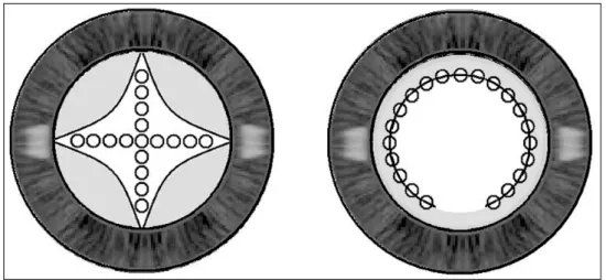
Figure 1. Two different open-
ing patterns of Nd:YAG laser posterior capsulotomy.Figure 2. One month after Neodymium:YAG laser posterior
capsulotomy performed with the modified circular opening procedure. This demonstrates that the inferior portion of cir- cular opening at 6 o’clock remains attached to the posterior capsule.차성 백내장, 원발성 또는 속발성 녹내장, 연령관련황반변 성등의 망막질환, 인공수정체의 고랑고정 또는 공막고정, 수술 중 후낭이 파열된 경우 그리고 인공수정체의 위치가 중심에서 이탈된 경우 등은 대상에서 제외하였다. 모든 환 자에서 개개인의 동의를 얻었으며 본원의 임상시험윤리위 원회(institutional review board, IRB)의 승인을 받았다.
대상 안(62안, 50명)을 무작위로 두 그룹으로 나누었다 (그룹 A와 B). 그룹 배정은 컴퓨터에서 산출한 무작위리스 트를 바탕으로 random block permutation 방법을 이용하였 고 대상환자들이 자신이 속한 그룹을 모르게 하였다. 그룹 A와 B는 삼백 예 이상의 시술 경험이 있는 숙련된 동일 시 술자에 의해 십자절개법과 변형 원형절개법을 이용한 Nd:YAG 레이저 후낭절개술을 각각 시행 받았다. Nd:YAG 레이저 후낭절개술을 시행하기 위해서 Alcaine (0.5%
proparacaine hydrochloride, Alcon, USA)으로 점안 마취 후 Standard contact lens (PEYMANN 12 mm YAG lens, Ocular instruments)를 사용하였고, 이중 조준 광선을 사용 하여 비교적 정확한 조준을 가능하게 하는 Q-switched Nd:YAG laser (Quantel medical®, France)로 후낭을 절개 하였다. 그룹 A는 첫 레이저를 후낭의 12시 쪽 중간 변연부 에 조사 후 3 mm에서 5 mm의 지름을 갖는 십자형의 후낭 절개를 시행 받았다. 그룹 B 역시 후낭의 12시 쪽 중간 변 연부에 먼저 레이저를 조사한 후에 인공수정체 중심으로부 터 2-2.5 mm의 거리를 유지하면서 5시와 7시 방향으로 원형의 절개를 진행했다. 결과적으로 5시에서 7시 부분은 레이저 조사를 받지 않아서 inverted-U 모양의 후낭 절개 가 되도록 하였다(Fig. 1, 2). 평균 레이저 에너지 강도는 그룹 A는 1.91 ±0.31 mJ, 그룹 B는 1.97 ±0.24 mJ였고, 평균 레이저 조사 수는 그룹 A는 20 ±5, 그룹 B는 25 ± 5였다. 레이져를 조사할 때 후낭으로 부터 약 0.1 mm 정도 뒤쪽으로 초점을 맞춤으로서 인공수정체 손상을 방지하기 위한 노력을 기울였다.
두 방법 간에 시술 후 시기능에 변화가 있는지를 연구하 기 위하여, 최대교정시력, 대비감도, 눈부심감도를 측정하 였다. 최대교정시력은 logMAR (logarithm of the mini- mum angle of resolution)로 변환하여 표기하였다. 대비감 도는 ACV (Visual Capacity Analyzer, L2 informatique, France)을 이용하여 photopic (100 cd/m2 using Landolt rings at 1.6 cpd and 4.2 cpd) 환경에서 측정하였다. 눈부 심 측정을 위해서는 200 lux의 빛을 시축의 20˚ 바깥쪽에 위치시키고 Landolt rings을 이용하여 1.6 cpd과 4.2 cpd에 서 각각 측정하였다.
시술 당일 산동하지 않은 상태에서 최대교정시력, 대비 감도, 눈부심감도를 포함하는 시술 전 시기능을 측정하였고 시술 후 1개월에 시기능을 다시 평가하였다. Nd:YAG 레이 저 후낭절개술 후 Flarex® (0.1% fluorometholone ace- tate, Alcon)를 하루에 4번씩 시술 안에 1주일간 점안하였 다. 시술과 관련된 합병증인 망막박리, 안압상승, 낭포성 황 반부종의 발생을 확인하기 위해서 시술 후 3개월과, 6개월
Table 1. Patient characteristics
Characteristics Group A
(cruciate opening)
Group B
(modified circular opening) p-value*
No. of eye 31 31
DM (n) 8 9
Age (yr) 62.2 ± 6.8 64.0 ± 4.2 0.092
Sex (M/F) 13/7 12/8 0.187
Intervals from cataract operation to laser capsuloctomy (mon) 12.3 ± 6.1 11.4 ± 7.0 0.458
Mean laser power (mJ) 1.91 ± 0.31 1.97 ± 0.24 0.102
Mean number of laser applications 20 ± 5 25 ± 5 0.261
Values are presented as number or mean ± SD.
DM = diabetes mellitus.
*Statistical significances between group A and B were tested by Mann-Whitney U-test.
Table 2. Changes in mean (± SD) IOP, floater, anterior chamber reactions and IOL pittings after Nd:Yag laser capsulotomy at post-
laser 1 monthCharacteristics
Group A (cruciate opening)
Group B
(modified circular opening) p-value
Prelaser (a) Postlaser (b) Prelaser (c) Postlaser (d) a-b* c-d* a-c† b-d†
IOP (mm Hg) 12.8 ± 4.8 13.3 ± 5.1 13.4 ± 2.9 13.9 ± 3.2 0.431 0.198 0.131 0.125
The presence of free floating capsular segments (number of patients)
0 3 0 0 0.052
Anterior chamber reactions (grade 0 to 4) 0 0.56 0 0.81 0.133
IOL pittings (three or more IOL pittings within 1.5 mm radius from IOL center)
0 4 0 0 0.047
SD = standard deviation; IOP = intraocular pressure; IOL = intraocular lens.
*Statistical significances between group A and B were tested by Paired t-test; †Statistical significances between group A and B were tested by Mann-Whitney U-test.
에 경과관찰을 하였다. Goldman applanation tonometer를 사용하여 안압을 측정하였고 세극등 현미경 검사를 통해 전방세포반응을 측정하였다. 인공수정체 패임은 시술 후 1 달째 측정하였는데, 인공수정체 중심부에서 1.5 mm 거리 에 3개 이상의 저명한 패임이 있는 경우 패임이 발생한 눈 으로 분류하였다. 시술 후 1주째 그룹 A의 한 명의 환자와 그룹 B의 두 명의 환자가 불완전한 후낭절개 때문에 Nd:YAG 레이저 후낭절개술을 다시 시행 받았다.
Nd:YAG 레이저 후낭절개술 후 최대교정시력, 대비감도, 눈부심감도 등을 Mann-Whitney Utest와 Paired-t test 로 비교하였다. p값이 0.05 미만일 때 그 값을 통계적으로 유의하다고 보았다.
결 과
Nd:YAG 레이저 후낭절개술을 시행할 당시 A와 그룹 B 의 평균 나이는 각각 62.2 ±6.8세와 64.0 ±4.2세였다. 백 내장 수술을 하고 난 후 Nd:YAG 레이저 후낭절개술을 시 행하기까지의 기간은 각각 12.3 ±6.1개월과 11.4 ±7.0개 월 이었고, 평균 수술 전 안압은 각각 12.8 ± 4.8 mmHg 과 13.4 ±2.9 mmHg이었다. 두 그룹 간에서 나이와 성별,
안압 및 백내장 수술과 Nd:YAG 레이저 후낭 절개술의 시 행 받기까지의 기간에 있어서 통계적으로 유의한 차이는 없었다(p=0.092, 0.187, 0.131 and 0.458, respectively) (Table 1). 두 그룹에서 시술 후 망막 박리, 안압 상승, 낭 포성 황반부종과 같은 합병증이 발생한 환자는 없었다.
Nd:YAG 레이저 후낭절개술 후에 안압, 자유롭게 부유하 는 후낭 절편의 유무, 전방 세포 반응에도 두 그룹 간의 유 의한 차이는 발견되지 않았다(p=0.125, 0.052 and 0.133, respectively). 그러나 인공 수정체 중심부로부터 1.5 mm 이내에 3개 이상의 패임이 발생한 빈도는 두 그룹 간에서 유의한 차이를 보였다(p=0.047)(Table 2).
Nd:YAG 레이저 후낭절개술 후 최대교정시력은 두 그룹 모두에서 유의하게 증가하였다(p=0.027 in group A and 0.033 in group B). 대비감도(p=0.039 in 1.6 cpd and 0.018 in 4.2 cpd in group A, 0.023 in 1.6 cpd and 0.021 in 4.2 cpd in group B)와 눈부심감도(p=0.031 in 1.6 cpd and 0.033 in 4.2 cpd in group A, 0.016 in 1.6 cpd and 0.049 in 4.2 cpd in group B) 역시 각각의 그룹에서 Nd:YAG 레이저 후낭절개술 후 유의하게 증가하였다.
Nd:YAG 레이저 후낭절개술 후 두 그룹 간의 대비감도와 눈부심감도의 유의한 차이는 없었다(Table 3).
Table 3. Changes in mean (± SD) BCVA, contrast sensitivity and glare sensitivity after Nd:Yag laser capsulotomy at postlaser 1 month
CharacteristicsGroup A (cruciate opening)
Group B
(modified circular opening) p-value
Prelaser (a) Postlaser (b) Prelaser (c) Postlaser (d) a-b* c-d* a-c† b-d† BCVA (log MAR) 0.632 ± 0.221 0.091 ± 0.021 0.571 ± 0.394 0.055 ± 0.025 0.027 0.033 0.191 0.281 Contrast threshold (degrees)
1.6 cpd 0.049 ± 0.86 0.031 ± 0.72 0.036 ± 0.13 0.019 ± 0.72 0.039 0.023 0.342 0.061
4.2 cpd 0.029 ± 0.12 0.019 ± 0.66 0.023 ± 0.31 0.012 ± 0.66 0.018 0.021 0.136 0.095
Glare threshold (dregrees)
1.6 cpd 0.063 ± 0.20 0.049 ± 0.52 0.041 ± 0.56 0.023 ± 0.29 0.031 0.016 0.277 0.069
4.2 cpd 0.030 ± 0.25 0.023 ± 0.31 0.032 ± 0.13 0.016 ± 0.61 0.033 0.049 0.209 0.071
SD = standard deviation; BCVA = best corrected visual acuity; CPD = cycle per degree.
*Statistical significances between group A and B were tested by Paired t-test; †Statistical significances between group A and B were tested by Mann-Whitney U-test.
Table 4. Changes in group A mean (± SD) BCVA, contrast sensitivity and glare sensitivity after Nd:Yag laser capsulotomy at post-
laser 1 month in group ACharacteristics Patients with IOL pittings* Patients without IOL pittings† p-value
Prelaser (a) Postlaser (b) Prelaser (c) Postlaser (d) a-b‡ c-d‡ a-c§ b-d§ BCVA (log MAR) 0.609 ± 0.126 0.112 ± 0.021 0.582 ± 0.394 0.053 ± 0.025 0.039 0.045 0.312 0.263 Contrast threshold (degrees)
1.6 cpd 0.043 ± 0.86 0.035 ± 0.72 0.032 ± 0.13 0.017 ± 0.72 0.025 0.009 0.362 0.023
4.2 cpd 0.025 ± 0.12 0.022 ± 0.66 0.024 ± 0.31 0.009 ± 0.66 0.031 0.039 0.175 0.029
Glare threshold (dregrees)
1.6 cpd 0.059 ± 0.20 0.052 ± 0.52 0.044 ± 0.56 0.021 ± 0.29 0.041 0.019 0.099 0.043
4.2 cpd 0.031 ± 0.25 0.025 ± 0.31 0.030 ± 0.13 0.015 ± 0.61 0.027 0.012 0.442 0.031
SD = standard deviation; BCVA = best corrected visual acuity; CPD = cycle per degree; IOL = intraocular lens.
*Three or more IOL pittings within 1.5-mm radius from IOL center; †Two or less IOL pittings within 1.5-mm radius from IOL center.
‡Statistical significances between group A and B were tested by Paired t-test; §Statistical significances between group A and B were tested by Mann-Whitney U-test.
그룹 A에서 Nd:YAG 레이저 후낭절개술 후 인공 수정체 중심부의 1.5 mm 이내의 3개 이상의 인공수정체 패임이 발생한 환자 군은 패임이 2개 이하로 발생한 환자 군과 비 교하여 최대교정시력에서 유의한 차이를 보이지 않았다 (p=0.263). 그러나 대비감도(p=0.023 in 1.6 cpd and 0.029 in 4.2 cpd)와 눈부심 감도(p=0.043 in 1.6 cpd and 0.031 in 4.2 cpd)는 유의한 차이를 보였다(Table 4).
고 찰
백내장 수술 후 수정체낭의 적도 부위에 남아 있는 수정 체상피세포가 후낭쪽으로 성장하면서 후발백내장이 발생하 게 된다.7후발백내장은 수정체 초음파 유화술의 장기적인 합병증으로서8-10만성적으로 수술 후 시력저하를 야기하는 주요 원인으로 알려져 있다.10-12후발백내장 발생을 예방하 기 위해서 새로운 약물이나 인공수정체 디자인, 수술 기법
등이13,14연구 중에 있음에도 불구하고 여전히 수정체 초음
파 유화술 후 10-50% 정도에서 발생되고 있다.8-10저명 하게 후발백내장이 있는 환자 군에서 Nd:YAG 레이저 후낭
절개술 후 시력 향상에 대한 많은 보고가 있었고,15-18대비 감도와 눈부심감도의 개선도 이전 연구에서 확인된 바 있
다.3,4,19Nd:YAG 레이저 후낭 절개술은 낭포성 황반 부종이
나 수술 후 안압상승, 인공수정체의 패임 같은 합병증을 야기할 수 있다.8,20-23또한 전방 유리체 표면의 손상과 후 유리체 박리가 발생할 경우 망막박리의 위험을 증가시킬 수 있는 것으로 알려져 있다.24 따라서 합병증을 최소화하 고 효율을 극대화 시키기 위한 후낭 절개술의 다양한 기법 이 고안되어 왔다. 십자절개법은 가장 많이 사용되는 기법 이다.6,25 Aslam and Dhillon26은 작은 절개(최소 1.5 mm의 지름)를 시행하여도 개구부가 깨끗하면 좋은 시력을 보장 할 수 있다고 하였고, 절개술의 지름과 시기능 개선과의 관 계는 유의하지 않다고 주장하였다. 그러나 실제 임상적으로 는 십자절개법을 통한 Nd:YAG 후낭절개술을 시행 받은 후 인공수정체 패임이나 동공에 노출된 절개면 때문에 주관적 인 시기능 감소를 호소하는 환자들을 접할 수 있었다. 따라 서 저자들은 십자절개법을 시행한 후 발생되는 여러 문제점 들을 극복하기 위해서 변형 원형절개법을 이용한 Nd:YAG 후낭절개술을 시행해 보았다. 변형 원형절개법은 십자절개
법과 비교하여 인공수정체 중심으로부터 3 mm 이내에는 직접적인 레이저 조사를 하지 않기 때문에 인공수정체 중 심부 패임의 위험도를 낮출 수 있고, 절개 경계부가 동공에 노출되는 위험도도 감소시킬 수 있을 것으로 기대되었다.
또한 당뇨망막병증과 주변부 망막변성 환자에서 형광안저 촬영과 안저사진을 검사할 때 남아있는 후낭으로부터 간섭 을 최소화하여 더 정확한 검사를 할 수 있는 장점도 가질 것으로 생각되었다. 고전적인 원형절개법과 비교해서는 사 용되는 총 레이저 에너지량이 적기 때문에 레이저 에너지 와 관련되는 합병증의 빈도를 낮출 수 있고 절개된 후낭 절 편에 의한 부유물증상도 적게 야기할 것으로 생각되었다.
본 연구에서 두 가지의 절개방법과 안압, 자유롭게 부유 하는 후낭 절편의 유무 그리고 전방세포반응 간의 유의한 상관성은 발견되지 않았다. 망막박리, 안압 상승, 낭포성 황 반부종과 같은 합병증은 두 그룹 모두에서 발생하지 않았 다. 또한 두 그룹 간에서 평균 레이저의 세기와 조사 수에 서도 유의한 차이는 없었다. 그룹 A에서 Nd:YAG 후낭절개 술 후 인공수정체 중심부에서 1.5 mm 이내에 패임의 발생 할 위험도가 그룹 B보다 유의하게 큰 것을 발견하였다. 그 룹 A에서 인공수정체 중심부로부터 1.5 mm 이내에 3개 이 상의 패임을 갖는 환자들이 패임이 2개 이하인 환자들과 비 교해서 유의하게 더 나쁜 대비감도와 눈부심 감도를 보였 다. 이것은 변형 원형절개법이 인공수정체 중심 1.5 mm 안 에 패임이 발생하지 않았기 때문에 십자절개법 보다 더 나은 대비감도와 눈부심감도를 얻을 수 있었을 것으로 생각된다.
하지만 두 방법 간에 통계적으로 유의한 차이는 없었다.
결론적으로 변형 원형절개법을 이용한 Nd:YAG 레이저 후낭절개술은 인공수정체 중심부의 패임을 줄이는 데 효율 적인 방법으로 생각된다.
참고문헌
1) Dangel ME, Kirkham SM, Phipps MJ. Posterior capsule opacifica- tion in extracapsular cataract extraction and the triple procedure: a comparative study. Ophthalmic Surg 1994;25:82-7.
2) Apple DJ, Mamalis N, Loftfield K, et al. Complications of intra- ocular lenses. A historical and histopathological review. Surv Ophthalmol 1984;29:1-54.
3) Tan JC, Spalton DJ, Arden GB. The effect of neodymium: YAG capsulotomy on contrast sensitivity and the evaluation of methods for its assessment. Ophthalmology 1999;106:703-9.
4) Magno BV, Datiles MB, Lasa MS, et al. Evaluation of visual func- tion following neodymium:YAG laser posterior capsulotomy.
Ophthalmology 1997;104:1287-93.
5) Weiblinger RP. Review of the clinical literature on the use of the Nd:YAG laser for posterior capsulotomy. J Cataract Refract Surg 1986;12:162-70.
6) Goble RR, O'Brart DP, Lohmann CP, Fitzke F, Marshall J. The role
of light scatter in the degradation of visual performance before and after Nd:YAG capsulotomy. Eye (Lond) 1994;8 ( Pt 5):530-4.
7) Georgopoulos M, Findl O, Menapace R, et al, Influence of intra- ocular lens material on regeneratory posterior capsule opacifica- tion after neodymium:YAG laser capsulotomy. J Cataract Refract Surg 2003;29:1560-5.
8) Schaumberg DA, Dana MR, Christen WG, Glynn RJ. A systematic overview of the incidence of posterior capsule opacification.
Ophthalmology 1998;105:1213-21.
9) Sterling S, Wood TO. Effect of intraocular lens convexity on poste- rior capsule opacification. J Cataract Refract Surg 1986;12:655-7.
10) Nishi O. Incidence of posterior capsule opacification in eyes with and without posterior chamber intraocular lenses. J Cataract Refract Surg 1986;12:519-22.
11) Frezzotti R, Caporossi A. Pathogenesis of posterior capsular opacification. Part I. Epidemiological and clinico-statistical data. J Cataract Refract Surg 1990;16:347-52.
12) Moisseiev J, Bartov E, Schochat A, Blumenthal M. Long-term study of the prevalence of capsular opacification following ex- tracapsular cataract extraction. J Cataract Refract Surg 1989;15:
531-3.
13) Emery J. Capsular opacification after cataract surgery and capsule.
Curr Opin Ophthalmol 1998;9:60-5.
14) Apple DJ, Solomon KD, Tetz MR, et al. Posterior capsule opacification. Surv Ophthalmol 1992;37:73-116.
15) Chambless WS. Neodymium:YAG laser posterior capsulotomy re- sults and complications. J Am Intraocul Implant Soc 1985;11:31-2.
16) Gardner KM, Straatsma BR, Pettit TH. Neodymium: YAG laser posterior capsulotomy: the first 100 cases at UCLA. Ophthalmic Surg 1985;16:24-8.
17) Stark WJ, Worthen D, Holladay JT, Murray G. Neodymium: YAG lasers. An FDA report. Ophthalmology 1985;92:209-12.
18) Wasserman EL, Axt JC, Sheets JH. Neodymium: YAG laser poste- rior capsulotomy. J Am Intraocul Implant Soc 1985;11:245-8.
19) Sunderraj P, Villada JR, Joyce PW, Watson A. Glare testing in pseudophakes with posterior capsule opacification. Eye (Lond) 1992;6 ( Pt 4):411-3.
20) Bath PE, Fankhauser F. Long-term results of Nd:YAG laser poste- rior capsulotomy with the Swiss laser. J Cataract Refract Surg 1986;12:150-3.
21) Krauss JM, Puliafito CA, Miglior S, et al. Vitreous changes after neodymium-YAG laser photodisruption. Arch Ophthalmol 1986;
104:592-7.
22) Newland TJ, McDermott ML, Eliott D, et al. Experimental neo- dymium:YAG laser damage to acrylic, poly(methyl methacrylate), and silicone intraocular lens materials. J Cataract Refract Surg 1999;25:72-6.
23) Ranta P, Tommila P, Immonen I, et al. Retinal breaks before and af- ter neodymium:YAG posterior capsulotomy. J Cataract Refract Surg 2000;26:1190-7.
24) Sheard RM, Goodburn SF, Comer MB, et al. Posterior vitreous de- tachment after neodymium:YAG laser posterior capsulotomy. J Cataract Refract Surg 2003;29:930-4.
25) Hu CY, Woung LC, Wang MC. Change in the area of laser posterior cap- sulotomy: 3 month follow-up. J Cataract Refract Surg 2001;27: 537-42.
26) Aslam TM, Dhillon B. Neodymium:YAG laser capsulotomy: a clinical morphological analysis. Graefes Arch Clin Exp Ophthalmol 2002;240:972-6.
=ABSTRACT=
The Effect of Two Different Opening Patterns of Neodymium:YAG Laser Posterior Capsulotomy on Visual Function
Youngkook Kim, MD, Jongseok Park, MD, PhD
Department of Ophthalmology, Eulji General Hospital, Eulji University School of Medicine, Seoul, Korea
Purpose: To investigate the effects of two different opening patterns in neodymium:yttrium-aluminum-garnet (Nd:YAG) la- ser posterior capsulotomy on visual function.
Methods: A randomized prospective study was conducted on 62 pseudophakic eyes from 50 patients with posterior capsu- lar opacification (PCO). Groups A and B underwent laser posterior capsulotomy with a cruciate opening and a modified cir- cular opening, respectively. The best corrected visual acuity (BCVA), contrast sensitivity, and glare sensitivity were meas- ured before and after laser posterior capsulotomy in both groups. The occurrences of intraocular lens (IOL) pittings were investigated and compared between the two groups.
Results: After laser capsulotomy, there were significant increases in BCVA, contrast and glare sensitivity in both groups and no significant differences between the two groups. However, group A had significantly more patients with three or more IOL pittings within a 1.5-mm radius from the IOL center (p = 0.047). In group A, these patients had significantly less contrast sensitivity (p = 0.023 in 1.6 cpd) and glare sensitivity (p = 0.043 in 1.6 cpd) than did patients with two or fewer IOL pittings.
Conclusions: Nd:YAG laser posterior capsulotomy using a modified circular opening can be considered an effective meth- od to manage PCO.
J Korean Ophthalmol Soc 2012;53(3):390-395
Key Words: Contrast sensitivity, Glare sensitivity, Neodymium:YAG laser posterior capsulotomy, Opening patterns
Address reprint requests to Jongseok Park, MD, PhD Department of Ophthalmology, Eulji General Hospital
#14 Hangeulbiseok-gil, Nowon-gu, Seoul 139-872, Korea
Tel: 82-2-970-8269, Fax: 82-2-970-8273, E-mail: [email protected]