pISSN: 0378-6471 eISSN: 2092-9374 http://dx.doi.org/10.3341/jkos.2013.54.9.1401
= 증례보고 =
PC기반 시야검사를 통한 효과적인 시선고정 유도방법 연구
이지형1⋅이백희1⋅박현지1⋅최정한3⋅유희천1,2⋅강자헌3
포항공과대학교 산업경영공학과1, 포항공과대학교 창의IT융합공학과2, 경희대학교 의학전문대학원 강동경희대학교병원3
목적: 본 연구는 시야 검사 시 피검사자의 시선고정을 시선고정 시표에 효과적으로 유도하기 위한 방법들을 제안하고 비교 평가하였다.
대상과 방법: 본 연구는 두 가지 시선고정용 시표 형태(dot, number)와 소리 신호 제공 여부(on, off)를 조합한 네 가지 시선고정 유도방 법(dot-on, dot-off, number-on, number-off)을 비교 평가하였다. 이들 시선고정 유도방법들은 PC기반 시야검사기를 사용하여 적용 되었으며, 정상인 14명을 대상으로 24-2 threshold 시야검사를 수행하였다. 본 연구는 시선고정 유도방법의 유효성은 시선고정오류율, 시표확인율, 그리고 주관적 만족도(7점 척도) 측면에서 분석되었다.
결과: 시선고정오류율은 dot-off조건(5.7%)과 number-on조건(6.4%)에서 상대적으로 낮게 나타났고, 시표확인율은 모든 조건에서 유 의한 차이 없이 높았으며(95-96%), 주관적 만족도는 dot-on조건(5.7점)과 number-on조건(5.4점)에서 다른 조건에 비해 유의하게 높 은 것으로 파악되었다.
결론: 본 연구에서는 기존 시야검사기에서 사용되는 dot-off조건에 비해 number-on방법이 시선고정오류율 및 주관적 만족도 측면을 함께 고려할 경우 효과적인 시선고정 유도방법으로 파악되었다.
<대한안과학회지 2013;54(9):1401-1406>
■Received: 2012. 10. 26. ■ Revised: 2013. 5. 22.
■Accepted: 2013. 8. 10.
■Address reprint requests to Ja Heon Kang, MD, PhD
Department of Ophthalmology, Kyung Hee University Hospital at Gangdong, #892 Dongnam-ro, Gangdong-gu, Seoul 134-727, Korea
Tel: 82-2-440-7760, Fax: 82-2-440-7756 E-mail: [email protected]
녹내장진단을 위한 기존 시야검사기(perimeter)는 크고 고가로 인하여 주로 전문의료기관에서 운용되고 있어 시야 검사에 대한 피검사자들의 접근성이 낮다. 예를 들어, Humphrey Field Analyzer II-750i (HFA; Carl Zeiss Meditec, USA)는 크기가 600×580×510 mm3이고, 무게 가 40 kg으로 인하여 충분한 설치공간이 필요하고 한 번 설 치되면 이동시키기가 쉽지 않다.1또한 국내의 경우 시야검 사기의 가격이 상당히 높아 종합병원 또는 안과전문병원 외에는 운용이 어려운 실정이다.1 따라서 소규모병원이나 개인안과에서 운용할 수 있는 적정 시야검사기 개발은 피 검사자의 시야검사 접근성을 향상시켜 녹내장 조기 진단에 효과적일 것이다.
시야검사 시 피검사자의 시선은 시야측정영역 중심에 위 치한 시선고정용 시표(fixation target)로 고정이 요구되는 데, 주변에 시야측정용 시표가 제시될 때 시선 고정이 부적
절하게 통제될 수 있어, 정확한 시야검사를 위해서는 시선고 정을 효과적으로 통제하는 방법이 필요하다. 기존 시야검사 기들(예: Humphrey Field Analyzer II-750i, Humphrey® matrix™, Octopus® 900 등)은 시선고정용 시표로 의미도 없고 변화도 없는 점(dot)을 사용하고 있어 시야검사 중 피 검사자가 시선을 지속적으로 고정하는데 한계가 있다.2-4 한편 기존 시야검사기는 시야검사의 신뢰성을 파악하기 위 하여 맹점(blind spot)에 시표 제시를 통한 시선고정오류율 (fixation error rate)측정이나 동공의 움직임을 추적하여 시선이탈을 모니터링하고 있다.5,6 그러나 정확하면서도 효 율적인 시야검사를 위해서는 맹점 시표확인 검사 및 동공 움직임 추적과 같은 사후분석과 더불어 시야검사 중 피검 사자 시선을 시선고정용 시표로 능동적으로 집중시키는 방 법의 개발이 필요하다.
본 연구는 피검사자의 시선을 시선고정용 시표에 능동적 으로유도하기 위한 방법을 개발하고 평가하였다. 시야검사 는 PC기반으로 개발된 시야검사기를 사용하여 정상인을 대 상으로 진행되었다. 본 연구는 두 가지 시표 형태와 소리 신 호제공 여부를 조합하여 고안된 네 가지 시선고정 유도방법 들(dot-on, dot-off, number-on, 그리고 number-off)에 대하여 시선고정오류율, 시표확인율, 그리고 주관적 만족도 측면에서 비교 분석하였다.
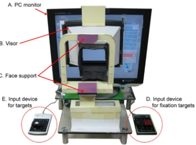
Figure 1. A prototype of PC-based visual field tester. (A) PC
monitor (19 inch, Flatron L1940 plus, LG Electronics, Korea;pixel pitch = 0.294 mm) for presenting targets for testing a visual field. (B) visor for shielding outside lights, helping con- centration during test, selecting an eye to be tested and main- taining the distance between the eye and the monitor. (C) face support for maintaining the position of the eye and supporting comfortable posture. (D) input device for fixation target (1 or 2) confirmation. (E) input device for target detection.
Table 1. Distribution of age and gender of participants in visual field testing
Gender 20s 30s 40s 50s 60s Total
Male 1 1 1 2 3 8
Female 1 1 1 3 - 6
Total (%) 2 (14.3) 2 (14.3) 2 (14.3) 5 (35.7) 3 (21.4) 14 (100)
Figure 2. The target locations of the visual field testing S/W
for the right eye. The visual field forms 24˚ superiorly, in- feriorly, temporally, and 30˚ nasally. The location of a blind spot was 1° superiorly and 16° temporally from the fixation target positioned at the center of a visual field area. The total number of target locations is 56 (1 for the fixation target, 54 for visual field targets, and 1 for blind spot).대상과 방법
시야검사는 교정시력이 1.0 이상의 시야장애가 없는 14 명(남성: 50.9 ±12.6세; 여성: 44.3 ± 12.8세)의 정상인 을 대상으로 수행되었다. 실험참여자는 강동경희대학교병 원에서 2008년부터 2010년까지 녹내장 피검사자 104명의 연령분포를 고려하여 Table 1과 같이 50대와 60대가 전체 의 50% 이상이 되도록 모집되었다.
본 연구의 시야검사는 Fig. 1과 같은 검사의 접근성 향상 을 위하여 개발된 PC기반 시야검사기를 사용하여 수행되었 다. PC기반 시야검사기는 PC monitor, 탈부착식 차광장치, 안면 지지장치, 시선고정용 시표확인 입력장치, 그리고 시 야측정용 시표 확인 입력장치로 구성되었다. 19 inch PC monitor (Flatron L1940 Plus, LG Electronics)는 시야측 정영역에 시선고정용 시표와 시야측정용 시표를 제시하는 데 사용되었으며(Fig. 1A), 탈부착식 차광장치는 외부 빛 을 차단하고 피검사자가 시야검사에 집중할 수 있는 환경 을 조성하기 위해 PC monitor에 부착되었다(Fig. 1B). 또 한 안면 지지장치는 피검사자의 이마와 턱을 고정시키기
위하여 사용되었고(Fig. 1C), 시선고정용 시표 확인 입력장 치는 실험참여자가 ‘1’ 또는 ‘2’로 제시되는 시선고정용 시 표를 입력하기 위해 사용되었다(Fig. 1D). 마지막으로 시 야측정용 시표 확인 입력장치는 실험참여자가 시표 확인 여부를 입력하는데 사용되었다(Fig. 1E).
본 연구의 시야검사방법으로는 망막의 중심와(fovea)를 중심으로 시야각 24°내의 중심시야(central vision)를 측정 하는 HFA의 24-2 threshold test가 적용되었다(Fig. 2).
본 연구에서 MS Visual C# 2010로 개발된 시야검사 S/W 는 Fig. 2와 같이 24° 영역내의 56개(시선고정용 시표 1개, 맹점시표 1개, 시야측정용 시표 54개) 위치에 시표를 제시 하도록 개발되었다.7-12본 연구의 시표 제시 횟수는 시선고 정용 시표와 시야검사용 시표 위치에 각 1회 그리고, 맹점 시표위치에 5회로 총 60회였다. 시표 제시 간격은 50대 미 만의 경우 1.5초(총90초) 그리고 50대 이상의 경우 2초(총 120초)로 설정되었다.
본 연구는 시선고정용 시표에 제시되는 두 가지 시표 형 태(dot, number)와 시표가 제시될 때의 소리 신호 제공 여 부(on, off)를 조합하여 dot-on, dot-off, number-on, 그 리고 number-off의 네 가지 시선고정 유도방법을 설정하 였다. 시선고정용 시표 형태로는 기존 시야검사기에서 사용 되고 있는 dot형태와 시선고정 유도를 위한 number형태(1
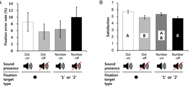
Figure 4. Results of visual field testing performance and subjective satisfaction. Dot-on condition: central target =
dot and sound = on; dot-off condition: central target = dot and sound = off; number-on condition: central target = 1 or 2 and sound = on; number-off condition: central target = 1 or 2 and sound = off. (A) Fixation error rate (mean± SE). (B) Subjective satisfaction by using 7-point scale (mean ± SE).
Figure 3. Experimental procedure. Four-step visual field test-
ing is conducted: S1 = Introduction; S2 = Identification of the testing distance, alignment between the eye and the fix- ation target on the screen, and fixation of the face on the face support; S3 = Visual field testing randomly by 4 conditions (dot-on, dot-off, number-on, and number-off), 1 minute break among each condition; S4 = Evaluation of subjective sat- isfaction (7-point scale).또는 2)가 사용되었으며, 소리 신호로는 시표가 제시됨과 동시에 ‘딩동’ 소리가 사용되었다. 예를 들면, number-on 조건은 시표가 제시될 때마다 시선고정용 시표로 ‘1’ 또는
‘2’가 제시되는 동시에 ‘딩동’하는 소리를 들려주는 방법이다.
시야검사는 Fig. 3과 같이 네단계 절차(S1: 실험 준비, S2: 시야검사 준비, S3: 시야검사, S4: 주관적 만족도평가) 로 수행되었다. 실험준비단계에서는 실험참여자에게 실험 에 대한 충분한 설명을 제공하고 실험동의서를 받았다. 시 야검사 준비단계에서는 실험참여자의 검사안(좌 또는 우) 을 선택한 후맹점시표가 보이지 않는 시야검사 거리를 파 악하였다. 시야검사단계에서는 설정된 시야검사 거리에서
무작위로 선정된 시선고정 유도방법에 대하여 시야검사가 수행되었다. 마지막으로, 주관적 만족도 평가단계에서는 시 야검사에 대한 만족도를 7점척도(매우 불만족: 1점, 불만 족: 2점, 약간 불만족: 3점, 보통: 4점, 약간 만족: 5점, 만 족: 6점, 매우 만족: 7점)로 평가하였다. 본 시야검사 실험 에서는 실험참여자에게 시선고정 유도방법 변경 시 1분의 휴식 시간과 검사안 변경 시 5분의 휴식을 제공하였다. 본 연구에서는 각 실험조건에서 총5회 제시되는 맹점시표에 대해 3회 이상 확인한 경우에는 부적합한 실험 수행으로 판 단하여 재실험을 수행하였다.13
본 연구는 시선고정 유도방법이 시선고정오류율, 시표확 인율, 그리고 주관적 만족도에 미치는 효과를 파악하기 위 하여 유의수준 0.05에서 ANOVA를 수행하였다. 통계적으 로 유의한 인자의 사후검정에는 다중 평균 비교를 위한 Fisher’s method가 사용되었다. 통계분석에는 Minitab (ver. 14; Minitab Inc., State Collage, Pa., USA)이 사용 되었다.
결 과
본 연구에서 제안한 네 가지 시선고정 유도방법들은 시 선고정오류율 측면에서 통계적으로 유의한 차이가 없는 것 으로 나타났으나, dot-off 조건과 number-on 조건에서 오 류율이 상대적으로 낮은 것으로 파악되었다(Fig. 4A). 평 균 시선고정 오류율은 dot-off 조건(5.7 ±2.3%)과 num- ber-on조건(6.4 ±2.5%)에서 유사하였으며, number-off
A B
Dot -off
Dot -on Number
-on Number
-off Dot
Dot -off
-on Number
-on Number -off
A B
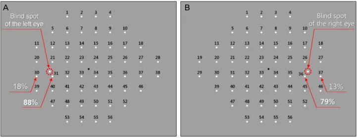
Figure 5. The rates of missing targets near the blind spots at the left eye (A) and right eye (B).
조건(10.0 ±3.0%)과 dot-on 조건(8.6 ±2.8%)에 비해 약 1.3-1.8배 낮으나 이들 조건들 간의 차이는 통계적으로 유의하지 않았다.
또한 네 가지 시선고정 유도방법들은 통계적으로 유의한 차 이가 없이 모두 평균 95-96%의 높은 시표확인율을 보이는 것으로 나타났다. 각 유도방법들에서 시표확인율은 dot-on 조건의 경우 95.9 ± 4.7%, dot-off 조건의 경우 95.3 ± 5.1%, number-on 조건의 경우 94.9 ±4.5%, number-off 조건의 경우 95.3 ± 4.3%로 파악되었다.
마지막으로 주관적 만족도 측면에서는 소리 신호를 제공 하는 조건들(dot-on, number-on)이 제공하지 않는 조건 들(dot-off, number-off)에 비해 통계적으로 유의하게 선 호되는 것으로 파악되었다(Fig. 4B). Dot-on 조건(5.7 ± 0.2점)과 number-on 조건(5.4 ± 0.2점)의 평균 주관적 만족도는 dot-off 조건(4.9 ±0.3점)과 number-off 조건 (4.8 ±0.2점) 보다 1.2배 정도 유의하게 높은 것으로 나타 났다(F[1, 108]=8.57, p=0.004)
고 찰
본 연구는 보다 정확하고 효율적인 시야검사를 위하여 시야검사 시 피검사자의 시선을 능동적으로 시선고정용 시 표에 고정하도록 유도하는 방법을 모색하였다. 기존 연구들 은 주로 시야검사의 효율성 향상을 위한 시표 제시 algo- rithm연구들과14-16 정확한 시야평가를 위한 진단 모형 개 발 연구들을17-19수행해 왔으나, 정확한 시야검사를 위하여 피검사자의 시선을 시선고정용 시표로 고정시키는 방법에 대한 연구는 거의 없었다. 본 연구는 피검사자의 주의를 집 중시키기 위하여 숫자라는 의미있는 정보를 시선고정용 시 표로 사용하였다. 본 연구는 시야검사 결과의 정확성 향상
을 위하여 시선고정용 시표로부터의 시선 이탈을 효과적으 로 방지할 수 있는 방법을 탐색한 연구이다.
시선고정오류율 이상대적으로 가장 낮은 방법으로는 dot-off 조건과 number-on 조건, 선호도가 가장 높은 방 법으로는 dot-on 조건과 number-on 조건이 선정되었는 데, 이들 두 가지 측면을 함께 고려할 경우 number-on 조건 이 최적인 것으로 파악되었다. 피검사자의 시선은 dot-off 조건(시선고정오류율=5.7%)과 number-on조건(6.4%)이 다른 조건에 비해 유사하게 시선고정오류율이 낮은 것으로 파악되었다. 주관적 만족도는 dot-on 조건(5.7점)과 number –on조건(5.4점)이 dot-off 조건(4.9점)과 number-off 조 건(4.8점)보다 상대적으로 높은 것으로 나타났다. 만족도 에 대한 사후 설문에 의하면 dot-on조건과 number-on조 건의 규칙적인 ‘딩동’소리 신호가 피검사자를 각성시키고 다음 시표에 대비할 수 있게 해주어 시야검사에 도움을 주 는 것으로 파악되었다. 이러한 피검사자의 설명은 알림음과 같은 청각 신호(auditory signal)가 인간의 주의 집중을 유 도하는데 효과가 있다는 연구20에 의하여 뒷받침될 수 있다.
시야검사 결과에 대한 사후분석 결과, 정상인이 맹점 주 변으로부터 3° 이내에 제시되는 시표에 대하여 좌우안 각 각 12%와 21% 정도만 확인할 수 있는 것으로 파악되었다 (Fig. 5). 이러한 낮은 시표확인률은 맹점과 3° 이내의 시 표가 반경이 7°인 맹점 영역에 포함되기 때문으로 추정된 다.21 기존 시야검사기를 조사한 결과, 맹점 주변으로부터 3° 이내에 제시되는 시표의 경우 검사는 수행되나 mean deviation (MD)과 같은 global index 계산 시에는 제외되 는 것으로 파악되었다. 따라서 시야검사의 정확성 및 효율 성 향상을 위하여 맹점에 근접한 시표들을 시야검사 시 사 용하지 않는 것이 추천된다.
본 연구는 선호되는 시선고정 유도방법을 시야검사의 정
확성과 만족도 측면에서 평가하였는데, 추후 시선고정 유도 방법을 시야검사 결과에 대하여 신뢰성 측면에서의 평가와 시표 제시 간격이 시선고정 유도 효과에 미치는 영향을 분 석하는 연구가 필요하다. 최적의 시선고정 유도방법은 본 연구에서 고려한 시선고정오류율과 더불어 false-positive error나 false-negative error와 같은 reliability index22를 분석하여 결정될 수 있다. 한편 본 연구에서 수행된 시야검 사시에는 사전 연구를 통해 파악된 연령 별로 서로 다른 시 표제시 간격(50대 미만: 1.5초; 50대 이상: 2초)이 적용되 었는데, 이와 같이 상이한 시표 제시 간격의 적용이 시선고 정 유도방법의 효과와 혼란(confounding)이 되지 않았는지 추후 면밀히 검토될 필요가 있다.
시야검사의 접근성 향상을 위해 개발되고 있는 PC기반 시야검사기는 임상에서 사용하고 있는 기존 시야검사기를 대체할 수 있는지에 대한 검증연구가 추후 필요하다. 기존 고가의 시야검사기는 주로 종합병원 또는, 전문안과병원에 서 운용되었던 반면, PC기반 시야검사기는 PC를 사용하기 때문에 저가로 보급될 수 있어 소규모 안과기관 및 다양한 공공기관(예: 보건소, 노인복지회관 등)에서 사용될 수 있 다. 이를 위해서는 PC기반 시야검사기의 시야검사 결과와 기존 시야검사기의 시야검사 결과를 비교 분석하여 임상적 유용성에 대한 검증이 필요하다. 한편 시야 검사 시 PC 모 니터를 사용할 경우 외부 조명 조건에 의하여 유발될 수 있 는 시야 검사에 대한 영향은 탈부착식 차광장치 설치를 통 하여 해결될 수 있다.
REFERENCES
1) Lee J, Lee B, Kim Y, et al. The effect of eye fixation control method on visual field testing. In Proceedings of the 2011 Fall Conference of Ergonomics Society of Korea 2011.
2) Henson DB, Evans J, Chauhan BC, Lane C. Influence of fixation accuracy on threshold variability in patients with open angle glaucoma. Invest Ophthalmol Vis Sci 1996;37:444-50.
3) Kim CH, Lee DC. Analysis of eye movement features in Korean recognition procedure. In Proceedings of the 1995 Spring Conference of Ergonomics Society of Korea 1995.
4) Lee J, Lee B, Kim Y, et al. Evaluation of an ergonomic gaze fix- ation induction method for effective visual field testing. In Proceedings of the 2012 Fall Conference of Ergonomics Society of Korea 2012.
5) Carl Zeiss Meditec Inc. Humphrey® Field Analyzer II-i series
User Manual. 2005; 92.
6) Anderson DR. Perimetry with and without automation, 2nd ed. St.
Louis: CV Mosby, 1987; 104.
7) Choi JA, Lee NY, Park CK. Interpretation of the Humphrey Matrix 24-2 test in the diagnosis of preperimetric glaucoma. Jpn J Ophthalmol 2009;53:24-30.
8) Fan X, Wu LL, Ma ZZ, et al. Usefulness of frequency-doubling technology for perimetrically normal eyes of open-angle glaucoma patients with unilateral field loss. Ophthalmology 2010;117:
1530-7.
9) Giuffré I. Frequency doubling technology vs. standard automated perimetry in ocular hypertensive patients. Open Ophthalmol J 2009;3:6-9.
10) Kogure S, Toda Y, Tsukahara S. Prediction of future scotoma on conventional automated static perimetry using frequency doubling technology perimetry. Br J Ophthalmol 2006;90:347-52.
11) Nomoto H, Matsumoto C, Takada S, et al. Detectability of glau- comatous changes using SAP, FDT, flicker perimetry, and OCT. J Glaucoma 2009;18:165-71.
12) Brusini P. Frequency doubling technology staging system 2. J Glaucoma 2006;15:315-20.
13) Dersu I, Wiggins MN. Understanding Visual Fields, Part II;
Humphrey Visual Fields. J Ophathalmic Med Technology 2006;2:1-10.
14) Bengtsson B, Olsson J, Heijl A, Rootzén H. A new generation of algorithms for computerized threshold perimetry, SITA. Acta Ophthalmol Scand 1997;75:368-75.
15) Turpin A, McKendrick AM, Johnson CA, Vingrys AJ. Development of efficient threshold strategies for frequency doubling technology perimetry using computer simulation. Invest Ophthalmol Vis Sci 2002;43:322-31.
16) Anderson AJ. Spatial resolution of the tendency-oriented peri- metry algorithm. Invest Ophthalmol Vis Sci 2003;44:1962-8.
17) Chan K, Lee TW, Sample PA, et al. Comparison of machine learn- ing and traditional classifiers in glaucoma diagnosis. IEEE Trans Biomed Eng 2002;49:963-74.
18) Moreno-Montañés J, Antón A, García N, et al. Glaucoma proba- bility score vs Moorfields classification in normal, ocular hyper- tensive, and glaucomatous eyes. Am J Ophthalmol 2008;145:360-8.
19) Wroblewski D, Francis BA, Chopra V, et al. Glaucoma detection and evaluation through pattern recognition in standard automated perimetry data. Graefes Arch Clin Exp Ophthalmol 2009;247:1517-30.
20) Wogalter M, Leonard S. Attention capture and maintenance. In:
Wogalter M, Dejoy D, Laughery K, et al. Warnings and risk com- munication, 4th ed. London: Taylor & Francis, 2005; chap. 7.
21) Fletcher W. The big blind spot syndromes. 1996 North American Neuro-Ophthalmology Society Annual Meeting. 1996;1:59-65.
22) Fasih U, Shaikh A, Shaikh N, et al. Evaluation of reliability of visu- al field examination in glaucoma patients. Pak J Ophthalmol 2009;
25:145-51.
=ABSTRACT=
A Study for Effective Gaze Fixation Induction Methods in PC-Based Visual Field Testing
Ji Hyoung Lee1, Baek Hee Lee1, Hyun Ji Park1, Jeong Han Choi, MD3, Hee Cheon You1,2, Ja Heon Kang, MD, PhD3
Department of Industrial and Management Engineering, POSTECH1, Pohang, Korea Department of Creative IT Excellence Engineering, POSTECH2, Pohang, Korea
Department of Ophthalmology, Kyung Hee University Hospital at Gangdong, Kyung Hee University College of Medicine3, Seoul, Korea
Purpose: The present study explored novel methods in visual field tests that actively induce the gaze of the examinee to the fixation target in the center vision and compared their effectiveness.
Methods: Four gaze induction methods (dot-on, dot-off, number-on, and number-off) were prepared by combining 2 types of fixation targets (dot and number) and 2 conditions of sound presence (on and off). The gaze induction methods were im- plemented to a PC-based visual field testing system and the 24-2 visual field testing protocol was administered to 14 par- ticipants without glaucoma. The performance of the gaze induction method was evaluated in terms of fixation error rate, target detection rate, and subjective satisfaction (7-point scale, 1 for least satisfied and 7 for most satisfied).
Results: The fixation error rates of dot-on (5.7%) and number-on (6.4%) were relatively lower than the other methods; the target detection rates of the induction methods were very high (95-96%) without significant differences, and the subjective satisfaction levels of dot-on (5.7) and number-on (5.4) were significantly higher than the other methods.
Conclusions: In the present study we determined number-on as the preferred effective gaze induction method compared to the conventional dot-off method when fixation error rates and subjective satisfaction were considered.
J Korean Ophthalmol Soc 2013;54(9):1401-1406 Key Words: Gaze induction, Glaucoma, Visual field test
Address reprint requests to Ja Heon Kang, MD, PhD
Department of Ophthalmology, Kyung Hee University Hospital at Gangdong
#892 Dongnam-ro, Gangdong-gu, Seoul 134-727, Korea
Tel: 82-2-440-7760, Fax: 82-2-440-7756, E-mail: [email protected]