Journal of Bacteriology and Virology 2016. Vol. 46, No. 4 p.288 – 294 http://dx.doi.org/10.4167/jbv.2016.46.4.288
Acrosorium polyneurum Extract Inhibits the LPS-Induced Inflammatory Response by Impairing the MAPK and NF-κB Pathways
Zahid Manzoor1,2, Irshad Ali1,2, Doobyeong Chae1,2 and Young-Sang Koh1,2*
1Department of Microbiology and Immunology, School of Medicine and Brain Korea 21 PLUS Program, Jeju National University, Jeju; 2Institute of Medical Science, Jeju National University, Jeju, Korea
Marine algae exhibit broad spectrum anti-bacterial and anti-inflammatory activities. Acrosorium polyneurum (A.
polyneurum) is a marine red alga and belongs to the family Delesseriaceae. The present research evaluates the anti- inflammatory effects of A. polyneurum extract (APE) on pro-inflammatory cytokine production. APE demonstrated substantial inhibitory effects on production of pro-inflammatory cytokine in bone marrow-derived macrophages (BMDMs).
APE pre-treatment in the lipopolysaccharide (LPS)-stimulated BMDMs exhibited a robust inhibitory effect on production of interleukin (IL)-12, IL-6 and tumor necrosis factor (TNF)-α. It revealed a robust inhibitory effect on phosphorylation of ERK1/2, JNK1/2 and p38. APE also showed remarkable inhibitory effect on phosphorylation and degradation of IκBα.
Furthermore, APE pre-treatment demonstrated substantial inhibition of LPS-induced production of nitric oxide and inducible nitric oxide synthase. Collectively, these data suggest that APE has a noteworthy anti-inflammatory property and deserve further studies concerning its potential use as a medicinal agent for inflammation-related disorders.
Key Words: Acrosorium polyneurum; Mitogen-activated protein kinase; NF-κB; Nitric oxide; Pro-inflammatory cytokine
INTRODUCTION
Pattern recognition receptors (PRRs) are basically highly conserved proteins, expressed by the cells of innate immune system, and involved in recognition of pathogen-associated molecular patterns (PAMPs), which are exclusively present in microorganisms (1~3). Among various PRRs, toll-like receptors (TLR) are well studied and play critical roles in recognizing various types of PAMPs and managing proper immune response (4~6).
TLRs are the main regulator of innate host immune
response against harmful stimuli (1). For example, TLR4 expressed in macrophages recognizes lipopolysaccharide (LPS) and results in activation of different signaling cascades, such as nuclear factor-κB (NF-κB) and mitogen-activated protein kinase (MAPK) pathway, leading to pro-inflammatory cytokine production (7).
Phytochemicals have been major bioresources and ex- plored well for medicinal use (8). Now scientists are ex- ploring natural products from marine bioresources. A large number of biomaterials for good bioactivities have been documented, but much still remains to be explored for high nutritional value and pharmaceutical potential (9). Red algae
288
Received: October 13, 2016/ Revised: October 21, 2016/ Accepted: October 25, 2016
*Corresponding author: Young-Sang Koh. Department of Microbiology and Immunology, Jeju National University School of Medicine, 102 Jejudaehakno, Jeju 63243, Korea.
Phone: +82-64-754-3851, Fax: +82-64-702-2687, e-mail: [email protected]
**This research was supported by the 2016 Scientific Promotion Program funded by Jeju National University.
○CCThis is an Open Access article distributed under the terms of the Creative Commons Attribution Non-Commercial License (http://creativecommons.org/license/by-nc/3.0/).
Communication
are known as oldest multicellular plants and most diverse eukaryotes (10). Red algae are the largest species among algae, providing a good resource for medicinal food and helpful in different ways in food industry. For example, car- rageenans are sulphated polysaccharides, extracted from red algae and widely used in the food industry for gelling or stabilizing activities. Several red algae are good source of food and some are used in preparing agars (11). Extracts from marine algae have been reported to exhibit broad spectrum anti-bacterial, anti-inflammatory and antioxidant activities (12, 13). Acrosorium polyneurum (A. polyneurum), a marine red alga, belongs to the family Delesseriaceae and is distri- buted on the seashores of Korea. During ongoing research to evaluate the biological effects of various marine alga ex- tracts, in the present study, A. polyneurum extract (APE) was studied for the anti-inflammatory properties for the first time.
The effect of APE has not been studied regarding its impact on primary murine macrophages. Thus, in the present study we report for the anti-inflammatory effects of APE on LPS- stimulated bone marrow-derived macrophages (BMDMs) and RAW 264.7 cells.
MATERIALS AND METHODS
Preparation of A. polyneurum extract
Thalli of A. polyneurum were collected on Jeju Island, Korea. The material for extraction was cleaned, dried at room temperature and fine powder was made by grinding.
The dried alga was extracted as described previously (13).
The evaporated ethanol extract was suspended in water.
Mice
Six-week-old female C57BL/6 mice were purchased from Orient Bio Inc. (Seongnam, Korea) for BMDMs. All animal procedures were approved by and performed according to the guidelines of the Institutional Animal Care and Use Committee of Jeju National University, Jeju, Korea (#2010- 0028).
Cell cultures and measurement of cytokine and nitric oxide production
Bone marrow cells were differentiated in DMEM (Gibco, Grand Island, NY, USA) medium containing macrophage colony-stimulating factor for BMDMs generation as de- scribed previously (14, 15). For BMDMs, on day 6 of in- cubation, the cells were harvested and seeded in 48-well plates at a density of 1 × 10⁵ cells/0.5 ml and, then, treated with the APE for 1 hour (h) before stimulation with LPS (10 ng ml-1). Supernatants were harvested 18 h after stimulation. The concentrations of murine interleukin (IL)- 12 p40, IL-6 and tumor necrosis factor (TNF)-α in the culture supernatants were determined by enzyme-linked immuno- sorbent assay (ELISA) (BD PharMingen, San Jose, CA, USA; R&D system, MN, USA), according to the manufac- turer's instructions. Production of nitric oxide (NO) in RAW- 264.7 cells treated with APE and stimulated with LPS was measured with Griess reagent system (Promega, Madison, WI, USA) as previously described (16).
Cell viability assay
To assess cell viability, the standard procedure of 3-(4,5- dimethyl-2,5 thiazolyl)-2,5 diphenyl tetrazolium bromide (MTT) assay was used as described previously (17).
Western blot analysis
Western blot analysis was performed using standard tech- niques as previously described (18, 19). Briefly, BMDMs and RAW264.7 cells were dispensed to 60-mm culture dishes (Nunc, Roskilde, Denmark) at 4 × 10⁶ cells per dish and cultured for 24 h at 37℃. The cells were pre-treated with or without APE (25 μg ml-1) for 1 h before treatment with LPS (10 ng ml-1) at the indicated time points. The cells were collected and, then, lysed in lysis buffer (PRO-PREP lysis buffer, iNtRON Biotechnology, South Korea). A protein sample (30 μg) was subjected to electrophoresis in 10%
SDS-polyacrylamide gels and transferred to a polyvinylidene fluoride membrane (Bio-Rad, Hercules, CA, USA). The membrane was incubated with 1/1,000-diluted rabbit poly- clonal antibodies that specifically recognize phospho-p44/
42 (p-ERK1/2), p44/42 MAPK, phospho-p38, p38 MAPK and phospho-SAPK/JNK, SAPK/JNK, IκBα (Cell Signaling Technology, Danvers, MA, USA), inducible nitric oxide synthase (iNOS) and β-actin (Santa Cruz Biotechnology, Santa Cruz, CA, USA). After washing, the membrane was incubated with a horseradish peroxidase-linked goat anti- rabbit IgG (Cell Signaling Technology), and immunoactive bands were detected as previously described (19).
Statistical analysis
All experiments were executed at least 3 times. The data are shown as the mean ± the standard deviation (SD) of 3 independent experiments. One-way ANOVA (SPSS pro- gram; IBM, Armonk, NY, USA) was used for comparison between treated and control groups. p < 0.05 was considered to be statistically significant.
RESULTS AND DISCUSSION
Effects of APE on pro-inflammatory cytokine pro- duction in LPS-stimulated BMDMs
Cytokines are required to regulate host responses to inflammation and macrophages are the key producers of various cytokines (1). BMDMs express TLR4 that recognizes LPS and leads to the production of cytokines (20). There- fore, we determined the anti-inflammatory activity of APE by testing its inhibitory effects on IL-12, IL-6 and TNF-α production in LPS-stimulated BMDMs. The maximal con- centration of APE for treating cells was assessed by MTT assay, and as a result, APE had no effect on the cell viability at indicated concentrations (data not shown). LPS induced a substantial increase of pro-inflammatory cytokine production in BMDMs. APE pre-treatment profoundly inhibited pro- inflammatory cytokine production in the LPS-stimulated BMDMs (Fig. 1). These results show that APE had an in- hibitory effect on production of cytokines in LPS-stimulated BMDMs. IL-12 has various critical immunoregulatory acti- vities and is a main cytokine in Th1-mediated autoimmune responses, therefore downregulation of unregulated IL-12 production by APE may have potential to ameliorate IL-12- related autoimmune diseases (21, 22). IL-6 and TNF-α have
important physiological roles, however; dysregulated pro- duction of these cytokines has been associated with various Figure 1. Inhibitory effects of Acrosorium polyneurum Extract (APE) on cytokine production in LPS-stimulated BMDMs. (A-C) Before stimulation with LPS (10 ng ml-1), BMDMs were treated with APE at various doses as shown for 1 h and cytokines levels were assessed by ELISA. ND, not detectable; APE, A. polyneurum extract. *p < 0.05, **p < 0.01 vs. APE-untreated cells in the pre- sence of LPS.
inflammatory diseases (23, 24). Thus in future, controlling the over production of IL-6 and TNF-α by APE might be
helpful in ameliorating inflammation-associated diseases such as autoimmune and autoinflammatory diseases.
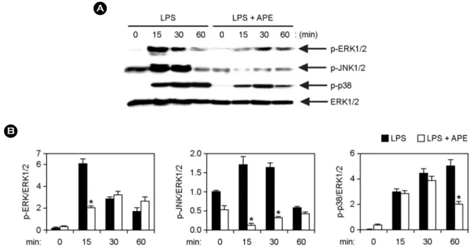
Figure 2. Inhibitory effects of APE on phosphorylation of MAPKs by LPS-stimulated BMDMs. (A) Cells were pre-treated with or without APE (25 μg ml-1) for 1 h before stimulation with LPS (10 ng ml-1). Total cell lysate was obtained at various time intervals as shown.
Western blot analysis was done on the cell lysate to evaluate phosphorylation of ERK1/2, JNK1/2 and p38. Total ERK1/2 MAPK was taken as the loading control. (B) Phosphorylation of MAPKs protein was quantified using scanning densitometry. *p < 0.05 vs. APE-untreated cells in the presence of LPS.
Figure 3. Inhibitory effects of APE on NF-κB activation by LPS- stimulated BMDMs. (A) Cells were treated as described in Fig. 2A, and Western blot analysis was performed.
(B) Scanning densitometry was per- formed as described in Fig. 2B. *p
< 0.05 vs. APE-untreated cells in the presence of LPS.
Effects of APE on the phosphorylation of MAPKs and activation of NF-κB by LPS-stimulated BMDMs
Recognition of LPS by TLR4 results in activation of different intracellular signaling pathways including MAPK and NF-κB, and leads to the production of cytokines (1, 25).
Henceforth, we studied the effects on MAPK phosphory- lation and NF-κB activation in LPS-stimulated BMDMs, with and without APE treatment by Western blot analysis (Fig. 2 and 3). Stimulation of BMDMs with LPS resulted in phosphorylation of ERK1/2, JNK1/2 and p38 MAPKs.
APE pre-treatment in the presence of LPS displayed robust inhibition of MAPKs phosphorylation (Fig. 2A, B). Together, these data suggest that APE can inhibit LPS-stimulated ERK1/2, JNK1/2 and p38 phosphorylation in BMDMs.
Stimulation of TLR4 leads to phosphorylation of IκB. The degradation of phosphorylated IκB results in translocation of NF-κB to the nucleus and facilitates its binding to the target promoter sites (1). Stimulation of BMDMs with LPS resulted in phosphorylation of IκBα (Fig. 3). In the presence of LPS, APE pre-treatment showed substantial inhibition of IκBα phosphorylation (Fig. 3A and B). Activation of NF-κB was also evaluated indirectly through the degra- dation of IκBα. LPS-stimulation resulted in degradation of IκBα within 15 min of stimulation (Fig. 3A and B). APE pre- treatment inhibited IκBα degradation, and as a consequence is likely to inhibit the activation of NF-κB in LPS-stimulated BMDMs (Fig. 3A and B). Collectively, these data propose that APE inhibited the NF-κB activation. Taken together, these findings advocate that inhibition of LPS-stimulated pro-inflammatory cytokines production by APE may asso- ciate with blockage of the MAPK and NF-κB-dependent pathway.
Effect of APE on the production of NO and iNOS in LPS-stimulated RAW264.7 cells
RAW 264.7 cells express TLR4 that recognizes LPS and leads to the production of NO (26, 27). Therefore, the effect of APE on production of NO in LPS-induced RAW 264.7 cells was tested with Griess assay. To measure the cell via- bility at the same time, colorimetric MTT assay was used,
and as a result, APE had no adverse effect on the viability of cells at the indicated doses (data not shown). LPS induced a significant increase of NO production in RAW264.7 cells.
APE pre-treatment strongly inhibited NO production in the Figure 4. Inhibitory effects of APE on the production of nitric oxide (NO) and inducible nitric oxide synthase (iNOS) in LPS- stimulated RAW264.7 cells. RAW264.7 cells were pre-treated or not treated with APE at various doses as shown for 1 h before stimulation with LPS (10 ng ml-1). (A) The NO production was investigated by Griess assay. (B) The protein levels of iNOS were measured by Western blot analysis and β-actin was used as the loading control. (C) For quantification of iNOS protein expression scanning densitometry was used and normalized by that of β-actin.
*p < 0.05, **p < 0.01 vs. APE-untreated cells in the presence of LPS.
LPS-stimulated RAW264.7 cells (Fig. 4A). We performed Western blot analysis to explore the effect of APE on the production of iNOS in LPS-stimulated RAW264.7 cells.
LPS-stimulation exhibited a strong increase in the iNOS production in RAW264.7 cells (Fig. 4B and C). However, APE pre-treatment significantly inhibited iNOS expression in LPS-stimulated RAW264.7 cells (Fig. 4B and 4C). Over- expression of iNOS has been linked with different diseases, including arthritis, septic shock and chronic inflammatory diseases (27). The present study proposes that APE has an inhibitory effect on the iNOS production and might be helpful in treating inflammatory diseases.
In conclusion, we established that APE had an inhibitory effect on the pro-inflammatory cytokines production by di- minishing MAPKs and NF-κB signaling pathways. APE also showed inhibition of NO production by down regulating iNOS expression and, thus, deserves further study regarding its potential use as a medicinal agent. In addition, these results suggest that APE might be helpful in the treatment of inflammation-associated diseases. Hence, further studies are mandatory regarding comprehensive analysis and detailed mode of actions of the pure active components of APE.
REFERENCES
1) Takeuchi O, Akira S. Pattern recognition receptors and inflammation. Cell 2010;140:805-20.
2) Manzoor Z, Koh YS. Mitogen-activated protein kinases in inflammation. J Bacteriol Virol 2012;42:189-95.
3) Koh YS. Nucleic acid recognition and signaling by Toll-like receptor 9: Compartment-dependent regulation.
J Bacteriol Virol 2011;41:131-2.
4)Efron PA, Tsujimoto H, Bahjat FR, Ungaro R, Debernardis J, Tannahill C, et al. Differential matur- ation of murine bone-marrow derived dendritic cells with lipopolysaccharide and tumor necrosis factor-α. J Endotoxin Res 2005;11:145-60.
5) Manzoor Z, Koo JE, Koh YS. Mitogen-activated protein kinase signaling in inflammation-related carcinogenesis.
J Bacteriol Virol 2014;44:297-304.
6) Yuk JM, Jo EK. Toll-like receptors and innate immunity.
J Bacteriol Virol 2011;41:225-35.
7) Akira S, Takeda K. Toll-like receptors signalling. Nat Rev Immunol 2004;4:499-511.
8) Bae W, Lim HK, Kim KM, Cho H, Lee SY, Jeong CS, et al. Apoptosis-inducing activity of marine sponge haliclona sp. extracts collected from kosrae in nonsmall cell lung cancer A549 cells. Evid Based Complement Alternat Med 2015;doi: 10.1155/2015/717959.
9)Lordan S, Ross RP, Stanton C. Marine bioactives as functional food ingredients: potential to reduce the in- cidence of chronic diseases. Mar Drugs 2011;9:1056 -100.
10)Athanasiadis A. Taxonomy of Rhodophyta with particular reference to Mediterranean species. Bocconea 2003;16:
193-8.
11)Liu J, Zhan X, Wan J, Wang Y, Wang C. Review for carrageenan-based pharmaceutical biomaterials: favour- able physical features versus adverse biological effects.
Carbohydr Polym 2015;121:27-36.
12)Vijayavel K, Martinez JA. In vitro antioxidant and antimicrobial activities of two Hawaiian marine limu:
Ulva fasciata (Chlorophyta) and Gracilaria salicornia (Rhodophyta). J Med Food 2010;13:1494-9.
13) Manzoor Z, Kim S, Chae D, Yoo ES, Kang HK, Hyun JW, et al. Sea lettuce (Ulva fasciata) extract has an inhibitory effect on pro-inflammatory cytokine produc- tion in CpG-stimulated bone marrow-derived macro- phages and dendritic cells. Food Sci Biotechnol 2013;
22:781-6.
14) Manzoor Z, Mathema VB, Chae D, Yoo ES, Kang HK, Hyun JW, et al. Extracts of the seaweed Sargassum macrocarpum inhibit the CpG-induced inflammatory response by attenuating the NF-κB pathway. Food Sci Biotechnol 2014;23:293-7.
15) Koo JE, Hong HJ, Dearth A, Kobayashi KS, Koh YS.
Intracellular invasion of Orientia tsutsugamushi activates inflammasome in ASC-dependent manner. PLoS ONE 2012;7:e39042.
16) Chae D, Manzoor Z, Kim SC, Kim S, Oh TH, Yoo ES, et al. Apo-9'-fucoxanthinone, isolated from Sargassum muticum, inhibits CpG-induced inflammatory response by attenuating the mitogen-activated protein kinase pathway. Mar Drugs 2013;11:3272-87.
17) Koo JE, Hong HJ, Mathema VB, Kang HK, Hyun JW, Kim GY, et al. Inhibitory effects of Carpinus tschonoskii
leaves extract on CpG-stimulated pro-inflammatory cyto- kine production in murine bone marrow-derived macro- phages and dendritic cells. In Vitro Cell Dev Biol Anim 2012;48:197-202.
18) Manzoor Z, Koo JE, Ali I, Kim JE, Byeon SH, Yoo ES, et al. 4-Hydroxy-2,3-dimethyl-2-nonen-4-olide has an inhibitory effect on pro-inflammatory cytokine produc- tion in CpG-stimulated bone marrow-derived dendritic cells. Mar Drugs 2016;doi:10.3390/md14050088 19) Manzoor Z, Mathema VB, Chae D, Kang HK, Yoo ES,
Jeon YJ, et al. Octaphlorethol A inhibits the CpG- induced inflammatory response by attenuating the mitogen-activated protein kinase and NF-κB pathways.
Biosci Biotechnol Biochem 2013;77:1970-2.
20) Hsu LC, Park JM, Zhang K, Luo JL, Maeda S, Kaufman RJ, et al. The protein kinase PKR is required for macro- phage apoptosis after activation of Toll-like receptor 4.
Nature 2004;428:341-5.
21)Ishii KJ, Koyama S, Nakagawa A, Coban C, Akira S.
Host innate immune receptors and beyond: making sense of microbial infections. Cell Host Microbe 2008;3:352 -63.
22) Bao L, Lindgren JU, van der Meide P, Zhu S, Ljunggren
HG, Zhu J. The critical role of IL-12p40 in initiating, enhancing, and perpetuating pathogenic events in murine experimental autoimmune neuritis. Brain Pathol 2002;
12:420-9.
23)Gado K, Domjan G, Hegyesi H, Falus A. Role of interleukin-6 in the pathogenesis of multiple myeloma.
Cell Biol Int 2000;24:195-209.
24)Gonzalez S, Rodrigo L, Martinez-Borra J, Lopez- Vazquez A, Fuentes D, Nino P, et al. TNF-alpha -308A promoter polymorphism is associated with enhanced TNF-alpha production and inflammatory activity in Crohn's patients with fistulizing disease. Am J Gastro- enterol 2003;98:1101-6.
25) Guha M, Mackman N. LPS induction of gene expression in human monocytes. Cell Signal 2001;13:85-94.
26)Nomura F, Akashi S, Sakao Y, Sato S, Kawai T, Matsumoto M, et al. Cutting edge: endotoxin tolerance in mouse peritoneal macrophages correlates with down- regulation of surface toll-like receptor 4 expression. J Immunol 2000;164:3476-9.
27) Clancy RM, Aminm AR, Abramsonm SB. The role of nitric oxide in inflammation and immunity. Arthritis Rheum 1998;41:1141-51.