Background: This study was designed to evaluate the clinical effectiveness of traditional Korean medicine treatment for lumbar spinal stenosis as assessed by radiological criteria.
Methods: This was an observational study of 122 patients who were diagnosed with lumbar spinal stenosis and admitted to Jaseng Hospital between January 2016 and June 2017. They were analyzed according to sex, age, cause of disease, disease stage, length of admission, type of stenosis, morphological grade, and dural sac cross-sectional area. All patients were treated with traditional Korean medicine. Patients were assessed with the Numeric Rating Scale (NRS), Oswestry Disability Index (ODI) and EQ-5D before and after treatment.
Results: Regarding the distribution of the factors analyzed, these were of note: more females than males (1:3.52); and highest proportions were age more than 70 years (37.70%), cause of lumbar spinal stenosis unknown (67.21%), and subacute stage (42.62%). Comparing before and after treatment, the NRS score for low back and pelvic pain decreased from 6.14 ± 1.71 to 4.28 ± 1.91 (p < 0.001), and the NRS score for radiating pain and numbness decreased from 6.27 ± 1.61 to 2.02 ± 1.54 (p < 0.001). ODI decreased from 46.86 ± 19.40 to 33.63 ± 18.66 (p < 0.001), and gait-related ODI decreased from 3.34 ± 1.23 to 2.80 ± 1.11 (p < 0.001). There were no statistically significant differences in improvement of the NRS, ODI, gait-related ODI, and EQ-5D for morphological grade and dural sac cross-sectional area.
Conclusion: Traditional Korean medicine is effective treatment for patients with lumbar spinal stenosis. Even in patients with severe radiological findings, it is possible to reduce pain and improve quality of life.
©2017 Korean Acupuncture & Moxibustion Medicine Society. This is an open access article under the CC BY- NC-ND license (http://creativecommons.org/licenses/by-nc-nd/4.0/).
Article history:
Submitted: July 14, 2017 Revised: July 29, 2017 Accepted: August 16, 2017 Keywords:
dural sac cross-sectional area, lumbar spinal stenosis, morphological grade, numeric rating scale, Oswestry disability index, traditional Korean medicine Original Article
Effects of Traditional Korean Medicine Treatment On Lumbar Spinal Stenosis and Assessing Improvement by Radiological Criteria:
An Observational Study
Hyun-Joong Kim
1,*, Sun-Ho Lee
1, Ji-Hoon Choi
1, Je-Heon Noh
1, Min-Young Kim
2, Jae-Won Jang
3, Do-Hyung Ha
41 Department of Acupuncture & Moxibustion Medicine, Daejeon Jaseng Hospital of Korean Medicine, Daejeon, Korea 2 Department of Korean Medicine Obstetrics and Gynecology, Daejeon Jaseng Hospital of Korean Medicine, Daejeon, Korea 3 Department of Korean Internal Medicine, Daejeon Jaseng Hospital of Korean Medicine, Daejeon, Korea
4 Department of Acupuncture & Moxibustion Medicine, Bundang Jaseng Hospital of Korean Medicine, Seongnam, Korea
ARTICLE INFO ABSTRACT
Journal of Acupuncture Research
Journal homepage: https://www.e-jar.org
*
Corresponding author.Department of Acupuncture & Moxibustion Medicine, Daejeon Jaseng Hospital of Korean Medicine, 58, Munjeong-ro, 48 Beon-gil, Seo-gu, Daejeon, Korea E-mail: [email protected]
https://doi.org/10.13045/jar.2017.02215 pISSN 2586-288X eISSN 2586-2898
©2017 Korean Acupuncture & Moxibustion Medicine Society, Published by E-Tree Publishing. This is an open access article under the CC BY-NC-ND license (http://creativecommons.org/licenses/by-nc-nd/4.0/).
Introduction
Lumbar spinal stenosis (LSS) is a disease that causes various symptoms such as back pain, lower back pain, and intermittent claudication. It is a clinical syndrome that occurs due to narrowing of the lumbar spine canal, lateral nerve root canal and intervertebral foramen [1]. The most common symptom of LSS is neurogenic intermittent claudication, which complicates the bilateral or unilateral discomfort of the hip and lower limbs. It is exacerbated when the lumbar spine is extended, and relaxed when the lumbar curve is flexed [2]. The incidence of LSS increases with age. As the proportion of elderly in the population increases, so
effective treatment for LSS is important.
LSS treatment in Western medicine can be divided into
conservative treatment and surgical treatment. Conservative
treatment includes stabilization, physical therapy, orthosis, oral
analgesic, and steroid injection. Surgical treatment includes
neuromuscular block and laminectomy [3]. In Korean medicine,
bee venom pharmacopuncture, chuna, acupuncture and moxa,
and herbal medicine are used. There are Korean medicine LSS
studies of bee venom pharmacopuncture therapy [4], acupuncture
and moxa therapy, chuna therapy [5], motion style treatment
[6], acupotomy therapy [7], and diarrhea-inducing treatment by
gamsui-mal [8]. There are also studies on the correlation between
bone mineral density and LSS [9], cross-sectional area of lumbar paraspinal muscles and walking ability in LSS patients [10], and clinical and radiological correlation between lumbar lordotic angle, lumbar intervertebral disc angle and LSS [11].
However, there are insufficient studies on the relationship between morphological grade and treatment of LSS patients. In this study, LSS patients treated with traditional Korean medicine were studied. The effects of such treatment on LSS and the differences in treatment effect according to morphological grade and dural sac cross-sectional area (DSCA) were assessed by radiological criteria.
Materials and Methods
This was an observational study of patients who were diagnosed with LSS and hospitalized and treated at Jaseng Hospital between January 1, 2016 and June 30, 2017. Patients were diagnosed with LSS on the basis of clinical symptoms, physical examination and magnetic resonance imaging (MRI).
Exclusion criteria were: serious diseases that can cause back pain (e.g., cancer, spinal infection); chronic diseases that may interfere with the interpretation of therapeutic outcomes or effectiveness of treatment (e.g., significant cardiovascular disease, diabetic neuropathy, fibromyalgia); progressive neurological deficit or severe neurological symptoms; inappropriate or unsafe conditions for acupuncture treatment (e.g., bleeding disorders, hemostatic disorders); currently taking immunosuppressants or psychosomatic drugs; pregnancy; resistance to X-ray, MRI or treatment diet; patients not followed-up; patients who did not meet the radiological diagnostic criteria for LSS (DSCA > 100 mm2, morphological grade < A4, anteroposterior diameter of spinal canal > 12 mm, and no canal or foraminal stenosis).
This study was approved by Jaseng Hospital’s institutional review board on July 5, 2017 (approval no. Jaseng 2017-07-004) as a retrospective statistical analytical study that did not record patients’ individual identifying information. At the time of admission, patients gave their consent for their academic data to be used in this study.
Data on the following were extracted from the collected medical records: sex; age; cause of disease (motivation for onset); disease stage; length of admission (days); number, type and location of stenoses; MRI findings (DSCA, morphological grade); location and intensity of pain; comorbidities; Numeric Rating Scale (NRS), Oswestry Disability Index (ODI) and EQ-5D Index before and after treatment.
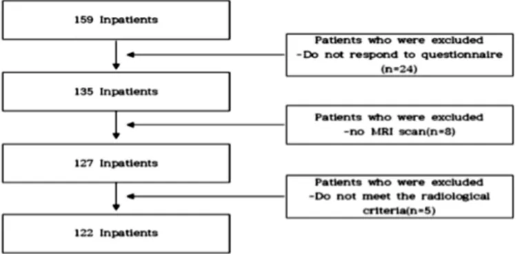
Of the 159 LSS inpatients, 122 patients were selected for study inclusion (Fig. 1).
Treatments
Patients were treated with acupuncture, pharmacopuncture, chuna therapy, herbal medicine therapy, and physical therapy.
Acupuncture treatment
The needles used for acupuncture were disposable stainless steel (0.30 × 40 mm; Dong Bang Medical Co. Ltd., Daecheon, Korea). Needles were inserted at the EX-B2 (Hwata Hyeopcheok) points [12], BL 23 (Shinsu) points, and Ashi points of the lumbar, pelvis, and lower limbs for 15 minutes twice a day. The depth of needle insertion was 20 mm or more. Acupuncture was combined with the use of an infra-red ray apparatus. Two Korean medicine doctors conducted the acupuncture treatment once each morning and afternoon from the day of admission.
Pharmacopuncture treatment
Based on physical examination and radiological imaging, 1 ml of Shinbaro pharmacopuncture was injected into the left EX-B2 and 1 ml was injected into the right EX-B2 (Hwata Hyeopcheok) points, at the most severe spinal level. Pharmacopuncture (from Jaseng Pharmacopuncture Medicinal Research Institute) was extracted by adding and removing medicines of Cheongpa-jeon [13], which has the efficacies of improving blood circulation, removing wind energy, and pain control ( 活血去風止痛 ), controlling dampness and eliminating swelling ( 化濕消腫 ), and strengthening muscle and bone ( 强筋骨 ). Before treatment, the area to be injected was disinfected with povidone (10% povidone-iodine) solution to prevent infection. Pharmacopuncture was then injected directly into the skin at a depth of about 3 cm using a disposable syringe (1 ml, 26G × 1.5 syringe; CPL Co. Ltd., Gyeonggi-do, Korea).
Pharmacopuncture treatment was conducted once every 2 days from the day of admission.
Chuna therapy
We performed the flexion–distraction technique, lumbar extension technique, lateral extension rotation technique, and lateral lumbar correction technique five to seven times a week from the day of admission.
Herbal medicine therapy
Herbal medicines (Jaseng Hospital prescriptions) were administered to patients with LSS. Cheongpa-jeon [13] and Cheongshinbaro-hwan were taken three times a day, 30 minutes after each meal, from the day of admission.
Physical therapy
Muscle meridian medium frequency therapy (interference current therapy), muscle meridian low frequency therapy (transcutaneous electrical nerve stimulation), microwave therapy, hot pack, traction therapy or manipulation were chosen according to patients’ conditions and performed five to seven times a week from the day of admission.
Disease stage classification
The stage of the disease was determined according to Kim et al’s method [14]. LSS was classified as: most acute stage, within 1 week of onset; acute stage, from 1 week to 1 month of onset; subacute stage, from 1 month to 6 months of onset; and chronic stage, 6 months or more from onset. Onset was based on the most recent incident of back, hip or leg pain.
Fig. 1. Flowchart showing how patients were selected for inclusion.
Stenosis type classification Canal stenosis
Canal stenosis is the narrowing of the central canal due to degenerative disc disease, and thickening and break away of the ligamentum flavum and facet joints. It mainly presents with bilateral pain without limited localization. Neurogenic intermittent claudication is mainly caused by cauda equina pressure due to canal stenosis.
Foraminal stenosis
This is stenosis caused by compression of the lateral recess or neural foraminal. One or more nerve roots are stimulated to localize the pain in the region. There are many cases of localized pain in the region, as well as leg pain, numbness and weakness in the lower extremities. Neuromuscular pressure is mainly applied to the nerve roots, and symptoms appear in the form of sciatica;
neurogenic intermittent claudication may also occur [15].
Radiological criteria
DSCA We used a medical imaging storage system (STARPACS;
INFINITT Healthcare Co. Ltd., Seoul, Korea) to measure DSCA on axial T1-enhanced MRI at disc level, the most severe LSS (Fig. 2).
To reduce error in measurement, the average of two measurements by two Korean medical doctors (each doctor made 1 measurement each) was taken. Patients with DSCA ≥ 100 mm
2were categorized into the mild–normal group. Those with DSCA more than 75 mm
2but less than 100 mm
2were categorized into the moderate group.
Those with DSCA ≤ 75 mm
2were categorized into the severe group [16].
Morphological grade
Using the same medical imaging storage system (STARPACS;
INFINITT Healthcare Co. Ltd. Seoul, Korea), we confirmed the morphological grade of the dural sac on axial T2-enhanced MRI at disc level, the most severe LSS (Fig. 3). Morphological grade was measured according to Schizas et al’s classification [17], summarized briefly below.
Grade A: No or only mild spinal LSS. On axial T2-enhanced
MRI, cerebrospinal fluid is clearly shown in the dural sac. Can be classified into four stages: A1 to A4. Stage A1 is when the rootlets are arranged in the dorsal spinal cord and take possession of less than half of the dural sac. Stage A2 is when the rootlets are arranged in the dorsal spinal cord; the rootlets touch the dura and spread in the form of a horseshoe. Stage A3 is when the rootlets are arranged in the dorsal side of the dural sac over half of the dural sac. Stage A4 is when the rootlets are arranged in the center and take possession of the majority of the dural sac.
Grade B: Moderate stage of LSS. The rootlets take possession of the whole of the dural sac, but the rootlets are still independent.
Some cerebrospinal fluid is shown. Grainy granules can be seen in the dural sac.
Grade C: Severe stage of LSS. There are no visible rootlets and no visible cerebrospinal fluid signal. There is a gray signal in the dural sac. Posterior epidural fat can be identified.
Grade D: Most severe (extreme) stage of LSS. Epidural fat (as observed in Grade C) and rootlets cannot be seen.
Numeric Rating Scale (NRS)
Of the different pain assessment methods available that express the degree of pain on a scale of 0 to 10, we chose the NRS to evaluate the degree of pain in our patients. Although it is a subjective indicator, the NRS is easy and simple to use. Zero stands for “painless”, 10 stands for “the most severe pain the patient can imagine”. The Visual Analog Scale is similar, but NRS does not require vision or mobility. Patients completed the NRS on both days of admission and discharge.
Oswestry Disability Index (ODI)
The ODI has 10 questions that are designed to measure the degree of disability in the daily life of patients with back pain.
The score for each item ranges from 0 to 5. The total score is calculated as twice the sum of the item scores [18]. We used the Korean version of the ODI, the reliability and validity of which has been verified [19]. Patients completed the ODI on both days of admission and discharge. We collected data on the fourth item on walking condition separately due to its relation with claudication—
the main symptom of LSS.
EQ-5D
The EQ-5D is a questionnaire that was developed in 1990 by the EuroQol group as a tool to assess five aspects of general health status: morbidity, self-care, usual activity, pain/discomfort, and anxiety/depression. The answer options for each question are:
“There are no problems at all”, “There are some problems”, and
“There are important problems”. As a result, we can define 35;243 possible health conditions. If you add two more states of death and loss of consciousness, then 245 health levels are possible [20]. The EQ-5D-5L version, which assesses health status at five levels, is published on the EuroQol website. In this study, we used the three- level scale (EQ-5D-3L); it has quality weightings for domestic (Korean) application, and it was used in the Korea National Health and Nutrition Examination Survey until 2013. The weighting formula used in the analysis was calculated based on the quality- weight estimation research report of the quality-of-life survey tool presented in the 2007 guidelines of the Korea Centers for Disease Control and Prevention for the use of primitive data [20].
EQ-5D index = 1 – (0.050 + 0.096 × M2 + 0.418 × M3 + 0.046 × SC2 + 0.136 × SC3 + 0.051 × UA2 + 0.208 × UA3 + 0.037 × PD2 +
Fig. 2. Measurement of dural sac cross-sectional area.
Fig. 3. Classification of morphological grade.
0.151 × PD3 + 0.043 × AD2 + 0.158 × AD3 + 0.050 × N3)
if LQ_1EQL = 1 & LQ_2EQL = 1 & LQ_3EQL = 1 & LQ_4EQL
= 1 & LQ_5EQL = 1 then EQ-5D = 1,
where M refers to athletic ability, SC to self-management, UA to everyday life, PD to pain/discomfort, and AD to anxiety/
depression. The number 2 (i.e., M2, SC2, UA2, PD2, AD2) means
“There is a little problem”, the number 3 (i.e., M3, SC3, UA3, PD3, AD3) means “There is a serious problem”. If it is applicable, then it substitutes 1. If it is not applicable, then it substitutes 0. N3 means to substitute 1 if there is more than one “serious problem”. In the Korean version of the EQ-5D, convergence and discriminant validity were confirmed in a validity and reliability study of the general population in Korea. The overall percent agreement between test–retest was 76–97%. The kappa coefficient was 0.24–
0.59, indicating that it has adequate reliability [21].
Statistical analyses
All statistical analyses were performed with IBM SPSS Statistics for Windows, Version 22.0 (IBM Corp., Armonk, NY, USA). Data are expressed as mean ± standard deviation and p values. Sex and age were included in the initial statistical analysis. Changes in NRS, ODI, gait-related ODI, and EQ-5D after treatment as compared to before were analyzed using paired t-test and Wilcoxon signed-rank test. Kruskal-Wallis test and one-way ANOVA were performed according to data after Shapiro-Wilk test as needed. A p value < 0.05 was considered statistically significant.
Results
Most of the patients were females (77.87%); mean age was 66.79
± 10.32 years. The youngest patient was 39 years old and the oldest was 90 years old. The majority of patients (59.02%) were elderly aged ≥ 65 years (Table 1).
In about two-thirds of patients, the reason for LSS was unknown (67.21%); the next most frequent causes were lumbar strain (9.01%) and overwork (7.38%) (Table 2).
Most patients had subacute (52/122, 42.62%) or chronic (40/122, 32.79%) LSS (Table 3).
Length of admission ranged from a minimum of 2 days to a maximum of 71 days; mean hospitalization period was 20.44 ± 15.19 days (Table 4).
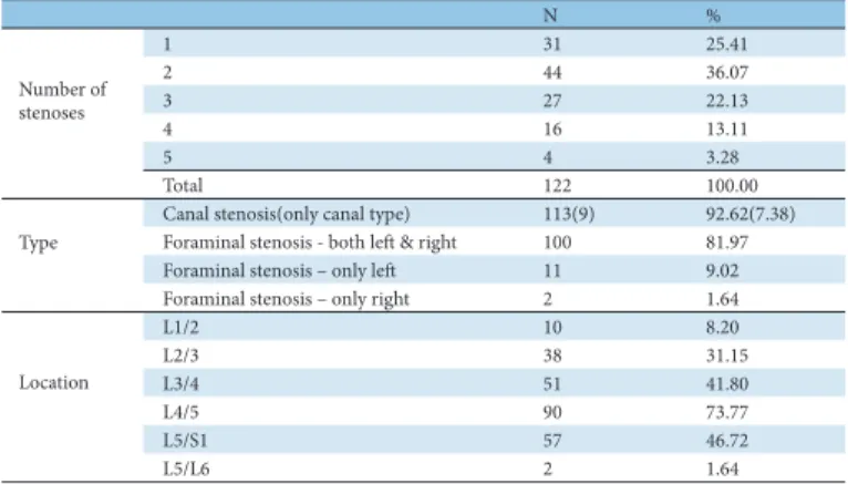
The number of stenoses in patients ranged from one to five.
LSS type was determined by considering MRI images and clinical symptoms. The number of patients who had canal stenosis and foraminal stenosis was the same (113/122, 92.62%). Nine patients (7.38%) had only canal stenosis and another nine patients (7.38%) had only foraminal stenosis, while 104 (85.25%) had both canal and foraminal stenoses. Among the patients with foraminal stenosis, most (81.97%) had both left and right foraminal stenoses.
The majority of patients (73.77%) had LSS in L4/5 (Table 5).
With regard to morphological grade, no patients were in Stage A1. About a quarter of patients (25.41%) were in Stages A2–
A4, while half (50.00%) already had Grade C LSS. Almost three- quarters of patients (72.13%) had DSCA ≤ 75 mm2, which indicates the most severe LSS. The narrowest DSCA was 25.16 mm2 and the widest was 176.36 mm2; mean DSCA was 66.68 ± 27.07 mm2 (Table 6).
The areas where patients experienced pain and numbness are shown in Table 7. Many had pain in more than one area, with most
Table 2. Distribution of Causes of LSS
N %
Reason unknown 82 67.21
Lumbar strain 11 9.01
Same motion in long period 5 4.10
Overwork 9 7.38
Trauma 5 4.10
Postoperative sequela 5 4.10
Etc 5 4.10
Total 122 100
Table 3. Distribution of LSS Disease Stage
N %
Most acute stage 14 11.48
Acute stage 16 13.11
Subacute stage 52 42.62
Chronic stage 40 32.79
Total 122 100
In this study, LSS was classified as:
• most acute stage, within 1 week of onset;
• acute stage, from 1 week to 1 month of onset;
• subacute stage, from 1 month to 6 months of onset;
• chronic stage, 6 months or more from onset.
Table 4. Distribution of Length of Admission
No. of days N %
0~5 17 13.93
6~10 17 13.93
11~15 21 17.21
16~20 15 12.30
21~25 18 14.75
26~30 11 9.02
31~35 9 7.38
36~40 3 2.46
41~45 3 2.46
46~50 1 0.82
51~ 7 5.74
Total 122 100
N %
1 31 25.41
2 44 36.07
3 27 22.13
4 16 13.11
5 4 3.28
Total 122 100.00
Canal stenosis(only canal type) 113(9) 92.62(7.38) Foraminal stenosis - both left & right 100 81.97
Foraminal stenosis – only left 11 9.02
Foraminal stenosis – only right 2 1.64
L1/2 10 8.20
L2/3 38 31.15
L3/4 51 41.80
L4/5 90 73.77
L5/S1 57 46.72
L5/L6 2 1.64
Number of stenoses
Type
Location
Table 5. Distribution of Number, Type and Location of Stenoses
N %
Male 27 22.13
Female 95 77.87
20~29 0 0.00
30~39 1 0.82
40~49 3 2.46
50~59 35 28.69
60~69 37 30.33
70~ 46 37.70
<65 50 40.98
≥65 72 59.02
122 100
Table 1. Distribution of Sex and Age Sex
Age
Total
efficacy, data were analyzed using the paired t-test and Wilcoxon signed-rank test according to normality (Tables 11–15). Also according to normality, Kruskal-Wallis test and one-way ANOVA were used to evaluate the differences according to morphological grade and DSCA (Table 16).
Evaluation of treatment
NRS To evaluate the progress and significance of treatment, changes in mean NRS scores for back pain, pelvic pain and lower extremity pain and numbness were separately measured before and after treatment.
We found that NRS score for low back and pelvic pain in the whole group of patients significantly decreased from 6.14 ± 1.71 before treatment to 4.28 ± 1.91 after treatment (p < 0.001). In addition, when analyzed according to sex, age, disease stage, and radiological criteria, significant decreases in NRS score were still seen (p < 0.001), except for morphological grade D where the improvement was not significant (p = 0.071) (Table 11).
For radiating pain and numbness to the lower extremities, NRS score in the whole patient group decreased from 6.27 ± 1.61 before treatment to 4.24 ± 1.76 after treatment (p < 0.001). Again, when
N %
A2~A4 31 25.41
B 26 21.31
C 61 50.00
D 4 3.28
Total 122 100.00
≤ 75mm2 88 72.13
> 75 mm2 and < 100 mm2 20 16.39
≥ 100mm2 14 11.48
Total 122 100.00
Table 6. Distribution of Morphological Grade and Dural Sac Cross-sectional Area (DSCA)
Morphological grade
DSCA
N %
Lumbar pain 110 90.16
Pelvic pain 97 79.51
Thigh pain and numbness 81 66.39
Calf pain and numbness 81 66.39
Foot pain and numbness 36 29.51
Table 7. Distribution of Pain and Numbness
N %
None 8 6.56
Herniated intervertebral disc 76 62.30
C- or T-Spine stenosis 3 2.46
Spondylolisthesis 4 3.28
Other musculoskeletal disorders 44 36.07
Other comorbidities (non-musculoskeletal disorders) 74 60.66 Table 8. Distribution of Comorbidities
Admission Discharge Improvement p-value*
A2~A4 31 0.065 0.004 0.014
B 26 0.011 0.061 0.007
C 59 0.061 0.041 0.000
D 4 0.272 0.161 0.406
A2~A4 25 0.091 0.057 0.033
B 20 0.224 0.035 0.006
C 50 0.072 0.086 0.001
D 3 0.000 † 0.000
A2~A4 31 0.629 0.271 0.144
B 26 0.735 0.011 0.121
C 61 0.544 0.000 0.182
D 4 0.724 0.387 0.491
A2~A4 31 0.021 0.009 0.010
B 26 0.005 0.016 0.004
C 61 0.001 0.000 0.000
D 4 0.161 0.406 0.272
A2~A4 31 0.138 0.000 0.052
B 26 0.000 0.000 0.021
C 61 0.000 0.000 0.000
D 4 0.096 0.040 0.300
*Using Shapiro-Wilk test. †3 patient’s NRS scores were equal. so they had been omitted.
Table 9. Test of Normality by Morphological Grade Morphological N Grade
NRS(lumbar & pelvic pain)
NRS(radiating pain &
numbness)
ODI
Gait-related ODI
EQ-5D
reporting pain in the lumbar and pelvic regions.
Eight patients (6.56%) had no comorbidities, while almost two- thirds (62.30%) had herniated intervertebral disc. A small number of patients (2.46%) had stenosis in the cervical or thoracic spine (Table 8).
The Shapiro-Wilk test was conducted on NRS scores for lumbar and pelvic pain, and radiating pain and numbness, ODI, gait- related ODI, and EQ-5D before and after treatment. The changes in each of these according to morphological grade and DSCA were tested for normality (Tables 9 & 10). These analyses included groups that had fewer than 30 patients, and should be considered carefully for this study purpose. After that, to evaluate treatment
Admission Discharge Improvement p-value*
≤ 75mm2 86 0.004 0.000 0.000
> 75 mm2 and < 100 mm2 20 0.107 0.059 0.161
≥ 100mm2 14 0.019 0.006 0.355
≤ 75mm2 73 0.036 0.021 0.000
> 75 mm2 and < 100 mm2 15 0.068 0.592 0.071
≥ 100mm2 10 0.025 0.108 0.445
≤ 75mm2 88 0.391 0.000 0.296
> 75 mm2 and < 100 mm2 20 0.528 0.916 0.726
≥ 100mm2 14 0.893 0.389 0.768
≤ 75mm2 88 0.000 0.000 0.000
> 75 mm2 and < 100 mm2 20 0.015 0.001 0.048
≥ 100mm2 14 0.003 0.058 0.529
≤ 75mm2 88 0.000 0.000 0.000
> 75 mm2 and < 100 mm2 20 0.013 0.085 0.009
≥ 100mm2 14 0.355 0.035 0.065
*Using Shapiro-Wilk test.
Table 10. Test of Normality by Dural Sac Cross-sectional Area (DSCA)
NRS(lumbar &
pelvic pain) NRS(radiating pain &
numbness) ODI Gait-related ODI EQ-5D
DSCA N
NRS
Admission Discharge Improvement
Male 26 5.81±2.08 3.81±2.25 2.00±1.37 p<0.001*
Female 94 6.23±1.59 4.40±1.80 1.83±1.44 p<0.001*
<65 50 5.84±1.66 3.74±1.56 2.10±1.68 p<0.001*
≥65 70 6.36±1.72 4.66±2.06 1.70±1.44 p<0.001*
Acute stage(Most
acute´) 30 6.17±1.58 4.17±2.23 2.00±1.51 p<0.001*
Subacute stage 51 6.06±1.78 4.12±1.85 1.94±1.67 p<0.001*
Chronic stage 39 6.23±1.74 4.56±1.74 1.67±1.44 p<0.001*
A2~A4 31 6.42±1.80 4.13±1.86 2.29±1.81 p<0.001†
B 26 6.35±1.70 4.50±2.53 1.85±1.43 p<0.001†
C 59 5.90±1.70 4.20±1.67 1.70±1.45 p<0.001†
D 4 6.25±0.96 5.00±1.41 1.25±1.26 p=0.071*
≤ 75mm2 86 6.19±1.71 4.44±2.00 1.74±1.50 p<0.001†
> 75 mm2
and < 100 mm2 20 6.10±1.92 3.90±1.80 2.20±1.74 p<0.001*
≥ 100mm2 14 5.93±1.39 3.79±1.48 2.14±1.56 p<0.001†
120 6.14±1.71 4.28±1.91 1.87±1.55 p<0.001*
Average NRS values are means ± standard deviation.
* paired t-test before and after NRS; significance at p<0.05.
† Wilcoxon signed-rank test before and after NRS; significance at p<0.05.
Table 11. Differences in Lumbar and Pelvic Pain Numeric Rating Scale (NRS) Before and After Treatment
Sex Age
Disease stage
Morphological grade
DSCA Total
N p-value
analyzed according to sex, age, disease stage, and radiological criteria, significant decreases in NRS score were still seen (p <
0.001), except for morphological grade D where the improvement was not significant (p = 0.065) and DSCA ≥ 100 mm2 where the improvement was borderline significant (p < 0.05) (Table 12).
The non-significant results for morphological grade D were likely due to the small number of patients.
ODI and gait-related ODI
To assess the degree of treatment progression and its significance, changes in mean ODI score after treatment as compared to before treatment (including for ODI Item 4) were analyzed. However, item 4 of the ODI was also separately analyzed due to its relation to claudication—the main symptom of LSS (Tables 13 & 14).
We found that ODI score in the whole group of patients significantly decreased from 46.86 ± 19.40 before treatment to 33.63 ± 18.66 after treatment (p < 0.001). ODI Item 4, which represents walking disorder, also significantly decreased from 3.34 ± 1.23 to 2.80 ± 1.11 in the whole patient group (p <
0.001). When analyzed according to sex, age, disease stage, and radiological criteria, the decreases were still significant, except for
morphological grade D (due to the small number of patients).
EQ-5D
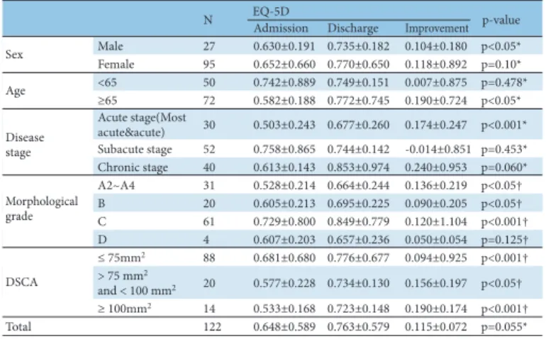
Change in quality of life before and after treatment was assessed by analyzing EQ-5D index values (Table 15).
There was only a slight increase from 0.648 ± 0.589 before treatment to 0.763 ± 0.579 after treatment observed in the whole patient group, which was not statistically significant (p = 0.055).
However, males (p < 0.05), elderly aged ≥ 65 years (p < 0.05), and those with acute-stage LSS (p < 0.001) showed significant improvements. When analyzed according to radiological criteria,
NRS
Admission Discharge Improvement
Male 22 6.14±1.28 4.09±1.63 2.05±1.89 p<0.001*
Female 76 6.30±1.70 4.29±1.80 2.01±1.44 p<0.001*
<65 38 6.13±1.63 3.87±1.63 2.26±1.61 p<0.001*
≥65 60 6.35±1.60 4.48±1.81 1.87±1.50 p<0.001*
Acute stage(Most
acute´) 22 5.86±1.64 4.00±1.85 1.86±1.39 p<0.001*
Subacute stage 43 6.28±1.68 4.14±1.95 2.14±1.73 p<0.001*
Chronic stage 33 6.52±1.48 4.55±1.42 1.97±1.40 p<0.001*
A2~A4 25 6.36±1.60 4.12±1.76 2.24±1.67 p<0.001*
B 20 5.90±1.74 3.95±2.28 1.95±1.61 p<0.001†
C 50 6.40±1.60 4.44±1.58 1.96±1.50 p<0.001*
D 3 5.67±1.16 4.00±0.00 1.67±1.16 p=0.065*
≤ 75mm2 73 6.32±1.60 4.32±1.78 2.00±1.51 p<0.001†
> 75 mm2
and < 100 mm2 15 6.73±1.83 4.33±1.99 2.40±1.84 p<0.001*
≥ 100mm2 10 5.20±0.79 3.60±1.17 1.60±1.27 p<0.05†
98 6.27±1.61 4.24±1.76 2.02±1.54 p<0.001*
Average NRS values are means ± standard deviation.
* paired t-test before and after NRS; significance at p<0.05.
† Wilcoxon signed-rank test before and after NRS; significance at p<0.05.
Table 12. Differences in Radiating Pain and Numbness Numeric Rating Scale (NRS) Before and After Treatment
Sex Age Disease stage
Morphological grade
DSCA Total
N p-value
ODI
Admission Discharge Improvement Male 27 42.82±21.88 26.17±18.44 16.65±17.67 p<0.05*
Female 95 48.01±18.60 35.75±18.26 12.26±13.49 p<0.001*
<65 50 42.86±18.37 29.99±15.95 12.87±14.51 p<0.001*
≥65 72 49.64±19.73 36.16±20.05 13.48±14.68 p<0.001*
Acute stage(Most
acute´) 30 53.79±22.24 34.89±20.51 18.90±18.32 p<0.001*
Subacute stage 52 42.82±19.64 32.05±18.53 10.76±13.61 p<0.001*
Chronic stage 40 46.93±15.39 34.74±17.67 12.19±11.47 p<0.001*
A2~A4 31 51.51±19.90 35.90±19.25 15.61±20.91 p<0.001*
B 26 47.56±22.44 34.99±19.46 12.56±13.33 p<0.001†
C 61 43.54±17.36 31.05±17.40 12.49±11.11 p<0.001†
D 4 57.11±20.12 46.61±26.29 10.50±11.97 p=0.089*
≤ 75mm2 88 46.50±19.28 34.77±20.03 11.72±12.09 p<0.001†
> 75 mm2
and < 100 mm2 20 45.94±20.17 31.56±14.09 14.38±11.82 p<0.001*
≥ 100mm2 14 50.48±20.11 29.41±15.15 21.06±26.58 p<0.05†
122 46.86±19.40 33.63±18.66 13.23±14.56 p<0.001*
ODI values are means ± standard deviation.
* paired t-test before and after ODI; significance at p<0.05.
† Wilcoxon signed-rank test before and after ODI; significance at p<0.05.
Table 13. Differences in Oswestry Disability Index (ODI) Before and After Treatment
Sex Age Disease stage
Morphological grade
DSCA Total
N p-value
Admission Discharge Improvement
Male 27 3.11±1.45 2.56±1.09 0.56±1.25 p<0.05*
Female 95 3.41±1.15 2.86±1.11 0.55±0.95 p<0.001*
<65 50 2.98±1.17 2.62±1.05 0.36±1.08 p<0.05*
≥65 72 3.60±1.20 2.92±1.14 0.68±0.96 p<0.001*
Acute stage(Most
acute´) 30 3.63±1.27 2.83±1.12 0.80±1.03 p<0.001*
Subacute stage 52 3.02±1.18 2.77±1.08 0.25±0.93 p<0.05*
Chronic stage 40 3.55±1.18 2.80±1.16 0.75±1.06 p<0.001*
A2~A4 31 3.52±1.21 2.84±1.10 0.68±1.42 p<0.05†
B 26 3.42±1.36 2.85±1.12 0.58±0.86 p<0.05†
C 61 3.18±1.16 2.72±1.11 0.46±0.85 p<0.001†
D 4 4.00±1.41 3.25±1.26 0.75±0.96 p=0.108*
≤ 75mm2 88 3.27±1.26 2.83±1.18 0.44±0.83 p<0.001†
> 75 mm2
and < 100 mm2 20 3.45±1.15 2.80±0.77 0.65±1.04 p<0.05†
≥ 100mm2 14 3.64±1.15 2.57±1.15 1.07±1.77 p<0.05†
122 3.34±1.23 2.80±1.11 0.55±0.09 p<0.001*
ODI (Item No. 4) values are means ± standard deviation.
* paired t-test before and after ODI (Item No. 4); significance at p<0.05.
† Wilcoxon signed-rank test before and after ODI (Item No. 4); significance at p<0.05.
Table 14. Differences in Gait-related Oswestry Disability Index (ODI) (Item No. 4) Before and After Treatment
Sex Age
Disease stage
Morphological grade
DSCA Total
Gait-related ODI
N p-value
Admission Discharge Improvement Male 27 0.630±0.191 0.735±0.182 0.104±0.180 p<0.05*
Female 95 0.652±0.660 0.770±0.650 0.118±0.892 p=0.10*
<65 50 0.742±0.889 0.749±0.151 0.007±0.875 p=0.478*
≥65 72 0.582±0.188 0.772±0.745 0.190±0.724 p<0.05*
Acute stage(Most
acute´) 30 0.503±0.243 0.677±0.260 0.174±0.247 p<0.001*
Subacute stage 52 0.758±0.865 0.744±0.142 -0.014±0.851 p=0.453*
Chronic stage 40 0.613±0.143 0.853±0.974 0.240±0.953 p=0.060*
A2~A4 31 0.528±0.214 0.664±0.244 0.136±0.219 p<0.05†
B 20 0.605±0.213 0.695±0.225 0.090±0.205 p<0.05†
C 61 0.729±0.800 0.849±0.779 0.120±1.104 p<0.001†
D 4 0.607±0.203 0.657±0.236 0.050±0.054 p=0.125†
≤ 75mm2 88 0.681±0.680 0.776±0.677 0.094±0.925 p<0.001†
> 75 mm2
and < 100 mm2 20 0.577±0.228 0.734±0.130 0.156±0.197 p<0.05†
≥ 100mm2 14 0.533±0.168 0.723±0.148 0.190±0.174 p<0.001†
122 0.648±0.589 0.763±0.579 0.115±0.072 p=0.055*
EQ-5D index values are means ± standard deviation.
* paired t-test before and after EQ-5D; significance at p<0.05.
† Wilcoxon signed-rank test before and after EQ-5D; significance at p<0.05.
Sex Age Disease stage
Morphological grade
DSCA Total
Table 15. Differences in EQ-5D Before and After Treatment EQ-5D
N p-value
Morphological Grade DSCA p-value
NRS(lumbar & pelvic pain) 0.452* 0.425*
NRS(radiating pain & numbness) 0.900* 0.490*
ODI 0.762† 0.076†
Gait-related ODI 0.890* 0.229*
EQ-5D 0.546* 0.288*
* Kruskal-Wallis test. † One-way ANOVA.
Table 16. Differences in Treatment Effect According to Radiological Criteria