ISSN 2234-3806 • eISSN 2234-3814
https://doi.org/10.3343/alm.2021.41.2.247 www.annlabmed.org 247
Ann Lab Med 2021;41:247-249
https://doi.org/10.3343/alm.2021.41.2.247
Letter to the Editor
Clinical Microbiology
A Case of Brain Abscess Caused by the Dematiaceous Mold Neoscytalidium dimidiatum in a Korean Man
Su Yeon Jo , M.D.1, Shinwon Lee , M.D.2, Kye-Hyung Kim , M.D.2, and Jongyoun Yi , M.D.3,4
1Department of Laboratory Medicine, Pusan National University Yangsan Hospital, Yangsan, Korea; 2Department of Internal Medicine, Pusan National University School of Medicine, Busan, Korea; 3Department of Laboratory Medicine, Pusan National University School of Medicine, Busan, Korea;
4Department of Laboratory Medicine, Pusan National University Hospital, Busan, Korea
Dear Editor,
Neoscytalidium dimidiatum is a rapidly growing dematiaceous mold mainly found in tropical and subtropical environments.
Primarily a plant pathogen, it commonly causes nail and skin infections in humans; however, very rarely it causes brain infec- tions [1-3]. Superficial N. dimidiatum infections have been re- ported in South America, Southeast Asia, India, and Africa [4].
Additionally, there are rare reports of deep-seated infections in immunocompromised patients, with poor prognoses [1, 2, 4, 5].
Only two brain abscess cases have been reported in India and Iran [2, 3]. In Korea, this fungus is non-endemic, and N. dimid- iatum human infections have not been reported. We describe a case of a brain abscess caused by N. dimidiatum in a Korean man who survived after prolonged antifungal therapy. The Insti- tutional Review Board of Pusan National University Hospital, Busan, Korea, exempted the approval of this study and the need for informed consent.
This study was conducted for 15 months, from hospitalization to treatment termination and final brain imaging follow-up. A 62-year-old man with progressive dysarthria and general weak- ness for two weeks was admitted to a tertiary university hospital in March 2017. He had no systemic symptoms, and his under- lying medical conditions included liver cirrhosis, past surgery for hepatocellular carcinoma (HCC), and diabetes. Three months
earlier, he had visited Thailand. Brain magnetic resonance im- aging (MRI) showed a 1.8-cm lesion; a brain abscess or HCC metastasis was suspected. Antibacterial agents were empirically administered. Stereotactic surgery was performed to aspirate the lesion, and pus was drained.
The aspirate was cultured on Sabouraud dextrose agar at 30°C.
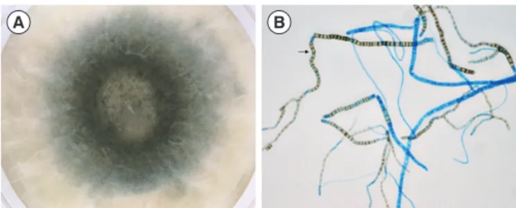
A mold was isolated and matured quickly within three days; col- onies were woolly and dark (Fig. 1A). Hyphae were septate and branched, and numerous rectangular or barrel-shaped arthro- conidia were observed, with no empty cells between them (Fig.
1B). Based on these morphological features, the isolate was presumably identified as N. dimidiatum [1].
To genotypically confirm the species, the internal transcribed spacer (ITS) regions were sequenced [6]. The ITS1-5.8S ribo- somal DNA-ITS2 sequence (GenBank accession no. MG028648) was analyzed using the International Society for Human and Animal Mycology (ISHAM) ITS database [6] and showed 99.6%
similarity to N. dimidiatum; Lasiodiplodia theobromae was the second most similar hit (91.0%). The D1/D2 region of the large subunit ribosomal RNA was also sequenced [7]. Phylogenetic analysis showed the ITS was better than D1/D2 at discriminat- ing Neoscytalidium species (Fig. 2). Matrix-assisted laser de- sorption/ionization time-of-flight mass spectrometry was not used for identification because the analysis could be inhibited
Received: January 28, 2020 Revision received: June 11, 2020 Accepted: September 19, 2020
Corresponding author: Jongyoun Yi, M.D., Ph.D.
Department of Laboratory Medicine, Pusan National University Hospital, 179 Gudeok-ro, Seo-gu, Busan 49241, Korea
Tel: +82-51-240-7417, Fax: +82-51-240-7413 E-mail: socioliberal@yahoo.co.kr
© Korean Society for Laboratory Medicine
This is an Open Access article distributed under the terms of the Creative Commons Attribution Non-Commercial License (https://creativecommons.org/licenses/by-nc/4.0) which permits unrestricted non-commercial use, distribution, and reproduction in any medium, provided the original work is properly cited.
1 / 1 CROSSMARK_logo_3_Test
2017-03-16 https://crossmark-cdn.crossref.org/widget/v2.0/logos/CROSSMARK_Color_square.svg
Jo SY, et al.
Brain Abscess Caused by Neoscytalidium dimidiatum
248 www.annlabmed.org https://doi.org/10.3343/alm.2021.41.2.247 by the melanin pigment of the isolate [7].
After the mold was isolated, the antimicrobial regimen was al- tered to intravenous amphotericin B (AMB). After a 4-week AMB therapy, the brain lesion showed an approximate 30% de- crease in size; however, a new 1.6-cm lesion appeared along the stereotactic aspiration tract. After two additional weeks of AMB therapy, the patient was switched to oral voriconazole and discharged. However, as the lesions remained unchanged ac- cording to the MRI conducted after 5-week voriconazole ther- apy, he was re-admitted and treated again with AMB for another
10 weeks; no further antifungals were subsequently adminis- tered. He was treated and followed-up for approximately 15 months in total. Follow-up MRIs showed steady and significant lesion improvement for one year. The dysarthria and general weakness had disappeared, and the brain abscess follow-up was concluded.
Our patient may have acquired the infection while visiting Thailand, and his immunocompromised state possibly contrib- uted to the brain infection caused by this fungus.
Deep N. dimidiatum infections show poor prognoses and have no standardized treatment [5]. In vitro susceptibility analy- ses of N. dimidiatum clinical isolates from superficial infections suggest that AMB and voriconazole have the highest activity and that voriconazole could constitute an alternative to AMB [8];
however, more deep-seated infections frequently show poor out- comes despite treatment with these drugs [9]. Posaconazole and caspofungin have less in vitro activity than voriconazole [8, 10]. Anidulafungin shows in vitro activity against some isolates, while micafungin demonstrates little activity [10].
We did not analyze antifungal susceptibility because it pro- vides only minimal inhibitory concentrations, which do not cor- relate with clinical outcomes [8]. Nevertheless, our case indi- cates that voriconazole was probably ineffective because the le- sions did not diminish, whereas AMB provided a gradual but obvious clinical effect. Previous brain abscess cases were fatal
A B
Fig. 1. Culture of the Neoscytalidium dimidiatum brain isolate on Sabouraud dextrose agar. (A) After incubation for three days, the isolate showed white woolly and dark brown to black pigmented colonies filling the agar. (B) Microscopic examination of the N.
dimidiatum isolate (×400). A cellophane tape mount with lactophe- nol cotton blue stain demonstrated septate hyphae and numerous rectangular arthroconidia (arrow). The arthroconidia and some hy- phae were brown.
Neoscytalidium dimidiatum UTHSCSA DI Neoscytalidium dimidiatum CBS 251.49 Neoscytalidium dimidiatum CBS 499.66 Neoscytalidium dimidiatum Author Neoscytalidium dimidiatum NDFB003 Neoscytalidium novaehollandiae CBS 122071 Neoscytalidium novaehollandiae WAC12691 Neoscytalidium orchidacearum CMU287 Neoscytalidium orchidacearum MFLUCC 12-0533 Botryosphaeria dothidea CMW8000
Botryosphaeria dothidea CBS110302 38
44 72
60 96
56 100
100
0.02 0.01 0.00
A Neoscytalidium orchidacearum CMU287
Neoscytalidium orchidacearum MFLUCC 12-0533 Neoscytalidium dimidiatum CBS 251.49 Neoscytalidium dimidiatum NDFB003 Neoscytalidium dimidiatum CBS 499.66 Neoscytalidium dimidiatum UTHSCSA DI Neoscytalidium novaehollandiae CBS 122071 Neoscytalidium novaehollandiae WAC12691 Neoscytalidium dimidiatum Author Botryosphaeria dothidea CMW8000 Botryosphaeria dothidea CBS110302 87
59
87
100
0.01 0.00
B
Fig. 2. Phylogenetic analysis of the isolate from this study and closely related Neoscytalidium spp. Botrysphaeria dothidea sequences were used as an outgroup. The isolate from this study is marked with ‘Author’ after the species name. (A) Internal transcribed spacer sequences.
(B) Sequences of the D1/D2 region of large subunit ribosomal RNA.
Jo SY, et al.
Brain Abscess Caused by Neoscytalidium dimidiatum
https://doi.org/10.3343/alm.2021.41.2.247 www.annlabmed.org 249
despite antifungal therapies [2, 3]. In contrast, our patient was eventually cured after early stereotactic drainage and 5-month antifungal therapy.
This is the first case of a brain infection by N. dimidiatum in Korea. Stereotactic surgery and genotypic identification using a reliable database such as ISHAM [6], rather than GenBank, could help accurately diagnose N. dimidiatum when an immu- nocompromised patient presents with brain lesions and a travel history to endemic countries. Prolonged AMB therapy can be effective in treating patients with this fungal infection, while the efficacy of voriconazole is uncertain.
ACKNOWLEDGEMENTS
None.
AUTHOR CONTRIBUTIONS
Conceptualization: JY. Data curation: JY, SYJ. Formal analysis:
JY. Methodology: JY, SYJ. Investigation: SL, KHK. Writing - origi- nal draft: SYJ. Writing - review & editing: JY, SL, KHK.
CONFLICTS OF INTEREST
No potential conflicts of interest relevant to this study are re- ported.
RESEARCH FUNDING
This work was supported by a 2-year research grant of Pusan National University.
ORCID
Su Yeon Jo https://orcid.org/0000-0001-7735-2897 Shinwon Lee https://orcid.org/0000-0001-7652-7093 Kye-Hyung Kim https://orcid.org/0000-0001-9682-9654 Jongyoun Yi https://orcid.org/0000-0001-9098-3765
REFERENCES
1. Larone DH. Medically important fungi: a guide to identification. 5th ed.
Washington, DC: ASM Press, 2011: 224.
2. Geramishoar M, Zomorodian K, Zaini F, Saadat F, Tarazooie B, Norouzi M, et al. First case of cerebral phaeohyphomycosis caused by Nat- trassia mangiferae in Iran. Jpn J Infect Dis 2004;57:285-6.
3. Mani RS, Chickabasaviah YT, Nagarathna S, Chandramuki A, Shivpra- kash MR, Vijayan J, et al. Cerebral phaeohyphomycosis caused by Scy- talidium dimidiatum: a case report from India. Med Mycol 2008;46:705- 11.
4. Tan DHS, Sigler L, Gibas CFC, Fong IW. Disseminated fungal infection in a renal transplant recipient involving Macrophomina phaseolina and Scytalidium dimidiatum: case report and review of taxonomic changes among medically important members of the Botryosphaeriaceae. Med Mycol 2008;46:285-92.
5. Machouart M, Menir P, Helenon R, Quist D, Desbois N. Scytalidium and scytalidiosis: what’s new in 2012? J Mycol Med 2013;23:40-6.
6. Irinyi L, Serena C, Garcia-Hermoso D, Arabatzis M, Desnos-Ollivier M, Vu D, et al. International Society of Human and Animal Mycology (ISHAM)-ITS reference DNA barcoding database—the quality con- trolled standard tool for routine identification of human and animal pathogenic fungi. Med Mycol 2015;53:313-37.
7. CLSI. Interpretive criteria for identification of bacteria and fungi by tar- geted DNA Sequencing. 2nd ed. CLSI MM18. Wayne, PA: Clinical and Laboratory Standards Institute. 2018.
8. Lacroix C and de Chauvin MF. In vitro activity of amphotericin B, itracon- azole, voriconazole, posaconazole, caspofungin and terbinafine against Scytalidium dimidiatum and Scytalidium hyalinum clinical isolates. J An- timicrob Chemother 2008;61:835-7.
9. Elinav H, Izhar U, Benenson S, Admon D, Hidalgo-Grass C, Polacheck I, et al. Invasive Scytalidium dimidiatum infection in an immunocompe- tent adult. J Clin Microbiol 2009;47:1259-63.
10. Madrid H, Ruíz-Cendoya M, Cano J, Stchigel A, Orofino R, Guarro J.
Genotyping and in vitro antifungal susceptibility of Neoscytalidium dimidiatum isolates from different origins. Int J Antimicrob Agents 2009;34:351-4.