Kienböck's disease is a form of avascular necrosis of the lunate bone with an unknown etiology.1-3) The Lichtman classification, based on plain radiography and magnetic resonance imaging (MRI) of the wrist, is commonly used in determining treatment options.4) According to the exis-
tence of carpal malalignment and osteoarthritic changes, Kienböck's disease can be divided into early stages (Licht- man stage I, II, and IIIA) and advanced stages (Lichtman stage IIIB and IV).5,6) In advanced stages, salvage proce- dures, such as proximal row carpectomy, limited carpal fusion, and wrist arthrodesis, have been recommended.1,6,7) However, these procedures inevitably reduce range of mo- tion (ROM) of the wrist joint, which is considered unde- sirable by many patients.3,8-10)
Radius osteotomies have shown favorable clinical results in early stages of Kienböck's disease,7,11-15) and some studies reported improvement of clinical symptoms in
Clinical Outcome of Lateral Wedge Osteotomy of the Radius in Advanced Stages of
Kienböck’s Disease
Young Ho Shin, MD, Jihyeung Kim, MD*, Hyun Sik Gong, MD*, Seung Hwan Rhee, MD*, Min Joon Cho, MD*, Goo Hyun Baek, MD*
Department of Orthopedic Surgery, Asan Medical Center, University of Ulsan College of Medicine, Seoul,
*Department of Orthopedic Surgery, Seoul National University College of Medicine, Seoul, Korea
Background: Radius osteotomies showed favorable clinical outcome in Kienböck's disease. However, few articles have been pub- lished on the long-term outcome of lateral wedge osteotomy of the radius in patients with advanced stage Kienböck's disease.
Methods: Eleven patients with Lichtman stage IIIB/IV Kienböck's disease (group A; mean follow-up period, 86.1 months; range, 48 to 163 months) and 14 patients with Lichtman stage IIIA Kienböck's disease (group B; mean follow-up period, 85.1 months; range, 49 to 144 months) underwent radial wedge osteotomy between August 2004 and August 2012. Radiological changes of the lunate and radiocarpal joint were compared between two groups after osteotomy. The wrist flexion/extension angle, grip strength, and Disabilities of the Arm, Shoulder and Hand (DASH) scores were evaluated preoperatively and at the final follow-up. The Nakamura Scoring System (NSSK) was used for comprehensive understanding of radiological and clinical outcomes.
Results: Nine patients of group A and 11 patients of group B showed radiological improvement in the lunate regarding sclerosis, cystic changes, or fragmentation. No patients showed progression of arthritic changes in radiocarpal and midcarpal joints. The wrist flexion/extension angle, grip strength, and DASH score were significantly improved in both groups after operation, but inter- group difference was not statistically significant at the final follow-up (p = 0.149, p = 0.267, and p = 0.536, respectively). The mean NSSK was 21.6 (range, 15 to 27) in group A and 21.8 (range, 15 to 26) in group B.
Conclusions: Radial wedge osteotomy yielded excellent radiological and functional outcomes in advanced stages of Kienböck's disease and these results were comparable to those of Lichtman stage IIIA disease. This technique could be a useful alternative to salvage procedures in the treatment of Lichtman stage IIIB/IV Kienböck's disease without severe radiocarpal arthritis.
Keywords: Osteonecrosis, Kienböck's disease, Radius osteotomy, Radial wedge osteotomy
Copyright © 2017 by The Korean Orthopaedic Association
This is an Open Access article distributed under the terms of the Creative Commons Attribution Non-Commercial License (http://creativecommons.org/licenses/by-nc/4.0) which permits unrestricted non-commercial use, distribution, and reproduction in any medium, provided the original work is properly cited.
Clinics in Orthopedic Surgery • pISSN 2005-291X eISSN 2005-4408 Received January 5, 2017; Accepted April 5, 2017
Correspondence to: Goo Hyun Baek, MD
Department of Orthopedic Surgery, Seoul National University College of Medicine, 101 Daehak-ro, Jongno-gu, Seoul 03080, Korea
Tel: +82-2-2072-3787, Fax: +82-2-764-2718 E-mail: ghbaek@snu.ac.kr
Lichtman stage IIIB and IV Kienböck's disease.6,16) Radial shortening osteotomy is known to reduce the actual stress across the radiolunate joint and shift the load to the ulnar side of the wrist joint.12,17-19) However, this joint leveling procedure is not a reasonable treatment option for patients with neutral or positive variance of the ulna. Instead, ra- dial closed wedge osteotomy, which decreases the radial inclination, could be used regardless of ulnar variance.6) In addition, this procedure could be combined with radial shortening in patients with negative ulnar variance.15)
Only few articles have been published on the long- term outcome of radial wedge osteotomy in Kienböck's disease, particularly among patients in advanced stages.
The purpose of this study was to evaluate the radiological and clinical outcomes of radial wedge osteotomy in pa- tients with Lichtman stage IIIB and IV Kienböck's disease and compare the results with those in patients with Licht- man stage IIIA Kienböck's disease.
METHODS
Patient Characteristics
This study is a retrospective case series analysis of prospec- tively gathered data. A total of 11 patients with Lichtman stage IIIB and IV Kienböck's disease (group A) were treat- ed by radial wedge osteotomy between August 2004 and August 2012 and followed for more than 3 years. Patients having severe arthritic changes at the radiocarpal joint with invisible joint space were excluded because they were considered candidates for salvage procedures.20) In the same period, fourteen consecutive patients with Lichtman stage IIIA Kienböck's disease (group B) were treated with the same procedure and followed for more than 3 years.
The medical records, radiographs, and evaluation data of these patients were reviewed. Two of the authors (YHS
and MJC) determined the preoperative Lichtman stage based on a review of simple radiographs and MRI scans.
In cases that cause confusion between stage IIIA and IIIB, 60° of radioscaphoid angle was used as a criterion for dif- ferentiation. The demographics are described in Table 1.
This study was approved by the Seoul National University College of Medicine/Seoul National University Hospital Institutional Review Board (No. H-1512-097-728).
Surgical Technique
All operations were performed by a single orthopedic surgeon (GHB) with use of volar Henry approach on the distal radius. Radial wedge osteotomy was performed by making a transverse cut and an oblique cut on the meta- diaphyseal junction of the distal radius, considering the thickness of the saw blade. In patients with positive and neutral ulnar variance, only the wedge osteotomy was done, whereas in patients with negative ulnar variance, ra- dial shortening was combined with the wedge osteotomy.
In all patients, the goal of corrected inclination angle was between 5° and 10°.15) In patients with negative ulnar vari- ance, the goal of radial shortening was to obtain a final variance of neutral to 2 mm positive18) while limiting radial length alteration to ≤ 3 mm regardless of ulnar variance.17) Two patients in group B, who had positive ulnar variance of more than 5 mm, had ulnar shortening osteotomy si- multaneously. All osteotomies were fixed with a plate and screws system: a five- or six-hole small dynamic compres- sion plate (Stryker Orthopedics, Mahwah, NJ, USA) in the earlier patients and a locking compression plate for the distal radius (Synthes, Oberdorf, Switzerland) in the later patients.
Postoperative Management and Outcome Evaluation Postoperatively, the patients were managed for the first 2
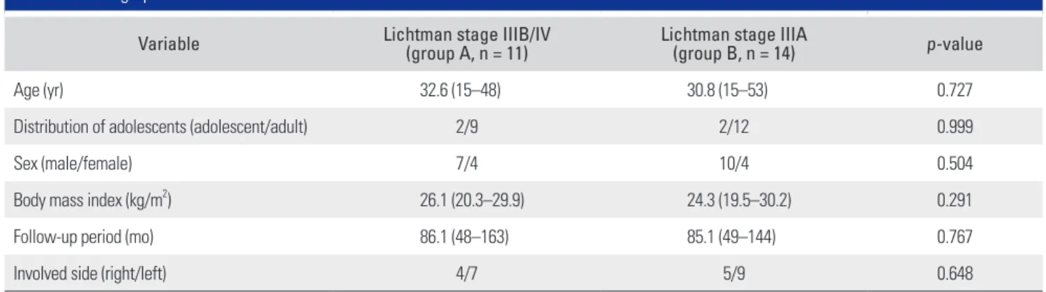
Table 1. Demographics of Enrolled Patients
Variable Lichtman stage IIIB/IV
(group A, n = 11) Lichtman stage IIIA
(group B, n = 14) p-value
Age (yr) 32.6 (15–48) 30.8 (15–53) 0.727
Distribution of adolescents (adolescent/adult) 2/9 2/12 0.999
Sex (male/female) 7/4 10/4 0.504
Body mass index (kg/m2) 26.1 (20.3–29.9) 24.3 (19.5–30.2) 0.291
Follow-up period (mo) 86.1 (48–163) 85.1 (49–144) 0.767
Involved side (right/left) 4/7 5/9 0.648
Values are presented as mean (range).
weeks with a sugar tong splint that was then converted to a removable short arm splint worn for an additional period of four to 8 weeks. Standard posteroanterior and lateral radiographs were taken immediately postoperatively and during the follow-up assessments at 2, 6, 9, and 12 weeks and 6 and 12 months postoperatively. Patients were then followed up annually.
Radiological changes of the lunate and radiocarpal joint were assessed with preoperative and final follow-up radiographs. Changes in lunate morphology and Lichtman stage and the presence or deterioration of degenerative changes at the radiocarpal and midcarpal joints were eval- uated by two of the authors (YHS and MJC). In addition, radiological measurements, including ulnar variance,21) radial inclination angle,13) radioscaphoid angle,5) carpal height ratio,22) and Stahl index23) were performed with preoperative and final follow-up radiographs. The images were assessed on a 21.3-inch liquid crystal display monitor (MDNG-6121; Barco, Kortrijk, Belgium) in portrait mode using picture archiving and communication system (PACS) software. These measurements were performed by two of the authors (YHS and MJC) and the mean value of the 2 measurements was used for analysis.
The flexion/extension angle of both wrists, grip strength of both hands, and score of Disabilities of the Arm, Shoulder, and Hand (DASH) questionnaire (scale of 0–100, with higher scores indicating greater disability) were assessed preoperatively and final follow-up to evalu- ate the clinical outcomes of radial wedge osteotomy. The ROM of the affected wrist and the contralateral side was measured using a goniometer. Grip strength was measured using the Jamar dynamometer (Asimow Engineering, Los Angeles, CA, USA) with the elbow flexed at 90° and the forearm in neutral position. The extent of wrist pain in activities of daily living was assessed using the visual analogue scale (VAS; 0 = no pain, 10 = severe pain) at the final follow-up. The Nakamura Scoring System (NSSK) for Kienböck's disease was used for comprehensive assess- ment of the clinical and radiological changes at the final follow-up.11) This scoring system consists of clinical as- sessments including pain, grip strength, change of flexion/
extension angle and radiological assessments including change of the lunate shape, carpal height ratio, and Stahl index. The NSSK score ranges from 0 to 30, with higher scores indicating more improvement after osteotomy: ex- cellent, 24–30; good, 18–23; fair, 12–17; and poor, 0–11.
Statistical Analysis
All of the statistical analyses were performed with IBM SPSS ver. 22.0 (IBM Co., Armonk, NY, USA), and p-value
< 0.05 was determined as the level of statistical signifi- cance. The Mann-Whitney U-test or Fisher exact test were used to compare the demographics, preoperative status and final follow-up outcomes of the two groups. Pre- and postoperative data were compared with use of the Wil- coxon signed-rank test within each group. Interobserver reliability of radiologic measurement was assessed with intraclass correlation coefficients (ICCs) derived from a two-way random effect model.24)
RESULTS
Analysis of Preoperative Status and Corrective Osteotomy Planning
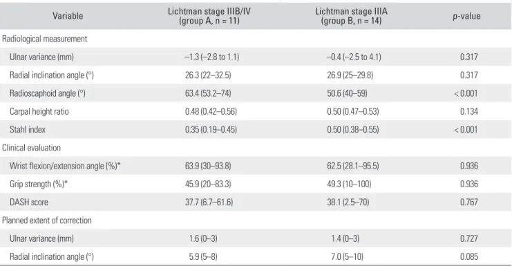
The preoperative radiological and clinical evaluations and corrective osteotomy planning are described in Table 2.
Ulnar variance, radial inclination angle, and carpal height ratio were not significantly different between two groups.
Ulnar variance was positive in 1, neutral in 2, and nega- tive in 8 patients of group A and positive in 2, neutral in 2, and negative in 10 patients of group B. Radioscaphoid angle and Stahl index were significantly different between two groups (p < 0.001, respectively). Clinical evaluations including the mean angle of wrist flexion/extension, grip strength, and DASH score were not significantly different between two groups. In addition, the planned correction of ulnar variance and radial inclination were not signifi- cantly different between two groups (p = 0.727 and p = 0.085, respectively).
Outcome Evaluation
The osteotomy site union was achieved in all patients.
There were no complications such as nonunion or infec- tion. Nine patients of group A and 11 patients of group B showed radiological improvement in the lunate regarding sclerosis, cystic changes, or fragmentation after operation:
sclerosis in 7, cystic changes in 5, and fragmentation in 8 patients of group A; and sclerosis in 10, cystic changes in 6, and fragmentation in 8 patients of group B. No progres- sion of the Lichtman stage was found in any patients, and no cases showed development and progression of arthritic changes of the radiocarpal and midcarpal joints at the final follow-up.
The radiological measurements and clinical evalu- ations at the final follow up are described in Table 3. The relationships between radiological parameters and preop- erative Lichtman stage were similar to those preoperatively observed. Ulnar variance and radial inclination angle were changed significantly after operation, but no significant differences were observed in the radioscaphoid angle, car-
Table 2. Preoperative Evaluation and Corrective Osteotomy Planning in Two Groups
Variable Lichtman stage IIIB/IV
(group A, n = 11) Lichtman stage IIIA
(group B, n = 14) p-value
Radiological measurement
Ulnar variance (mm) –1.3 (–2.8 to 1.1) –0.4 (–2.5 to 4.1) 0.317
Radial inclination angle (°) 26.3 (22–32.5) 26.9 (25–29.8) 0.317
Radioscaphoid angle (°) 63.4 (53.2–74) 50.6 (40–59) < 0.001
Carpal height ratio 0.48 (0.42–0.56) 0.50 (0.47–0.53) 0.134
Stahl index 0.35 (0.19–0.45) 0.50 (0.38–0.55) < 0.001
Clinical evaluation
Wrist flexion/extension angle (%)* 63.9 (30–93.8) 62.5 (28.1–95.5) 0.936
Grip strength (%)* 45.9 (20–83.3) 49.3 (10–100) 0.936
DASH score 37.7 (6.7–61.6) 38.1 (2.5–70) 0.767
Planned extent of correction
Ulnar variance (mm) 1.6 (0–3) 1.4 (0–3) 0.727
Radial inclination angle (°) 5.9 (5–8) 7.0 (5–10) 0.085
Values are presented as mean (range).
DASH: Disabilities of the Arm, Shoulder and Hand.
*Percentage of the affected to the normal contralateral hand.
Table 3. Radiological and Clinical Evaluations at Final Follow-up
Variable Lichtman stage IIIB/IV
(group A, n = 11) Lichtman stage IIIA
(group B, n = 14) p-value
Radiological measurement
Ulnar variance (mm) 1.1 (0–2.2) 1.0 (0–3.6) 0.403
Radial inclination angle (°) 21.1 (18.1–27.4) 21.0 (17.1–24.0) 0.609
Radioscaphoid angle (°) 61.8 (50.3–79.1) 51.8 (41.0–62.0) 0.004
Carpal height ratio 0.50 (0.44–0.58) 0.50 (0.46–0.54) 0.572
Stahl index 0.34 (0.25–0.41) 0.50 (0.34–0.55) < 0.001
Clinical evaluation
Wrist flexion/extension angle (%)* 84.5 (50–100) 80.3 (43.8–95.5) 0.149
Grip strength (%)* 79.8 (50–100) 84.8 (50–100) 0.267
DASH score 10.6 (0–28.3) 14.2 (0–38.3) 0.536
Pain VAS 1.2 (0–3) 1.1 (0–5) 0.687
NSSK score 21.6 (15–27) 21.8 (15–26) 0.893
DASH: Disabilities of the Arm, Shoulder and Hand, VAS: visual analogue scale, NSSK: Nakamura Scoring System.
*Percentage of the affected to the normal contralateral hand.
pal height ratio, and Stahl index between preoperative and final follow-up measurements both groups. The interob- server reproducibility of radiological measurements was excellent with an ICC of 0.951 for the ulnar variance, 0.868 for the radial inclination angle, 0.804 for the radioscaphoid angle, 0.978 for the carpal height ratio, and 0.988 for the Stahl index.
The mean angle of wrist flexion/extension, grip strength, and DASH score were significantly improved in both groups, but no significant difference was noted be- tween two groups at the final follow up (p = 0.149, p = 0.267 and p = 0.536, respectively). The mean pain VAS score during activities of daily living at the final follow-up was 1.2 (range, 0 to 3) in group A and 1.1 (range, 0 to 5) in group B. Four wrists in group A and 7 wrists in group B were as- ymptomatic. The mean NSSK was 21.6 (range, 15 to 27) in group A and 21.8 (range, 15 to 26) in group B at the final follow-up, showing no statistically significant difference between groups (p = 0.893): 3 were rated excellent, 7 good, and 1 fair in group A; and 4 were rated excellent, 9 good, and 1 fair in group B (Figs. 1 and 2).
DISCUSSION
Radial wedge osteotomy that decreases the radial incli- nation is known to reduce the pressure on the lunate by increasing the contact area between the diseased lunate and radius, shifting the load to the radial side of the wrist joint.6,11) The present study shows that radial wedge oste- otomy yielded excellent radiological and functional out-
comes in the treatment of advanced stages of Kienböck's disease and these results were comparable to those of stage IIIA Kienböck's disease.
Radial osteotomies have shown favorable clinical results in the treatment of Kienböck's disease, but these procedures could not restore the morphology of the col- lapsed lunate and the changed carpal alignment.7,11-15) Thus, the efficacy of radial osteotomies in advanced stages of Kienböck's disease has been controversial and salvage procedures have been recommended in these stages.8,10) However, salvage procedures, such as proximal row car- pectomy, limited carpal fusion, and wrist arthrodesis reduce the wrist ROM.3,8-10) In addition, these salvage pro- cedures could not halt the secondary arthritic changes in the radiocarpal and other joints around the wrist, despite symptomatic relief.9,10,25,26) Radial osteotomy is a relatively simple extra-articular procedure, and surgery itself does not influence the flexion/extension of the wrist joint. In addition, other procedures involving articular structures could be attempted later period of the disease without concern for previous operation’s scar adhesion.13,27)
Several studies have reported on the role of radial osteotomy in the treatment of advanced stage Kienböck's disease. Iwasaki et al.6) compared the results between radial shortening osteotomy and radial wedge osteotomy in Li- chtman stage IIIB and IV Kienböck’s disease patients. All patients showed good or excellent clinical results regard- less of procedures and 25% of patients (5 of 20 patients) showed radiological improvement of the lunate. However, they evaluated patients with a modified form of NSSK
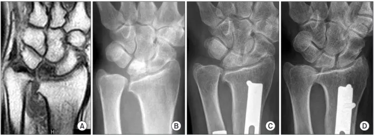
A B C D
Fig. 1. The preoperative T1-weighted magnetic resonance imaging (A) and posteroanterior (PA) radiograph (B) of a 51-year-old woman show stage IIIA Kienböck's disease in the left wrist. (C) The 5-year postoperative PA radiograph shows a decreased sclerotic lesion in the lunate. (D) The PA radiograph taken at the last visit at postoperative 11 years shows that the morphology of the lunate became nearly normalized. The bony trabecular pattern inside the lunate was similar to surrounding carpal bones. No progression of arthritic changes was visible in the radiocarpal and midcarpal joints.
that excluded the radiological evaluation, and the average follow-up period was rather short (29 months). Matsui et al.18) reported the results of radial shortening osteotomy and Soejima et al.16) reported the results of radial wedge osteotomy in advanced stages. Despite achievement of satisfactory clinical results in all patients, they denied the role of radial osteotomy for radiographic improvement of the lunate. Mozaffarian et al.8) reported poor results of ra- dial shortening osteotomy in patients with advanced stage Kienböck's disease (stage IV), but they did not evaluate the morphologic changes of the lunate.
We think that the excellent radiological and func- tional outcomes of our patients could be explained by the high proportion of patients receiving combined radial shortening and lateral closed wedge osteotomy. More than two third of patients in each group received combined os- teotomy as all of them showed negative ulnar variance. It is known that combined radial osteotomy could provide the benefits of 2 procedures simultaneously15) and prevent the complications related to each radial osteotomy. Concerns about overcorrection of bony morphology of the radiocar- pal and distal radioulnar joints could be reduced by use
of this technique. By controlling the shortening length of the radius under 3 mm, development of incongruence of the distal radioulnar joint and iatrogenic ulnar impaction syndrome could be avoided. By reducing the extent of cor- rection of the radial inclination angle under ten degrees, several known complications of radial wedge osteotomy, such as secondary radioscaphoid arthritic changes due to excessive load redistribution,6) compensatory excessive ul- nar deviation of the wrist,15) and development of limitation of forearm rotation,11) could be avoided.
The Lichtman classification system is based on radiological findings and does not reflect the clinical situ- ations.5) We think that the similar preoperative clinical presentation between group A and B could be associated with this limitation of the staging system. In addition, the preoperative Lichtman stage did not have any influence on the clinical and radiological outcomes of the operation in this study. The radioscaphoid angle of the wrist and preexisting arthritic changes of the radiocarpal joint were not improved significantly after radial wedge osteotomy despite clinical improvement of symptoms. This can be interpreted that carpal malalignment including scaphoid
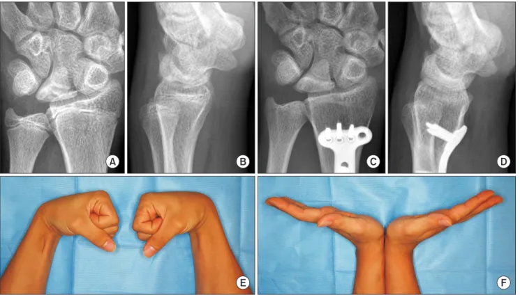
A B C D
E F
Fig. 2. The preoperative posteroanterior (PA) radiograph (A) and lateral radiograph (B) of a 15-year-old man show stage IIIB Kienböck's disease in the left wrist. Six-year postoperative PA radiograph (C) and lateral radiograph (D). Radiological improvement and height restoration were observed in the diseased lunate. No development of arthritic changes was visible in the radiocarpal and midcarpal joints. The photographs of the wrist in active volar flexion (E) and in active dorsiflexion (F) show a similar range of motion of the wrist joint compared to the contralateral side.
malrotation did not change after surgery. Our findings are compatible with those of previous studies on radial osteotomy reporting poor correlation between radiologic parameters and clinical outcomes.3,7,12)
Our study has several limitations. First, the number of patients was small. Second, 2 kinds of different plate and screw systems were used for osteotomy site fixation.
Thus, there is a need for larger prospective multi-center studies, incorporating objective measures for radial wedge osteotomy with other procedures.
In conclusion, radial wedge osteotomy yielded excel- lent radiological and functional outcomes in the treatment of advanced stage Kienböck's disease and these results were comparable to those of stage IIIA Kienböck's disease.
This procedure would be a useful alternative to the salvage procedures in the treatment of Lichtman stage IIIB and IV Kienböck's disease without severe radiocarpal arthritis.
CONFLICT OF INTEREST
No potential conflict of interest relevant to this article was reported.
ACKNOWLEDGEMENTS
We thank Joo Hyun Oh for her consultation for statistical analysis and English proofreading.
REFERENCES
1. Schuind F, Eslami S, Ledoux P. Kienbock's disease. J Bone Joint Surg Br. 2008;90(2):133-9.
2. Danoff JR, Cuellar DO, O J, Strauch RJ. The management of Kienbock disease: a survey of the ASSH membership. J Wrist Surg. 2015;4(1):43-8.
3. Innes L, Strauch RJ. Systematic review of the treatment of Kienbock's disease in its early and late stages. J Hand Surg Am. 2010;35(5):713-7.e1-4.
4. Lichtman DM, Mack GR, MacDonald RI, Gunther SF, Wil- son JN. Kienbock's disease: the role of silicone replacement arthroplasty. J Bone Joint Surg Am. 1977;59(7):899-908.
5. Goldfarb CA, Hsu J, Gelberman RH, Boyer MI. The Licht- man classification for Kienbock's disease: an assessment of reliability. J Hand Surg Am. 2003;28(1):74-80.
6. Iwasaki N, Minami A, Oizumi N, Suenaga N, Kato H, Min- ami M. Radial osteotomy for late-stage Kienbock's disease:
wedge osteotomy versus radial shortening. J Bone Joint Surg Br. 2002;84(5):673-7.
7. Weiss AP, Weiland AJ, Moore JR, Wilgis EF. Radial shorten- ing for Kienbock disease. J Bone Joint Surg Am. 1991;73(3):
384-91.
8. Mozaffarian K, Namazi H, Namdari A. Radial shortening osteotomy in advanced stages of Kienbock disease. Tech Hand Up Extrem Surg. 2012;16(4):242-6.
9. Rhee PC, Lin IC, Moran SL, Bishop AT, Shin AY. Scapho- capitate arthrodesis for Kienbock disease. J Hand Surg Am.
2015;40(4):745-51.
10. Nakamura R, Horii E, Watanabe K, Nakao E, Kato H, Tsu- noda K. Proximal row carpectomy versus limited wrist ar- throdesis for advanced Kienbock's disease. J Hand Surg Br.
1998;23(6):741-5.
11. Nakamura R, Tsuge S, Watanabe K, Tsunoda K. Radial wedge osteotomy for Kienbock disease. J Bone Joint Surg Am. 1991;73(9):1391-6.
12. Rodrigues-Pinto R, Freitas D, Costa LD, et al. Clinical and radiological results following radial osteotomy in patients with Kienbock's disease: four- to 18-year follow-up. J Bone Joint Surg Br. 2012;94(2):222-6.
13. Wada A, Miura H, Kubota H, Iwamoto Y, Uchida Y, Kojima T. Radial closing wedge osteotomy for Kienbock's disease:
an over 10 year clinical and radiographic follow-up. J Hand Surg Br. 2002;27(2):175-9.
14. Watanabe T, Takahara M, Tsuchida H, Yamahara S, Kiku- chi N, Ogino T. Long-term follow-up of radial shortening osteotomy for Kienbock disease. J Bone Joint Surg Am.
2008;90(8):1705-11.
15. Garcia-Elias M, An KN, Cooney WP, Linscheid RL. Lat- eral closing wedge osteotomy for treatment of Kienbock's disease: a clinical and biomechanical study of the optimum correcting angle. Chir Main. 1998;17(4):283-90.
16. Soejima O, Iida H, Komine S, Kikuta T, Naito M. Lat- eral closing wedge osteotomy of the distal radius for ad- vanced stages of Kienbock's disease. J Hand Surg Am.
2002;27(1):31-6.
17. Trumble T, Glisson RR, Seaber AV, Urbaniak JR. A biome- chanical comparison of the methods for treating Kienbock's disease. J Hand Surg Am. 1986;11(1):88-93.
18. Matsui Y, Funakoshi T, Motomiya M, Urita A, Minami M, Iwasaki N. Radial shortening osteotomy for Kienbock disease: minimum 10-year follow-up. J Hand Surg Am.
2014;39(4):679-85.
19. Muramatsu K, Ihara K, Kawai S, Doi K. Ulnar variance and the role of joint levelling procedure for Kienbock's disease.
Int Orthop. 2003;27(4):240-3.
20. Gong HS, Chung MS, Lee YH, Lee S, Lee JO, Baek GH.
Arthroplasty for advanced Kienbock's disease using a radial bone flap with a vascularised wrapping of pronator quadra- tus. J Bone Joint Surg Br. 2006;88(5):623-8.
21. Gelberman RH, Salamon PB, Jurist JM, Posch JL. Ulnar variance in Kienbock's disease. J Bone Joint Surg Am. 1975;
57(5):674-6.
22. Youm Y, McMurthy RY, Flatt AE, Gillespie TE. Kinemat- ics of the wrist: I. an experimental study of radial-ulnar deviation and flexion-extension. J Bone Joint Surg Am.
1978;60(4):423-31.
23. Stahl F. On lunatomalacia (Kienboock's disease) a clinical
and roentgenological study, especially on its pathogenesis and the late results of immobilization treatment. Lund, Swe- den: H. Ohlsson; 1947. 133.
24. Shrout PE, Fleiss JL. Intraclass correlations: uses in assessing rater reliability. Psychol Bull. 1979;86(2):420-8.
25. Luegmair M, Saffar P. Scaphocapitate arthrodesis for treat- ment of late stage Kienbock disease. J Hand Surg Eur Vol.
2014;39(4):416-22.
26. Croog AS, Stern PJ. Proximal row carpectomy for advanced Kienbock's disease: average 10-year follow-up. J Hand Surg Am. 2008;33(7):1122-30.
27. Kakinoki R, Yamakawa T, Nakayama K, Morimoto Y, Naka- mura T. Treatment of progressive necrosis of the lunate bone (Kienbock disease) after unsuccessful radial osteotomy.
Scand J Plast Reconstr Surg Hand Surg. 2007;41(5):267-71.