THE KOREAN JOURNAL OF HEMATOLOGY O R I G I N A L A R T I C L E
Clinical outcomes of Torque teno virus-infected thalassemic patients with and without hepatitis C virus infection
Samin Alavi
1, Zohreh Sharifi
2, Ali Kord Valeshabad
3, Kazem Nourbakhsh
1, Bibi Shahin Shamsian
1, Mohammad Taghi Arzanian
1, Alieh Safarisharari
1, Masoumeh Navidinia
41Pediatric Hematology-Oncology Department and Pediatric Infectious Research Center, Shahid Beheshti Medical University, Mofid Childrens' Hospital, 2Iranian Blood and Transfusion Organization, 3Student's Scientific Research Center (SSRC), Tehran University of Medical Sciences (TUMS), 4Pediatric Infectious Research Center, Shahid Beheshti Medical University, Tehran, Iran
p-ISSN 1738-7949 / e-ISSN 2092-9129 DOI: 10.5045/kjh.2011.46.2.123 Korean J Hematol 2011;46:123-7.
Received on April 22, 2011 Revised on June 9, 2011 Accepted on June 13, 2011
Background
Although a marked proportion of thalassemic patients acquire Torque teno virus (TTV) through blood transfusion, its clinical importance is unclear. This study was designed to investigate the clinical importance of TTV infection in thalassemic patients with and with- out hepatitis C virus (HCV) co-infection in Iran.
Methods
In this case-control study, 107 thalassemic patients on chronic transfusion and 107 healthy individuals were selected. According to HCV and TTV infection status (detected by semi-nested PCR), patients were categorized into 4 groups: TTV and HCV negative, TTV positive, HCV positive, and TTV and HCV positive. Blood ferritin, alanine amino- transferase (ALT), and aspartate aminotransferase (AST) levels in these 4 groups were assessed.
Results
Approximately half of the thalassemic patients (50.5%) and 27.1% of controls had TTV infection. Thalassemic patients had a greater chance of TTV infection compared to the control group with a sex-adjusted OR of 4.13 (95% CI=2.28-8.13). The increased levels of ALT, AST, and ferritin in the TTV and HCV-infected group were not significantly different from those in the TTV and HCV negative group. Co-infection with TTV and HCV did not significantly increase ALT, AST, and ferritin levels compared to infection with TTV alone.
Conclusion
Although common in thalassemic patients, TTV infection appears to have a negligible role in increasing the severity of liver disease, even when co-infection with HCV occurs.
Key Words Torque teno virus (TTV), Hepatitis C virus (HCV), Thalassemia
Correspondence to Ali Kord Valeshabad, M.D.
Medical Dormitory of TUMS, North Kargar Avenue, Tehran, Iran, MA 1439957181, P.O. Box: 13185-1678, Tehran, Iran
Tel: +9821-66439463 Fax: +9821-66919206
E-mail: [email protected]
Ⓒ 2011 Korean Society of Hematology
INTRODUCTION
Due to their dependency on blood transfusions, thalasse- mic patients are prone to transfusion-associated hepatitis, as a consequence of transfusion-related iron overload and exposure to viruses, which may cause hepatitis. The incidence of transfusion-related hepatitis in these patients has been substantially reduced since the implementation of blood screening for hepatitis B virus (HBV) and hepatitis C virus (HCV) nucleic acids and antibodies. However, a considerable proportion of thalassemic patients have raised levels of serum alanine aminotransferase (ALT), without a known causative
hepatitis virus [1, 2].
Initially, hepatitis G virus (HGV) was presumed to be the causative agent of transfusion-associated non-A-C-hep- atitis; however, this was not confirmed in subsequent studies.
In 1997, Nishizawa and co-workers isolated a novel single-s tranded DNA virus, designated TT virus (TTV), from patients with non A-E transfusion-acquired hepatitis, and which ap- peared to be associated with non A-G post-transfusion hep- atitis [3]. This virus was renamed Torque teno virus and was classified as a species of the Anellovirus genus in an unassigned family that is most closely related to the Circoviridae [4]. TTV infects the general population and particularly those at risk of parenteral exposure [5-8].
Transfusion-dependent patients, such as those with tha- lassemia, are more prone to carry TTV and most infections (80%) are of mixed genotypes [2, 9]. The prevalence of TTV in thalassemic patients varies in different studies and seems to be dependent on the diagnostic technique. Kondili et al. used two different sets of primers to detect TTV prevalence in 37 pediatric and young adult patients with thalassemia.
The first primer set found TTV in 73% of thalassemic patients, while the second set found TTV DNA in 100% of the patients [10]. TTV infection can be detected by polymerase chain reaction (PCR), in situ hybridization, and specific antibodies to TTV [11]. Most TTV infections appear to occur parent- erally but a substantial proportion of asymptomatic in- dividuals never exposed to blood-borne agents and the high prevalence of TTV, even among healthy children, implicate the fecal-oral route of transmission as being potentially im- portant [5, 12-14].
However, studies have not yet been able to confirm TTV as a primary cause of post-transfusion hepatitis, since most TTV-positive (TTV+) patients remain asymptomatic, and those progressing towards chronic liver disease are invariably co-infected with either HBV or HCV [5]. On the other hand, considering the fact that thalassemic patients are prone to acquiring various genotypes of TTV as a consequence of their life-long dependence on repeated blood transfusions, the importance of elucidating the potential role of this virus, if any, in development of clinical disease is highly important [2]. Hence, although a significant proportion of thalassemic patients acquire TTV by transfusion and some have co-in- fection with HCV, it remains uncertain if HCV and TTV co-infection leads to more severe hepatitis compared to TTV infection alone [15-20]. The clinical outcome of TTV in thalassemic patients is controversial, and there have been no previous studies in this regard in Iran. Thus, the current study is designed to investigate the prevalence and clinical importance of TTV infection in thalassemic patients with and without HCV co-infection in Iran.
MATERIALS AND METHODS
In this case-control study, a total of 107 thalassemic pa- tients, who received regular blood transfusions at the Thalassemia Center of Mofid Childrens' Hospital, Tehran, Iran, were selected as the patient group. One hundred and seven healthy volunteers were categorized as the control group. First, TTV DNA was assessed for all the patients and controls, and according to the HCV and TTV infection test results, cases were categorized into four groups: both TTV and HCV negative (TTV-HCV-), TTV positive alone (TTV+), HCV positive alone (HCV+), and TTV and HCV positive (TTV+HCV+). Blood ferritin, AST, and ALT levels in these 4 groups were assessed in order to determine the clinical outcome of TTV and HCV infections. In standard donor-screening tests, all the samples were negative for HIV, human T cell leukemia viruses, and HBV. Informed consent for serological studies was obtained from the patients or
their parents if the patients were under 18 years of age.
The study protocol was approved by the ethics committee of Tehran University of Medical Sciences (TUMS).
1. Virological and biochemical laboratory tests
Serum ALT and AST levels were measured using an auto- mated analyzer and values higher than 50 and 40 IU/L, respectively, were considered to be abnormal. Serum ferritin was determined by ELISA (IMx ferritin assay; Abbott Divi- sion Diagnostici, Rome, Italy) and because of its high plasma levels in thalassemic patients, values higher than 3,000 ng/mL were considered to be hyperferritinemia. Anti-HCV status was determined by a commercially available second gen- eration enzyme-linked ELISA (hepatitis C II; Abbott Labora- tories, North Chicago, IL, USA) and hepatitis B surface anti- gen (HBsAg) was determined by radioimmune assay (Abbot Laboratories, North Chicago, IL, USA). HCV RNA was de- tected by RT-PCR as described previously [21-23].
2. Isolation and determination of TTV DNA by PCR
Semi-nested PCR was used to detect serum TTV DNA.
Specifically, serum DNA purified from an equivalent of 7 μL of serum was amplified using the following PCR protocol in 9600 thermal cycler (Perkin-Elmer, Emeryville, CA, USA);
1 cycle at 95oC for 9 min, 35 cycles at 94oC for 30 s, 58oC for 30 s, 72oC for 45 s, 1 cycle at 72oC for 7 min. The reaction mix contained 30 pmol of each primer (sense NG059:
5'-ACA GAC AGA GGA GAA GGC AAC ATG-3', antisense NG063: 5'-CTG GCA TTT TAC CAT TTC CAA AGT-3') and 2.5 U of AmpliTaq Gold (Perkin-Elmer, Emeryville, CA) in a 50 μL reaction volume. Under the same conditions, the second round of PCR was performed using a semi-nested primer set (sense NG061; 5'-GGC AAC ATG TTA TGGATA GAC TGG-3', antisense NG063) and 5 μL of the amplification product. Multiple positive and negative controls were in- cluded in each PCR assay. PCR products were analyzed on 2% agarose gel with ethidium bromide staining. For all pos- itive samples of TTV DNA, separate assays were performed and sequences of PCR products were confirmed by automated sequencing on ABI 373 sequencer (Perkin-Elmer, Foster City, CA, USA).
3. Statistical analysis
Results were reported as the mean±standard deviation (SD) for quantitative variables and percentages for categorical variables. We categorized AST, ALT and ferritin levels into normal and abnormal groups using the clinical ranges de- scribed above. The groups were compared using Student’s t-tests for continuous variables and chi-square tests (or Fisher’s exact test, if required) for categorical variables.
Statistical significance was based on two-sided design-based tests with P≤0.05 being considered significant. All statistical analyses were performed using SPSS version 13 (SPSS Inc., Chicago, IL, USA) for Windows.
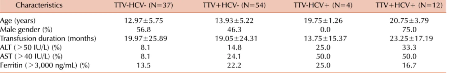
Table 1. Demographic and clinical data of 107 thalassemic patients according to TTV and HCV infection.
Characteristics TTV-HCV- (N=37) TTV+HCV- (N=54) TTV-HCV+ (N=4) TTV+HCV+ (N=12)
Age (years) 12.97±5.75 13.93±5.22 19.75±1.26 20.75±3.79
Male gender (%) 56.8 46.3 0.0 75.0
Transfusion duration (months) 19.97±25.89 19.05±24.31 13.75±15.37 23.25±17.19
ALT (>50 IU/L) (%) 8.1 14.8 25.0 33.3
AST (>40 IU/L) (%) 8.1 24.1 50.0 50.0
Ferritin (>3,000 ng/mL) (%) 13.5 22.2 25.0 16.7
Abbreviations: TTV, torque teno virus; HCV, Hepatitis C virus; ALT, alanine-aminotransferase; AST, aspartate-aminotransferase.
Table 2. The mean plasma levels of ALT, AST, and ferritin in 54 TTV-infected thalassemic patients of differing age, sex, and transfusion duration.
Characteristics ALT (IU/L) P AST (IU/L) P Ferritin (ng/mL) P
Age <15 years (N=28) 33.4±25.2 0.36 30.28±19.85 0.79 1,596.8±680.8 0.08
≥15 years (N=26) 43.4±50.4 32.42±23.57 4,042.8±7,205.3
Sex
Female (N=29) 30.2±15.7 0.11 26.79±11.85 0.08 2,024.6±1,726.2 0.25
Male (N=25) 47.5±54.4 37.16±28.27 3,644.5±7,281.3
Transfusion duration
<12 months (N=22) 34.0±19.6 0.40 30.82±16.52 0.79 2,133.1±1,729.6 0.35
RESULTS
In this study, 107 thalassemic patients (51.4% male) with a mean age of 14.61±5.96 years and an average transfusion duration of 19.64±23.60 months were selected as the patient group. A representative sample of 107 healthy volunteers (33.6% male) with a mean age of 13.13±6.37 years were selected as the control group. According to their HCV and TTV infection results, the patient group was divided into four subgroups (TTV-HCV-, TTV+, HCV+, and TTV+HCV+).
TTV infection was found in 27.1% of controls. Nearly two- thirds (65.4%) of thalassemic patients carried a virus of which approximately half (50.5%) had TTV infection alone and 11.2% had both TTV and HCV infection. HCV infection alone was found in 4 patients (3.7%).
Demographic and clinical data of the 4 groups is shown in Table 1. Seventy-five percent of TTV+HCV+ patients and 46.3% of TTV+ patients were male. Patients who were co-in- fected with TTV and HCV were older when compared to those infected with TTV alone (20.8±3.8 vs. 14.0±5.8 years, P<0.001) and tended to have been undergoing blood trans- fusions for longer (23.3±17.2 vs. 19.1±24.3 months, P=0.58), although this difference was not statistically significant.
Abnormal values of serum ALT (>50 IU/mL) and AST (>40 IU/mL) levels were more often found in TTV+HCV+ patients compared to the TTV+HCV- group (33.3% vs. 14.8% and 50.0% vs. 24.1%, respectively). This is in contrast to the incidence of hyperferritinemia (>3,000 ng/mL), which was observed more often in the TTV+HCV- group than in TTV+HCV+ patients, although was not statistically signifi- cant. An age >15 years, gender, or transfusion duration
of >12 months were not correlated with the incidence of infection with TTV alone. However, there was a significant relationship between older age (>15 years) and TTV-HCV co-infection (OR=1.86, 95% CI=1.30-2.65), although a similar relationship was not found between gender or transfusion duration (>12 months) and TTV-HCV co-infection.
In order to evaluate the influence of age, sex, and trans- fusion duration on clinical outcome in TTV+ patients, we compared mean plasma levels of ALT, AST, and ferritin in these patients under these categories (Table 2). However, we found no significant differences in these plasma bio- chemical markers with increased age, transfusion duration, or gender (P>0.05).
Thalassemic patients had a greater chance of TTV infection in comparison to the control group, with a pure OR of 3.93 (95% CI=2.16-7.13). This relationship was also con- firmed even when we considered sex as a probable confound- ing factor (OR=4.13, 95% CI=2.28-8.13), with males being significantly more prevalent among TTV patients than unin- fected controls (51.4% vs. 33.6%, respectively, P=0.012). In Table 3, the mean plasma levels of ALT, AST, and ferritin in the TTV-HCV-, TTV+HCV-, TTV-HCV+ and TTV+HCV+
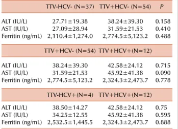
groups are compared. The increased levels of ALT, AST, and ferritin observed in the TTV+HCV- group did not differ statistically from those of the TTV-HCV- group. Further- more, co-infection with TTV and HCV did not significantly increase ALT, AST, or ferritin levels when compared with infection with either TTV or HCV alone.
Table 3. Comparison of the mean plasma levels of ALT, AST, and ferritin in TTV-HCV-, TTV+HCV-, TTV-HCV+, and TTV+HCV+
groups.
TTV-HCV- (N=37) TTV+HCV- (N=54) P ALT (IU/L) 27.71±19.38 38.24±39.30 0.158 AST (IU/L) 27.09±28.94 31.59±21.53 0.410 Ferritin (ng/mL) 2,110.4±1,274.0 2,774.5±5,123.2 0.488
TTV+HCV- (N=54) TTV+HCV+(N=12) ALT (IU/L) 38.24±39.30 42.58±24.12 0.715 AST (IU/L) 31.59±21.53 45.92±41.38 0.090 Ferritin (ng/mL) 2,774.5±5,123.2 2,324.3±2,473.7 0.778
TTV-HCV+(N=4) TTV+HCV+(N=12) ALT (IU/L) 38.50±14.27 42.58±24.12 0.75 AST (IU/L) 34.25±12.55 45.92±41.38 0.595 Ferritin (ng/mL) 2,532.5±1,445.5 2,324.3±2,473.7 0.888
DISCUSSION
The potential role and clinical importance of TTV infection in liver diseases, as a transfusion-transmitted agent in tha- lassemic patients and other transfusion-dependent diseases, has been discussed since the discovery of the virus in 1997.
The prevalence of TTV varies among thalassemic patients from different nations. Approximately half of the patients (50.5%) and more than a quarter of controls (27.1%) were found to have TTV infection. Hsu et al. also found 27.0%
of 122 healthy children in Taiwan to be infected with TTV [12]. In our study, thalassemic patients had a greater chance of TTV infection in comparison with the control group with a common OR of 4.31 (95% CI=2.28-8.13). This could reflect the greater importance of the parenteral route for virus trans- mission that has been noted in previous studies [5, 12, 24].
However, among infected subjects in the control group were healthy young children without any transfusion history or known diseases, suggesting a non-parenteral route of trans- mission for TTV. Epidemiological studies suggest that the main routes of TTV infection are parenteral, oral-fecal, and possibly salivary [14].
The prevalence of TTV among thalassemic patients varies in different studies and has been reported to be as high as 100% [10, 25-27], depending on diagnostic technique and specimen type. Chen et al. used peripheral blood mono- nuclear cells, plasma, saliva, and urine samples from 50 tha- lassemic patients for TTV detection. They found that all 50 patients had TTV in one or more specimens and that 16.0% of patients were positive for all specimen types [28].
In the current study, abnormal levels of ALT and AST were observed in a considerable proportion of TTV+ patients (14.8% and 24.1%, respectively) and in TTV+HCV+ patients (33.3% and 50.0%, respectively). Moreover, abnormal levels of these markers were also found in 8.1% of patients without TTV or HCV infection. The reason for this is not clear but
might be explained by liver disease associated with trans- fusion-related iron overload, the presence of undiagnosed TTV genotypes, or other blood-borne agents. Studies by Hu and co-workers confirm our findings that there is a higher incidence of abnormal ALT and AST levels in TTV+ patients (33.6% and 36.7%, respectively) and TTV+HCV+ patients (56.6% and 70.0%, respectively) [24]. They concluded that ALT, AST, and ferritin levels were invariably lower in TTV- patients than in TTV+ patients and that there are significantly elevated ferritin and ALT levels with TTV-HCV co-infection compared to TTV infection alone [24]. In the current study, TTV+HCV- patients tended to have increased levels of ALT and AST compared to the TTV-HCV- group, as did TTV+
HCV+ patients compared to the TTV+HCV- group, although these differences were not statistically significant. Some re- cent studies also demonstrated that co-infection with TTV and HCV in thalassemic patients does not alter the plasma profile of biochemical markers when compared with TTV infection alone [26]. The same pattern was also found for TTV+HCV+ patients compared with those infected with HCV alone. However, due to the low prevalence of HCV+
patients (4 patients), these findings should be considered with caution.
Assessing the severity of liver disease solely by measure- ments of plasma transaminase levels is inadequate. Histologi- cal data are also necessary, possibly with an estimated dura- tion of virus infection. However, due to the lack of histo- logical studies of patient tissues in the current study, we were not able to address this issue. In our study, to enhance cost-effectiveness ALT, AST, and ferritin levels were only measured in the control group. Regardless of whether TTV is a cause of liver disease in thalassemic patients, pathogenic mechanisms of the virus need to be rapidly elucidated in order to develop new strategies to prevent transmission and for therapeutic intervention. Targeted longitudinal studies of TTV in the future will be helpful in this regard [29].
On the basis of our study, it can be concluded that TTV, despite being widely distributed among thalassemic patients, appears to have a negligible role in increasing the severity of liver disease, even when co-infection with HCV occurs.
REFERENCES
1. Okamoto H, Mayumi M. TT virus: virological and genomic char- acteristics and disease associations. J Gastroenterol 2001;36:519- 29.
2. Chen BP, Rumi MG, Colombo M, et al. TT virus is present in a high frequency of Italian hemophilic patients transfused with plas- ma-derived clotting factor concentrates. Blood 1999;94:4333-6.
3. Nishizawa T, Okamoto H, Konishi K, Yoshizawa H, Miyakawa Y, Mayumi M. A novel DNA virus (TTV) associated with elevated transaminase levels in posttransfusion hepatitis of unknown etiology. Biochem Biophys Res Commun 1997;241:92-7.
4. Hino S. TTV, a new human virus with single stranded circular DNA genome. Rev Med Virol 2002;12:151-8.
5. Poovorawan Y, Tangkijvanich P, Theamboonlers A, Hirsch P.
Transfusion transmissible virus TTV and its putative role in the etiology of liver disease. Hepatogastroenterology 2001;48:256- 60.
6. Cossart Y. TTV - a virus searching for a disease. J Clin Virol 2000;17:1-3.
7. Simmonds P. Transfusion virology: progress and challenges.
Blood Rev 1998;12:171-7.
8. Blejer JL, Salamone HJ. Is TT virus (TTV) a true hepatitis virus cause? Medicina (B Aires) 2000;60:631-8.
9. Okamoto H, Takahashi M, Nishizawa T, et al. Marked genomic heterogeneity and frequent mixed infection of TT virus demon- strated by PCR with primers from coding and noncoding regions.
Virology 1999;259:428-36.
10. Kondili LA, Pisani G, Beneduce F, et al. Prevalence of TT virus in healthy children and thalassemic pediatric and young adult patients. J Pediatr Gastroenterol Nutr 2001;33:629-32.
11. Liweń I, Januszkiewicz-Lewandowska D, Nowak J. TT vi- rus-characteristics, occurrence and routes of transmission. Przegl Epidemiol 2002;56:91-9.
12. Hsu HY, Ni YH, Chen HL, Kao JH, Chang MH. TT virus infection in healthy children, children after blood transfusion, and children with non-A to E hepatitis or other liver diseases in Taiwan. J Med Virol 2003;69:66-71.
13. Davidson F, MacDonald D, Mokili JL, Prescott LE, Graham S, Simmonds P. Early acquisition of TT virus (TTV) in an area en- demic for TTV infection. J Infect Dis 1999;179:1070-6.
14. Yzèbe D, Xueref S, Baratin D, Boulétreau A, Fabry J, Vanhems P.
TT virus. A review of the literature. Panminerva Med 2002;44:
167-77.
15. Tokita H, Murai S, Kamitsukasa H, et al. High TT virus load as an independent factor associated with the occurrence of hep- atocellular carcinoma among patients with hepatitis C virus-re- lated chronic liver disease. J Med Virol 2002;67:501-9.
16. Meng XW, Komatsu M, Goto T, et al. Clinical significance of TT virus in chronic hepatitis C. J Gastroenterol Hepatol 2001;16:
202-8.
17. Cleavinger PJ, Persing DH, Li H, et al. Prevalence of TT virus in-
fection in blood donors with elevated ALT in the absence of known hepatitis markers. Am J Gastroenterol 2000;95:772-6.
18. Yuki N, Kato M, Masuzawa M, et al. Clinical implications of co- infection with a novel DNA virus (TTV) in hepatitis C virus car- riers on maintenance hemodialysis. J Med Virol 1999;59:431-6.
19. Charlton M, Adjei P, Poterucha J, et al. TT-virus infection in North American blood donors, patients with fulminant hepatic failure, and cryptogenic cirrhosis. Hepatology 1998;28:839-42.
20. Zein NN, Arslan M, Li H, et al. Clinical significance of TT virus infection in patients with chronic hepatitis C. Am J Gastroenterol 1999;94:3020-7.
21. Ni YH, Chang MH, Lue HC, et al. Posttransfusion hepatitis C virus infection in children. J Pediatr 1994;124:709-13.
22. Lai MW, Chang MH, Hsu HY. Non-A, non-B, non-C hepatitis: its significance in pediatric patients and the role of GB virus-C. J Pediatr 1997;131:536-40.
23. Chen HL, Chang MH, Ni YH, Hsu HY, Kao JH, Chen PJ. Hepatitis G virus infection in normal and prospectively followed post- transfusion children. Pediatr Res 1997;42:784-7.
24. Hu YW, Al-Moslih MI, Al Ali MT, et al. Clinical outcome of fre- quent exposure to Torque Teno virus (TTV) through blood trans- fusion in thalassemia patients with or without hepatitis C virus (HCV) infection. J Med Virol 2008;80:365-71.
25. Sampietro M, Tavazzi D, Martinez di Montemuros F, et al. TT virus infection in adult beta-thalassemia major patients. Haematologica 2001;86:39-43.
26. Ozyürek E, Ergünay K, Kuskonmaz B, et al. Transfusion-trans- mitted virus prevalence in Turkish patients with thalassemia.
Pediatr Hematol Oncol 2006;23:347-53.
27. Erensoy S, Sayiner AA, Türkoğlu S, et al. TT virus infection and genotype distribution in blood donors and a group of patients from Turkey. Infection 2002;30:299-302.
28. Chan PK, Chik KW, Li CK, et al. Prevalence and genotype dis- tribution of TT virus in various specimen types from thalassaemic patients. J Viral Hepat 2001;8:304-9.
29. Dhenain M, Boulétreau A, Bourguignon F, et al. The TT virus: re- view of the literature. Clin Invest Med 2000;23:355-65.