Introduction
Linear measurements are mainly used to assess the thickness and height of the alveolar crest as part of the preoperative evaluation for implant treatment, to measure the distance between anatomical landmarks in orthodon- tics and orthognathic surgery, and to estimate the size of pathological jaw lesions. The accuracy and reliability of linear measurements in cone-beam computed tomography (CBCT) images has been established in several published scientific studies,1,2-11 including studies where they have been obtained from CBCT-derived models.2,12
Ostensibly, in imaging of the oral and maxillofacial
complex, spatial resolution is an important parameter of image quality that appears to be of special interest for dental applications. As available spatial resolution also determines the accuracy with which anatomic detail can be measured, it also affects important procedures, such as the planning of dental implants or cephalometric analysis.
The spatial resolution of an image is directly influenced by the voxel size, which is the smallest unit in CBCT ima-
ges.13-16 To improve spatial resolution, the voxel size must
be reduced,17 and the CBCT images must be measured accurately. However, spatial resolution is affected by mul- tiple technical factors that make it difficult to predict from simple clues or to estimate from technical parameters.13 The presence of an increasing number of CBCT units on the market presents a need to understand and select from the various technical parameters.
The impact of variation in voxel size on CBCT-based
Intraobserver and interobserver reproducibility in linear measurements on axial images obtained by cone-beam computed tomography
Nathália Cristine da Silva1, Maurício Barriviera2, José Luiz Cintra Junqueira1, Francine Kühl Panzarella1, Ricardo Raitz1,*
1São Leopoldo Mandic Research Center, Department of Oral Radiology, College of Dentistry, São Paulo, SP, Brazil
2Department of Oral Radiology, College of Dentistry, Catholic University of Brasilia, São Paulo, SP, Brazil
ABSTRACT
Purpose: This study was performed to investigate the intra- and inter-observer variability in linear measurements with axial images obtained by PreXion(PreXion Inc., San Mateo, USA) and i-CAT(Imaging Sciences International, Xoran Technologies Inc., Hatfield, USA) CBCT scanners, with different voxel sizes.
Materials and Methods: A cylindrical object made from nylon with radiopaque markers(phantom) was scanned by i-CAT and PreXion 3D devices. For each axial image, measurements were taken twice in the horizontal(distance A-B) and vertical(distance C-D) directions, randomly, with a one-week interval between measurements, by four oral radiologists with five years or more experience in the use of these measuring tools.
Results: All of the obtained linear measurements had lower values than those of the phantom. The statistical analysis showed high intra- and inter-observer reliability(p=0.297). Compared to the real measurements, the measurements obtained using the i-CAT device and PreXion tomography, on average, revealed absolute errors ranging from 0.22 to 0.59mm and from 0.23 to 0.63mm, respectively.
Conclusion: It can be concluded that both scanners are accurate, although the linear measurements are underesti- mations, with no significant differences between the evaluators.(Imaging Sci Dent 2017; 47: 11-5)
KEY WORDS: Cone-Beam Computed Tomography; Dimensional Measurement Accuracy; Reproducibility of Results
Copyright ⓒ 2017 by Korean Academy of Oral and Maxillofacial Radiology
This is an Open Access article distributed under the terms of the Creative Commons Attribution Non-Commercial License(http://creativecommons.org/licenses/by-nc/3.0) which permits unrestricted non-commercial use, distribution, and reproduction in any medium, provided the original work is properly cited.
Imaging Science in Dentistry·pISSN 2233-7822 eISSN 2233-7830 Received May 23, 2016; Revised October 2, 2016; Accepted October 31, 2016
*Correspondence to : Prof. Ricardo Raitz
Rua Heitor Penteado, 1832, 101A, São Paulo, SP, Brazil, CEP 05438-300 Tel) 55-11-35675208, Fax) 55-11-35675208, E-mail) [email protected]
diagnostics has been discussed.17 As the PreXion CBCT device has a small voxel size and few studies18,19 have evaluated it, this study investigated the intraobserver and interobserver variability in linear measurements with axi- al images obtained by PreXion(PreXion Inc., San Mateo, CA, USA) and i-CAT(Imaging Sciences International, Xoran Technologies Inc., Hatfield, PA, USA), with differ- ent voxel sizes.
Materials and Methods
The phantom used for this study was based on that of a previous study.6 It was made from nylon, a slightly radi- opaque material with a low thermal expansion coefficient, in a cylindrical shape(5cm in diameter and 10cm in length). It had 4 channels, each 1mm in depth, arranged 90° apart on the longitudinal axis of the cylinder, and 10 channels, each with a depth of 1mm, arranged transversely 10mm apart.
The dimensional calibration of the phantom was per- formed using the PML-0024 measuring procedure with a universal measuring machine(Mitutoyo Sul Americana Ltda, Suzano, São Paulo, Brazil)(Fig. 1), at room tem- perature(20.0±0.5°C). The diameter of the phantom(the cylinder) was verified 4 times in 10 sections, as identified in the standard, in 2 coordinates, A/B and C/D, generating a report containing the resulting averages. The measure- ment uncertainty was U =0.01, based on a combined standard uncertainty, multiplied by a coverage factor of k=2.3 for a confidence level of approximately 95%. The average values obtained by dimensional calibration of the phantom were considered the real measurements(the gold standard).
To identify the “sections” of the phantom selected for
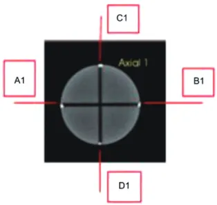
the experiment, zinc oxide and eugenol dotted portions were added at each channel intersection(Fig. 2), and the letters A, B, C, and D and the numbers 1 through 10 were applied for identification(Fig. 3).6
After obtaining the real measurements, the phantom was scanned in the i-CAT device, following an image ac- quisition protocol of a 6-cm×16-cm field of view(FOV), 20 seconds, and a 0.2-mm voxel size. In the PreXion de- vice, the phantom was scanned following an image ac- quisition protocol of 5-cm×5-cm FOV, 19 seconds, and a 0.1-mm voxel size. Thereafter, multiplanar reformatting was performed, thereby obtaining axial sections with a thickness of 1.0mm. The data were saved in the Digital Imaging and Communications in Medicine format using Prexion3DViewer software version 2.4.1.20(PreXion Inc., San Mateo, CA, USA) to measure the diameter of each axial image.
The diameter of the phantom was measured in sections, using 2 coordinates(An/Bn and Cn/Dn) with numbers from 2 to 7, in the depth of the channel, that is, between
Fig. 1. The universal measuring machine.
Fig. 2. Phantom image with the coordinates A1, B1, C1, and D1.
Fig. 3. Identifying coordinates of the phantom.
the “points” marked with zinc oxide and eugenol. This measurement was performed using a software tool. For each axial image, measurements were made in either hori- zontal direction(the A-B distance) and in either vertical direction(the C-D distance), twice, with a 1-week interval, by 4 examiners(4 oral radiologists) with 5 years or more experience in the use of these measuring tools. All post- processing procedures were carried out using the same software, Prexion3DViewer version 2.4.1.20, and the im- ages were visualized on a 24-inch LG E 2241 LCD moni- tor with a matrix resolution of 1920×1080. The values of the 2 repetitions for each coordinate were entered into an Excel spreadsheet(Microsoft, Seattle, WA, USA), sepa- rated by examiner, by scanner, by section, and by distance (A-B and C-D).
Statistical analysis was applied to the collected data using SPSS Statistics for Windows,(version 20.0; IBM Corp., Armonk, NY, USA). Intraclass correlation coeffi- cient(ICC) calculations were used to verify intraobserver and interobserver agreement and to compare the accuracy (reproducibility) of the i-CAT and PreXion CBCT with the real measurements(the gold standard). The linear mea- surements(mm) were subjected to descriptive analysis to present the absolute and relative errors(in %) of the measurements performed on the acquisitions made with both CBCT machines in relation to the gold standard. The Student t-test was used to compare the scans to each other regarding the linear measurements. The significance level used for all tests was 5%(α=.05).
Results
The ICC calculations revealed that the intra-observer agreement was excellent(ICC=1.000) when comparing the linear measurements performed at the 2 image evalu- ation stages, for both those obtained with the i-CAT scan- ner and for those with the PreXion scanner, for the 4 ex- aminers.
The inter-observer agreement when using the i-CAT or PreXion scanner was excellent(ICC=1.000) in all com- parisons between the 4 examiners.
Table 1 presents the descriptive analysis, in terms of the mean, standard deviation, and absolute and relative errors (%) of the linear measurements(mm) of the cylindrical object(the phantom) obtained with the use of the i-CAT and PreXion computed tomography scanners.
The measurements obtained in tomographic acquisition using the i-CAT device, on average, showed absolute er- rors ranging from 0.22mm to 0.59mm when compared with the real measurements, which were underestimated.
The linear measurements taken from acquisitions made with the PreXion scanner also underestimated the linear dimensions, with absolute errors ranging from 0.23mm to 0.63mm.
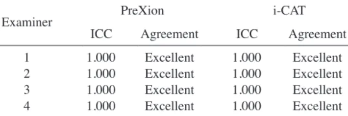
For the ICCs, it was found that with both scanners (i-CAT and PreXion), the accuracy of the linear measure- ments compared to the real measurements(the gold standard) was excellent(Table 2). When comparing the CBCTs to each other, no significant difference was found (p=.297).
Discussion
The use of CBCT technology to evaluate structures of the oral and maxillofacial complex increases the accuracy
Table 1. The means standard deviations and absolute and relative errors(%) of the linear measurements(mm) of the cylindrical object, ac- cording to each examiner
Examiner i-CAT PreXion
Mean Absolute error* Relative error** Mean Absolute error* Relative error**
12 34
48.23(0.11) 48.59(0.19) 48.22(0.10) 48.25(0.12)
-0.58(0.20) -0.22(0.26) -0.59(0.20) -0.56(0.23)
-1.19(0.40) -0.45(0.53) -1.21(0.40) -1.15(0.47)
48.23(0.13) 48.58(0.14) 48.18(0.09) 48.41(0.12)
-0.53(0.17) -0.23(0.21) -0.63(0.20) -0.40(0.23)
-1.08(0.34) -0.47(0.42) -1.29(0.41) -0.82(0.46)
*: Obtained by subtracting the measurements derived from the use of each scanner from those designated as the gold standard, **: Obtained by the ratio of the absolute errors and the gold standard measures
Table 2. Agreement between the linear measurements obtained with the use of i-CAT and PreXion CBCT scanners and the real measurements(gold standard), according to each examiner
Examiner PreXion i-CAT
ICC Agreement ICC Agreement 12
34
1.000 1.000 1.000 1.000
Excellent Excellent Excellent Excellent
1.000 1.000 1.000 1.000
Excellent Excellent Excellent Excellent ICC: intra-class correlation coefficient.
and reliability of the measurements, because of its ability to manipulate the resulting digital data volumes across 3 different views(sagittal, coronal, and axial) and to sel- ectively contrast, emphasize, and reduce the images to view certain anatomical structures.20 This study evaluated whether voxel size affected the intra-observer and inter- observer variability of the linear measurements obtained from the axial images of 2 CBCT devices.
In our study, the intra-observer and inter-observer agree- ment values, when using the i-CAT or PreXion scanner, were excellent in all comparisons of the 4 examiners (ICC=1.000), as shown in Tables 1 and 2. High interob- server and intraobserver CBCT reliability was reported by Moshfeghi et al.11 and Kamburoğlu et al.7, while in the study by Nikneshan et al.1, reproducibility for all orienta- tions was greater than 0.9.
In a comparison of the different devices, it was found that there was no significant difference(p=.297) despite the difference between the voxel sizes(0.1mm for the PreXion and 0.2mm for the i-CAT). Lukat et al.21 have already reported this result and emphasized that high spa- tial resolution allows for the thorough detection of subtle structures, such as small changes in the cortical or the buccal bone of the temporomandibular joint.3,5,22
Measurements for preoperative implant site assessment taken from CBCT images should be considered accurate when the error was less than 1mm.4,15 Owing to the me- chanical construction of CBCT machines, which require a relatively slow trajectory of the source image detector unit around the patient’s head, even slight patient move- ment seems almost inevitable. Given the small voxel sizes applied in the machines, a slight movement of more than 1 voxel will lead to errors in reconstruction.13 Therefore, during clinical application, one should not expect greater accuracy than half a millimeter at best. If in doubt, users should avoid sub-millimeter accuracy and preferably add some margin of error to their planning.
Table 1 shows the values of the measurements obtained using the i-CAT apparatus, on average, which produced absolute errors ranging from 0.22mm to 0.59mm com- pared with the actual measurements, which were under- estimated. As for the PreXion device, the measurements were lower than the actual linear dimensions, with ab- solute error varying between 0.23mm and 0.63mm, in accordance with the results of the study from Waltrick et al.14 that reported mean absolute errors of 0.23±0.2mm using voxel sizes of 0.2mm, 0.3mm, and 0.4mm.
Using ICCs, it was found that the accuracy of the linear measurements versus the gold standard was excellent for
both devices(Table 2), although all linear measurements were underestimated by the examiners, corroborating the results of several studies described in the literature.4,6,7,9-11,23
In addition, thin bony structures of less than 0.5mm in size are not relevant in actual clinical practice, which compromises the reliability of the CBCT images3,10 once areas without bone are considered. This lack of precision leading to values that underestimate the real measurements may be explained by the partial volume effect.24-27 This artifact results when the voxel reproduces an average val- ue between the true values for 2 objects(e.g. tooth and surrounding air) with distinctive density. Therefore, the interpretation becomes questionable.
One of the most common voxel sizes used in orthodon- tics is 0.3mm, although it should be used with caution if the goal is to assess small variations in bone thickness. A smaller voxel size would be more appropriate for these studies when it is possible to minimize the influence of partial volume averaging.27,28 Reducing the voxel size results in an increased dose of radiation, which has an ef- fect on the biological risk of X-ray exposure, especially in children.28
In the present study, known and calibrated measure- ments from a geometric phantom from a previous study were used as the gold standard.6 This provides the advan- tage of high reproducibility without exposure of living tissue. However, it was not possible to reproduce some physical phenomena that occur in anthropometric and clinical trials, such as attenuation of the radiation beam, dispersed radiation, and noise.
High-resolution protocols provide images rich in detail and are associated with high levels of X-radiation; thus, further clinical studies are necessary. In conclusion, the intraobserver and interobserver variability and the accu- racy of linear measurements in axial CBCT images were excellent. There were no differences in terms of the per- formance of the CBCT scanners.
Acknowledgements
The authors wish to thank Mr. Carlos Kazuo Suetake for providing technical information and Mr. Abmael Henrique dos Santos and Mr. Bruno S. J da Silva for assistance with the phantom calibration at Mitutoyo Sul American Ltda.
References
1. Nikneshan S, Aval SH, Bakhshalian N, Shahab S, Moham- madpour M, Sarikhani S. Accuracy of linear measurement
using cone-beam computed tomography at different recon- struction angles. Imaging Sci Dent 2014; 44: 257-62.
2. Wikner J, Hanken H, Eulenburg C, Heiland M, Gröbe A, As- saf AT, et al. Linear accuracy and reliability of volume data sets acquired by two CBCT-devices and an MSCT using vir- tual models: a comparative in-vitro study. Acta Odontol Scand 2016: 74; 51-9.
3. Leung CC, Palomo L, Griffith R, Hans MG. Accuracy and reliability of cone-beam computed tomography for measuring alveolar bone height and detecting bony dehiscences and fenestrations. Am J Orthod Dentofacial Orthop 2010; 137(4 Suppl): S109-19.
4. Ganguly R, Ruprecht A, Vincent S, Hellstein J, Timmons S, Qian F. Accuracy of linear measurement in the Galileos cone beam computed tomography under simulated clinical condi- tions. Dentomaxillofac Radiol 2011; 40: 299-305.
5. Patcas R, Markic G, Müller L, Ullrich O, Peltomäki T, Kellen- berger CJ, et al. Accuracy of linear intraoral measurements using cone beam CT and multidetector CT: a tale of two CTs.
Dentomaxillofac Radiol 2012; 41: 637-44.
6. Panzarella FK, Junqueira JL, Oliveira LB, de Araújo NS, Costa C. Accuracy assessment of the axial images obtained from cone beam computed tomography. Dentomaxillofac Radiol 2011; 40: 369-78.
7. Kamburoğlu K, Kolsuz E, Kurt H, Kiliç C, Özen T, Paksoy CS. Accuracy of CBCT measurements of a human skull. J Digit Imaging 2011; 24: 787-93.
8. Sabban H, Mahdian M, Dhingra A, Lurie AG, Tadinada A.
Evaluation of linear measurements of implant sites based on head orientation during acquisition: an ex vivo study using cone-beam computed tomography. Imaging Sci Dent 2015;
45: 73-80.
9. Sheikhi M, Dakhil-Alian M, Bahreinian Z. Accuracy and reli- ability of linear measurements using tangential projection and cone beam computed tomography. Dent Res J(Isfahan) 2015;
12: 271-7.
10. Thönissen P, Ermer MA, Schmelzeisen R, Gutwald R, Metzger MC, Bittermann G. Sensitivity and specificity of cone beam computed tomography in thin bony structures in maxillofacial surgery-A clinical trial. J Craniomaxillofac Surg 2015; 43:
1284-8.
11. Moshfeghi M, Tavakoli MA, Hosseini ET, Hosseini AT, Hos- seini IT. Analysis of linear measurement accuracy obtained by cone beam computed tomography(CBCT-NewTom VG).
Dent Res J(Isfahan) 2012; 9(Suppl 1): S57-62.
12. Maroua AL, Ajaj M, Hajeer MY. The accuracy and reproduc- ibility of linear measurements made on CBCT-derived digital models. J Contemp Dent Pract 2016; 17: 294-9.
13. Brüllmann D, Schulze RK. Spatial resolution in CBCT ma- chines for dental/maxillofacial applications-what do we know today? Dentomaxillofac Radiol 2015; 44: 20140204.
14. Waltrick KB, Nunes de Abreu Junior MJ, Corrêa M, Zastrow MD, Dutra VD. Accuracy of linear measurements and visibil- ity of the mandibular canal of cone-beam computed tomog- raphy images with different voxel sizes: an in vitro study. J Periodontol 2013; 84: 68-77.
15. Torres MG, Campos PS, Segundo NP, Navarro M, Crusoé-Re-
bello I. Accuracy of linear measurements in cone beam com- puted tomography with different voxel sizes. Implant Dent 2012; 21: 150-5.
16. Watanabe H, Honda E, Tetsumura A, Kurabayashi T. A com- parative study for spatial resolution and subjective image characteristics of a multi-slice CT and a cone-beam CT for dental use. Eur J Radiol 2011; 77: 397-402.
17. Spin-Neto R, Gotfredsen E, Wenzel A. Impact of voxel size variation on CBCT-based diagnostic outcome in dentistry: a systematic review. J Digit Imaging 2013; 26: 813-20.
18. Menezes RF, Araújo NC, Santa Rosa JM, Carneiro VS, Santos Neto AP, Costa V, et al. Detection of vertical root fractures in endodontically treated teeth in the absence and in the presence of metal post by cone-beam computed tomography. BMC Oral Health 2016; 16: 48.
19. Khoury HJ, Andrade ME, Araujo MW, Brasileiro IV, Kramer R, Huda A. Dosimetric study of mandible examinations per- formed with three cone-beam computed tomography scanners.
Radiat Prot Dosimetry 2015; 165: 162-5.
20. Dalmau E, Zamora N, Tarazona B, Gandia JL, Paredes V. A comparative study of the pharyngeal airway space, measured with cone beam computed tomography, between patients with different craniofacial morphologies. J Craniomaxillofac Surg 2015; 43: 1438-46.
21. Lukat TD, Perschbacher SE, Pharoah MJ, Lam EW. The ef- fects of voxel size on cone beam computed tomography imag- es of the temporomandibular joints. Oral Surg Oral Med Oral Pathol Oral Radiol 2015; 119: 229-37.
22. Liedke GS, Spin-Neto R, da Silveira HE, Schropp L, Stavro- poulos A, Wenzel A. Factors affecting the possibility to detect buccal bone condition around dental implants using cone beam computed tomography. Clin Oral Implants Res(in press).
23. Shokri A, Khajeh S. In vitro comparison of the effect of different slice thicknesses on the accuracy of linear measure- ments on cone beam computed tomography images in implant sites. J Craniofac Surg 2015; 26: 157-60.
24. Glover GH, Pelc NJ. Nonlinear partial volume artifacts in x-ray computed tomography. Med Phys 1980; 7: 238-48.
25. Baumgaertel S, Palomo JM, Palomo L, Hans MG. Reliability and accuracy of cone-beam computed tomography dental measurements. Am J Orthod Dentofacial Orthop 2009; 136:
19-28.
26. Sun Z, Smith T, Kortam S, Kim DG, Tee BC, Fields H. Effect of bone thickness on alveolar bone-height measurements from cone-beam computed tomography images. Am J Orthod Den- tofacial Orthop 2011; 139: e117-27.
27. Maret D, Telmon N, Peters OA, Lepage B, Treil J, Inglèse JM, et al. Effect of voxel size on the accuracy of 3D recon- structions with cone beam CT. Dentomaxillofac Radiol 2012;
41: 649-55.
28. Ganguly R, Ramesh A, Pagni S. The accuracy of linear mea- surements of maxillary and mandibular edentulous sites in cone-beam computed tomography images with different fields of view and voxel sizes under simulated clinical conditions.
Imaging Sci Dent 2016; 46: 93-101.