• 검색 결과가 없습니다.

This study aimed to evaluate the result of various treatment on exposed surface of implant.

N/A
N/A
Protected

Academic year: 2021

Share "This study aimed to evaluate the result of various treatment on exposed surface of implant."

Copied!
10
0
0

로드 중.... (전체 텍스트 보기)

전체 글

(1)

Dept. of Oral and Maxillofacial Surgery, Section of dentistry, Seoul National University Bundang Hospital, Korea

Abstract

Purpose: The implant surface would be exposed during healing period by several causes as marginal bone resorption and inadequate drilling. Implant surface exposure can cause accumulation of plaque and microbials around implant and can develop peri-implantitis. Therefore, surgeon should be fully aware of adequate management of exposed surface of implant.

This study aimed to evaluate the result of various treatment on exposed surface of implant.

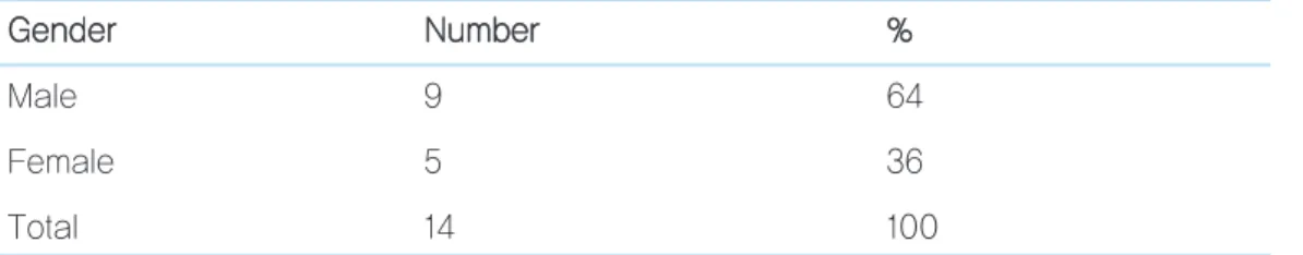
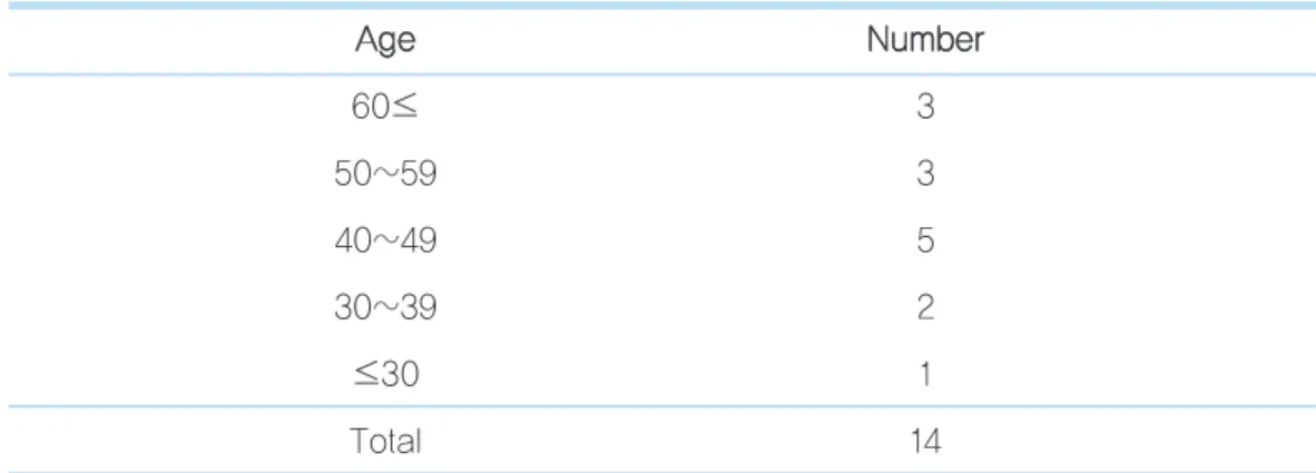
Material and methods: A retrospective chart review and radiologic analysis were conducted of 25 fixtures among 14 patients(male 9, female 5) during period from June 2007 to January 2008. We evaluated implant survival rate, soft tissue index and marginal bone loss after functional loading.

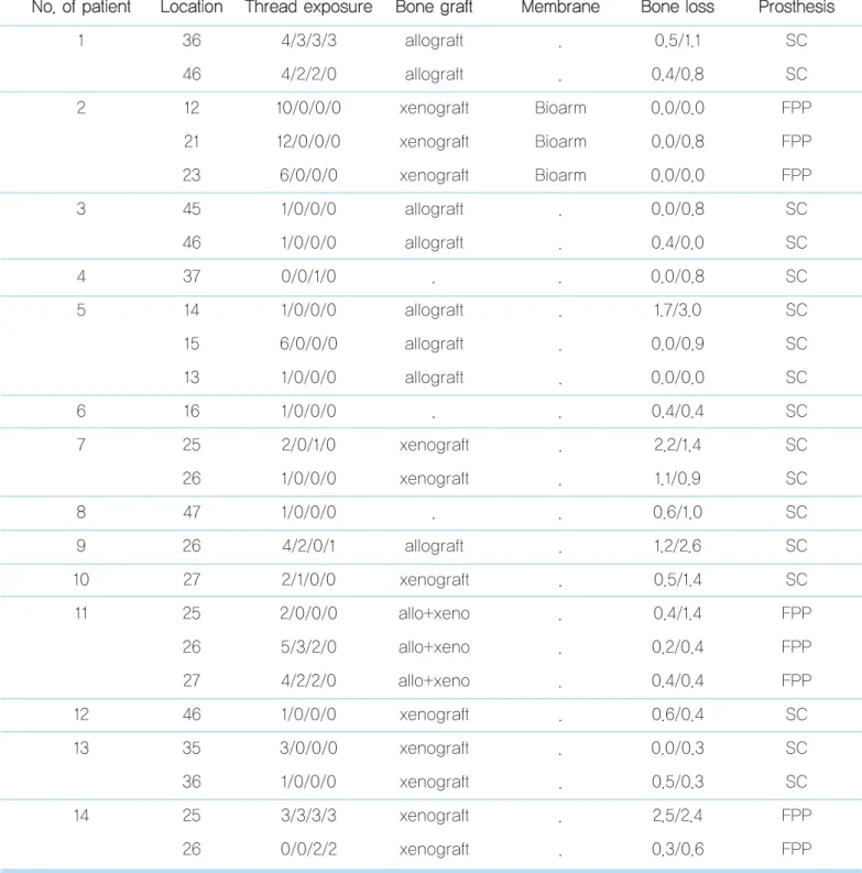
Results: In second surgery, measurement of implant thread exposure was 3.04mm ± 2.97 on buccal, 0.36 ± 0.9mm on lin- gual, 0.64 ± 1.11mm mesial, 0.64 ± 1.03mm distal in average. In 25 cases, bone graft was done on exposed area in 22 cases.

Allograft mixed with alloplast was used in 3 cases. Xenograft was used in 11 cases. And alloplast was used in 8 cases. The cases showed 100% survival rate and functioned stably during follow-up period.

Conclusion: We suggest that implant with exposed surface can function successfully with adequate treatment.

Key Words: thread exposure, bone graft

Treatment of implant with exposed threads during second surgery

Ji-Young Lee, Young-Kyun Kim

2차 수술 시 노출된 임프란트 나사산에 대한 처치

이지영, 김영균

분당서울대학교 병원 치과 구강악안면외과

(2)

적절한 위치의 임프란트 식립, 불량한 골질 및 골 양등은 식립된 임프란트 표면의 조기 노출의 원 인이 될 수 있다. 또한 일차 수술 시 임프란트 표 면이 노출되거나 골 접촉이 부족했던 경우 등에서 임프란트 의 변연골 흡수가 가속화 되어 조기 노출이 야기 될 수 있다.

이러한 조기 노출이 반드시 임프란트 실패와 연결되는 것은 아니지만, 치태 및 세균의 침착이 심화될 수 있고 지속적인 골 흡수와 임프란트 주위염으로 이어질 가능성이 높다.

1)

따라서 술자는 노출된 임프란트 표면에 대한 적절한 처치 및 관리에 대해 숙지 할 필요가 있다. 본 연구에서는 2차 수술 시 노출된 임프란트 나사산에 대한 다양한 처치 증례 및 임프란트 생존 률, 보철 후 골 흡수 등에 대한 결과를 보고하고 이차 수술 시 노출된 나사산의 처치 방법 및 그 적응증에 대해 고찰 해 보고 자 한다.

2007년 6월 부터 2008년 1월 까지 분당서울대학교병원 치 과에서 1인의 치과의사에게 임프란트 시술을 받은 후 2차 수

에서 2차 수술 시 나사산 노출이 관찰 되었다. 대상은 남자가 9명, 여자 5명이었으며 환자들의 나이는 21세부터 66세까지 로 평균 46.5세였다(Table 1, 2). 총 6명의 환자가 수술에 영 향을 미칠 수 있는 전신 질환을 보유하였으며 이 중 2명의 환 자는 2가지 이상의 전신질환을 보유하고 있었다. 전신 질환은 고혈압, 당뇨병, 심장질환, 갑상선질환, 간질환 등이었고 모 두 내과적으로 잘 조절 되었다. 흡연가는 1명이 있었다(Table 3). 평균 경과 관찰 기간은 13개월이었고 보철 후 6개월 이상 지난 증례들을 대상으로 하였다. 1차 수술 후 치유기간은 최 소 3개월에서 최대 5개월로 평균 4개월이었고, 2차 수술 후 보철물 제작까지의 기간은 최소 17일에서 최대 5개월로 평균 2개월 이었다. 2mm 이상 나사산이 노출된 증례에서 골이식 을 시행하였으며 1mm 노출된 증례는 골이식을 시행하지 않 았으나 예외적으로 협측에서 나사산이 노출된 증례는 골이식 을 시행하였다.

증례별로 사용된 임프란트의 길이 및 직경, 각 부위별 노출 된 정도, 최종 보철물 장착 후 주변조직 상태, 술 후 합병증, 노출된 부위의 처치 방법 등을 평가하였다. 그리고 1차 수술, 2차 수술, 보철 6개월 후 방사선 사진을 촬영하여 임프란트 변연골의 흡수 정도를 평가하였다. 변연골의 흡수는 평행 촬 영법으로 촬영된 치근단 방사선 사진상 임프란트 shoulder에 서부터 근원심의 치조정 부위 중 뚜렸하게 방사선 불투과성

Table 1. Gender distribution G

Ge en nd de err N Nu um mb be err % %

Male 9 64

Female 5 36

Total 14 100

Ji-Young Lee et al: Treatment of implant that have exposed threads during second surgery. Implantology 2009

(3)

original article

이 관찰되는 부위까지의 높이를 수직으로 골높이를 측정하였 으며 근원심의 높이를 구하여 그 평균을 계산하였다. 방사선 사진 상 각 임프란트의 높이를 측정하여 확대율을 고려하여 변연골 흡수량을 계산하였다. 그리고 1차 수술 후 골흡수량과 보철 기능 후의 골 흡수량의 차이를 비교하여 기술 하였다.

이차 수술 시 임프란트의 각 부위별 노출량은 수술 시 피판 거상 후 치주 프루브(periodontal probe)를 사용하여 협,설, 근, 원심측에서 측정하였다.

사용된 임프란트 종류 및 직경과 길이는 Table 4, 5와 같다.

3 증례에서는 노출부위에 특별한 처치를 시행하지 않았으며 22 증례에서 노출된 부위에 골이식재를 사용하였다. 골이식 이 시행된 증례들 중 3 증례에서 흡수성 차단막이 사용되었 다. 동종골은 oragraft

(LifeNet,U.S.A) orthoblast

II

Table 2. Age distribution A

Ag ge e N Nu um mb be err

60≤ 3

50~59 3

40~49 5

30~39 2

≤30 1

Total 14

Ji-Young Lee et al: Treatment of implant that have exposed threads during second surgery. Implantology 2009

Table 3. Type of Systemic Disease S

Sy ys stte em miic c d diis se ea as se e a an nd d s sm mo ok kiin ng g N Nu um mb be err

Hypertension 2

Hypertension+Diabetes mellitus 2

Liver disease 1

Endocrine, disease 1

Total 6

Ji-Young Lee et al: Treatment of implant that have exposed threads during second surgery. Implantology 2009

(4)

Table 5. Length and Diameter of Implant

IIm mp plla an ntt lle en ng gtth h((m mm m)) N Nu um mb be err D Diia am me ette err((m mm m)) N Nu um mb be err

8~8.5 2 3.4~3.8 6

10~10.5 7 4~4.3 6

11~11.5 5 4.5~4.8 7

12 4 5~5.3 6

13 4

14 3

Total 25 25

Ji-Young Lee et al: Treatment of implant that have exposed threads during second surgery. Implantology 2009

Implantium 14

Oneplant 2

Total 25

• GS II (Osstem, Korea), Implantium (Dentium, Korea)

• Oneplant (Warantec, Korea)

Ji-Young Lee et al: Treatment of implant that have exposed threads during second surgery. Implantology 2009

Table 6. Type of Graft Material B

Bo on ne e g grra afftt m ma atte erriia all N Nu um mb be err((IIm mp plla an ntt))

Allograft 8

Xeno 11

Xeno+Allograft 3

Total 22

• Xeno: Xenogenic bone Allo: Allogenicbone

Ji-Young Lee et al: Treatment of implant that have exposed threads during second surgery. Implantology 2009

(5)

original article

Table 7. Overview of data N

No o.. o off p pa attiie en ntt L Lo oc ca attiio on n T Th hrre ea ad d e ex xp po os su urre e B Bo on ne e g grra afftt M Me em mb brra an ne e B Bo on ne e llo os ss s P Prro os stth he es siis s

1 36 4/3/3/3 allograft . 0.5/1.1 SC

46 4/2/2/0 allograft . 0.4/0.8 SC

2 12 10/0/0/0 xenograft Bioarm 0.0/0.0 FPP

21 12/0/0/0 xenograft Bioarm 0.0/0.8 FPP

23 6/0/0/0 xenograft Bioarm 0.0/0.0 FPP

3 45 1/0/0/0 allograft . 0.0/0.8 SC

46 1/0/0/0 allograft . 0.4/0.0 SC

4 37 0/0/1/0 . . 0.0/0.8 SC

5 14 1/0/0/0 allograft . 1.7/3.0 SC

15 6/0/0/0 allograft . 0.0/0.9 SC

13 1/0/0/0 allograft . 0.0/0.0 SC

6 16 1/0/0/0 . . 0.4/0.4 SC

7 25 2/0/1/0 xenograft . 2.2/1.4 SC

26 1/0/0/0 xenograft . 1.1/0.9 SC

8 47 1/0/0/0 . . 0.6/1.0 SC

9 26 4/2/0/1 allograft . 1.2/2.6 SC

10 27 2/1/0/0 xenograft . 0.5/1.4 SC

11 25 2/0/0/0 allo+xeno . 0.4/1.4 FPP

26 5/3/2/0 allo+xeno . 0.2/0.4 FPP

27 4/2/2/0 allo+xeno . 0.4/0.4 FPP

12 46 1/0/0/0 xenograft . 0.6/0.4 SC

13 35 3/0/0/0 xenograft . 0.0/0.3 SC

36 1/0/0/0 xenograft . 0.5/0.3 SC

14 25 3/3/3/3 xenograft . 2.5/2.4 FPP

26 0/0/2/2 xenograft . 0.3/0.6 FPP

• Bone defect: Buccal/Lingual/Mesial/Distal

• Bone loss: Mesial/ Distal

• SC; single crown, FPP; fixed partial prosthesis

• Unit of data is millimeter(mm)

(6)

이종골은 BioOss

( Geistlich Biomaterials, german), Biocera

(Oscotec, korea)제품이 사용되었다(Table 6). 임 프란트 보철물은 단일 수복관이 17 증례, 부분 고정성 보철 수 복물이 8 증례였다. 2차 수술 시 평균 나사산 노출량은 협측 3.04±2.97mm, 설측 0.36±0.9mm 근심 0.64±1.11mm 원심 0.64±1.03mm 였다. 2차 수술 후 모든 증례에서 술 후 합병증은 나타나지 않았다. 보철 6개월 이후 치조골 흡수량은 최대 2.5mm에서 최소 0.0mm 로 평균 0.08 ±0.09mm로 나타났다. 모든 증례에서 관찰 기간 동안 보철물은 안정적으 로 기능하였다(Table 7).

<증례>

44세 남자 환자로 상악 좌측 구치부 결손부 수복을 주소로 본원 치과에 내원하였다. 치유에 영향을 미칠 수 있는 전신 병 력은 없었으며 흡연 경력도 없었다. #25, 26, 27 부위에 상악 동 거상술과 골유도재생술을 동반하여 임프란트를 식립하였 다. 4개월의 치유기간 후 이차 수술을 시행하였다. 이차 수술 시 임프란트 표면이 노출된 양은 Table 8과 같다. 노출된 나 사산은 동종골(orthoblast II)과 이종골(biocera)을 이용하여 골이식을 시행하였다(Fig. 1 A~G). 2개월의 치유 기간 후 부 분 고정성 보철물로 수복하였다. 보철 기능 6개월 후 변연골 흡수량과 임프란트 주변 연조직의 상태는 Table 8과 같으며 보철물은 안정적으로 기능하고 있다.

Lingual thread exposure 0 0 0

Mesial thread exposure 0 3 2

Distal thread exposure 0 2 2

Bone loss(mesial) 0.4 0.2 0.4

Bone loss(distal) 1.4 0.4 0.4

Attached gingival width 0 1 1

Plague index 1 1 1

Gingival index 2 0 0

• Unit of data is millimeter(mm)

Ji-Young Lee et al: Treatment of implant that have exposed threads during second surgery. Implantology 2009

(7)

original article

A B

C D

A. Intraoral view before second surgery. B. Intraoral view of exposed implant thread (area of #25,26,27).

C. Intraoral view of bone graft on exposed thread.

D. Periapical view after first surgery.

E F

E. Periapical view after second surgery. F. Periapical view of post-prosthesis.

(8)

Fig. 1. The sinus elevation with GBR and simultaneous implant were performed on #25, 26, 27 of the 44 year male patient. 2nd surgery was performed after 4 month healing period. The amount of thread exposure was on table 8. The thread exposure was treated with bone graft using allograft(Orthoblast II

) mixed with xenograft(Biocera

). After 2 month healing period, partial fixed prosthesis was placed. The implants showed 100% survival rate and functioned stably during follow-up period.

Ji-Young Lee et al: Treatment of implant that have exposed threads during second surgery. Implantology 2009

본 연구에서는 이차 수술 시 노출된 나사산의 처치 방법 및 그 적응증에 대해 고찰 해 보고, 2차 수술 시 노출된 임프란트 나사산에 대한 다양한 처치 증례 및 임프란트 생존률, 보철 후 골 흡수 등에 대한 임상 결과를 보고하고자 하였다.

임프란트 식립 후 이차 수술 시 임프란트 표면이 노출되는 원인은 다음과 같다.

2)

1) 임프란트가 드릴링한 깊이만큼 식립되지 못한 경우 2) 의도적으로 얕게 식립하고 골유도재생술을 시행하였으

나 실패한 경우

3) 골질이 치밀한 부위에서 tapping, countersinking을

하지 않은 상태에서 무리하게 임프란트를 식립한 경우 4) 치조정의 협설측 폭경이 협소한 경우에 임프란트 식립

후 골 개조과정 중에 협측 치조골이 흡수되어 나사산이 노출될 수 있다.

5) 임프란트 주변에 골이식을 시행하였으나 창상이 조기에 노출된 경우

6) 술 후 감염

7) 외과적 외상에 의한 치조정골의 흡수

이런 원인에 의해 2차 수술 시 노출된 임프란트 표면의 처 리에 있어서 1~2mm 정도 소량 노출된 경우와 그 이상 노출 된 경우로 나누어 생각 해 볼 수 있다. 1~2mm 정도 소량 노 출된 경우에 있어서는 심미성이 요구되는 전치부를 제외하고 는 특별한 처치를 시행하지 않아도 무방하다. 골이식을 하거 나 차폐막의 사용 시 판막의 과도한 긴장으로 인한 창상열개

(9)

의 발생율이 5~20%임을 고려할 때

2)

, 소량의 노출 부위를 피 개하기 위해 부가적인 골이식을 시행하는 것은 오히려 감염 등 술 후 합병증이 발생 할 가능성을 높일 수 있다고 사료된 다. 이에 관하여 Lekholm 등

1)

은 임프란트의 노출량이 많지 않은 경우 골유도재생술과 같은 부가적 처치는 필요하지 않 으며 철저한 구강위생 관리가 가장 중요하다고 보고 하였다.

그러나 2mm 이상 나사산이 노출된 경우는 상방의 연조직이 자극 받으며 염증 및 이차적 골흡수가 유발 될 가능성이 있기 때문에 노출 양상에 따라 단순 골이식, 골유도재생술, 골 성형 술 등을 고려해 볼 수 있다. 3mm 이상의 수직 결손부의 경우 결손부의 연조직을 제거 후 임프란트 표면에 골이식을 시행 후 차단막을 피개하여 재골유착을 기대해 볼 수 있다. 그리고 3mm 미만의 경우 골형성술을 통해 치태의 침착을 방지하는 구조를 형성하거나, 결손부의 골이식, 결손부 인접골을 이용 한 wedge osteotomy 등의 술식을 시행 할 수 있다.

2)

또한 Lekholm 등

1)

은 1차 수술 시 임프란트 표면이 일부 노 출된 경우와 1년 경과 후 2번째 나사산까지 노출된 Branemark 임프란트들에 대한 5년 후향적 연구를 시행한 결과 노출된 나사산은 심각한 연조직 문제점과 진행성 골흡 수를 유발하지 않았음을 확인하였다. 반면 Ericsson, Persson

3,4,5)

등은 한번 오염된 turned implant 표면에서 재 골유착이 이루어지는 것은 불가능하거나 매우 어려울 수 있 다고 보고하였다. Wetzel 등

6)

의 개 실험 연구에서도 적절한 임프란트 주위염 치료 후에 경조직 결손의 60~80%가 골조 직으로 채워지긴 하였지만 재골유착이 이루어진 양은 절대적 으로 부족하였다고 보고하였다. 그런데 이차수술을 통해 피 판을 거상한 후 노출된 임프란트 표면은 임프란트 주위염에 이환된 표면과 차이가 있을 것으로 사료된다. 이차 수술을 통 해 노출된 임프란트 표면은 상대적으로 오염이 적기 때문에 골이식을 시행하면 골전도에 의한 골치유 및 재골유착의 가 능성이 더 높을 것으로 사료된다. 또한 동물실험에서 의도적 으로 치태 침착에 의한 임프란트 주변의 골결손을 유발한 후

철저히 제거하고 임프란트 표면에 대한 항독소 처리를 시행 한 후 골이식을 시행하여 일부에서 이전의 오염되었던 임프 란트 표면과 재 골유착이 일어났다는 보고가 있다.

7)

본 연구에서는 평균 3.04 ± 2.97mm로 협측 나사산 노출 량이 다른 부위에 비해 가장 많았다. 이는 임프란트 식립 시 협측 피질골 판이 다른 부위에 비해 얇은 경우가 많은 것에 기 인한다고 사료된다. 특히 치조능이 얇은 경우에는 설측 치조 골 높이까지 임프란트를 식립하는 경우가 많으며 이 때 협측 나사산 노출이 불가피한 경우들이 많다는 연구가 있다.

2)

본 연구는 후향적으로 이루어져 임프란트의 종류 및 길이 폭경, 부위 등이 표준화되지 못하였으며 골이식술을 시행하 였을 경우 노출된 임프란트 표면과의 재 골유착의 확인이 불 가능하고 단지 방사선학적으로만 평가되었다는 한계점이 있 다. 또한 방사선 사진으로는 노출된 협측 나사산에 대한 처치 후의 평가가 어렵다는 한계점이 있다. 따라서 증례 등을 보완 한 추가 연구가 이루어질 예정이다.

본 연구의 모든 증례들은 100%의 임프란트 생존률을 보였 으며 보철 후 안정적으로 기능하였다. 임프란트의 표면이 조 기에 노출되는 경우에 있어서 노출량에 따라 적절하게 처치 하고 철저한 술 후 구강 위생 관리를 시행한다면 비교적 좋은 결과를 얻을 수 있을 것으로 사료된다.

1. Lekholm U, Sennerby L, Roos J, Becker W. Soft tissue and marginal bone conditions at osseointegrated implants that have exposed threads: A 5-year

original article

(10)

372.

4. Ericsson I, Persson LG, Berglundh T et al. The effect of antimicrobial therapy on peri-implantitis lesions. An experimental study in the dog.

Clin Oral Implants res. 1996; 7: 320-328.

5. Ericsson I, Persson LG, Berglundh T et al. The effect of antimicrobial

Oral Implants Res. 1999; 10: 111-119.

7. Jovanovic SA, Kenney EB et al. The regenerative potential of plaque- induced peri-implant bone defects treated by a submerged membrane technique: an experimental study. Int J Oral Maxillofac Implants. 1993;

8: 70-83.

교신저자 : 김영균

경기도 성남시 분당구 구미동 300 분당서울대학교병원 치과 구강악안면외과

Tel: 82-31-787-7541 Fax: 82-31-787-4068 e-mail: kyk0505@freechal.com

원고접수일: 2009. 10. 5

1차수정일: 2009. 11. 2

게재확정일: 2009. 11. 4

수치

Table 1.   Gender distribution G
Table 2.   Age distribution A Ag gee N Nu um mb be err 60≤ 3 50~59 3 40~49 5 30~39 2 ≤30 1 Total 14
Table 5.   Length and Diameter of Implant
Table 7.   Overview of data N
+2

참조

관련 문서

The purpose of this study is to evaluate the effects of heat treatment on the load-deflection properties and transitional temperature range(TTR) of

Purpose: This study was designed to evaluate the clinical characteristics and the treatment of colonoscopic perforation and the availability of

Finite element stress analysis of prosthesis using wide two implant versus standard three implant on mandibular

This study was performed to confirm the effect of the stress distribution on short implant supporting bone according to horizontal bone loss using a three-dimensional

The purpose of this study was to evaluate surface changes and abrasion of the implant fixture and TiN coated abutment screw after repeated delivery and

With the increase of the dental implant procedure, there are many cases of Maxillary Sinus Floor Elevation Surgery(MSFES). Additionally, its side effect has

Determining optimal surface roughness of TiO₂blasted titanium implant material for attachment, proliferation and differentiation of cells derived from

From result of this study, we could confirm that if we alternate existing clarifiers type of emission water treatment facilities into PCF of which site