www.jpis.org
pISSN 2093-2278 eISSN 2093-2286 Copyright © 2012 Korean Academy of PeriodontologyThis is an Open Access article distributed under the terms of the Creative Commons Attribution Non-Commercial License (http://creativecommons.org/licenses/by-nc/3.0/).
Surface characteristics and osteoblastic cell response of alkali-and heat-treated titanium-
8tantalum-3niobium alloy
Bo-Ah Lee1,†, Choong-Hee Kang2,†, Mong-Sook Vang2, Young-Suk Jung1, Xing Hui Piao1, Ok-Su Kim1, Hyun-Ju Chung1, Young-Joon Kim1,*
1Department of Periodontology, Dental Research Institute, Chonnam National University School of Dentistry, Gwangju, Korea
2Department of Prosthodontics, Chonnam National University School of Dentistry, Gwangju, Korea
Purpose: The aim of the present study was to evaluate the biological response of alkali- and heat-treated titanium-8tantalum- 3niobium surfaces by cell proliferation and alkaline phosphatase (ALP) activity analysis.
Methods: Commercial pure titanium (group cp-Ti) and alkali- and heat-treated titanium-8tantalum-3niobium (group AHT) disks were prepared. The surface properties were evaluated using scanning electron microscopy, energy dispersed spectros- copy and X-ray photoelectron spectroscopy (XPS). The surface roughness was evaluated by atomic force microscopy and a profilometer. The contact angle and surface energy were also analyzed. The biological response of fetal rat calvarial cells on group AHT was assessed by cell proliferation and ALP activity.
Results: Group AHT showed a flake-like morphology microprofile and dense structure. XPS analysis of group AHT showed an increased amount of oxygen in the basic hydroxyl residue of titanium hydroxide groups compared with group cp-Ti. The surface roughness (Ra) measured by a profilometer showed no significant difference (P>0.05). Group AHT showed a lower contact angle and higher surface energy than group cp-Ti. Cell proliferation on group AHT surfaces was significantly higher than on group cp-Ti surfaces (P<0.05). In comparison to group cp-Ti, group AHT enhanced ALP activity (P<0.05).
Conclusions: These results suggest that group AHT stimulates osteoblast differentiation.
Keywords: Alkaline phosphatase, Cell adhesion, Cell proliferation, Surface properties, Titanium alloy.
INTRODUCTION
Titanium and titanium alloys are among the most popular materials for dental and orthopedic implants due to their bio- compatibility, excellent resistance to corrosion, and good me- chanical properties [1]. One of the key factors for success with load-bearing dental implants is prompt, long-lasting implant stability. In most cases this stability relies on a high degree of implant osseointegration [2]. There have been ongoing efforts
to improve the osseointegration capability of titanium im- plants by enhancing the osteoconduction on their surfaces using surface morphology and chemistry [3-5].
A variety of techniques have been developed to produce microrough titanium surfaces that promote bone ingrowth and fixation between the implants and bone. Surface blast- ing, acid-etching and combinations of the two are widely used methods for modification of the surface topography. These modified surfaces demonstrate enhanced bone apposition Received: Nov. 9, 2012; Accepted: Nov. 13, 2012
*Correspondence: Young-Joon Kim
Department of Periodontology, Chonnam National University School of Dentistry, 77 Yongbong-ro, Buk-gu, Gwangju 500-757, Korea E-mail: [email protected], Tel: +82-62-530-5648, Fax: +82-62-530-5649
†These authors contributed equally to this study.
values on biomechanical testing [3,4]. In addition to the sur- face topography, surface chemistry is another key variable for peri-implant bone apposition. Kokubo et al. [4] introduced an alkaline- and heat-treated titanium surface that provides strong bone-bonding with high bone affinity. After the alka- line and heat treatments, titanium-based metals form bone- like apatite in simulated body fluid (SBF), which has ion con- centrations nearly equal to human body fluids [5]. This phe- nomenon also occurs on the surfaces of bioactive glass and glass ceramics.
Osteoblasts are cells that respond to their substrate and rely heavily on signals to maintain the osteoblast phenotype. If insufficient signals are provided by the substrate, a fibroblast phenotype develops [6]. Other inflammatory mediators also regulate cell activity, as do other molecules such as osteopro- tegerin and the receptor activator of nuclear factor kappa B ligand [7].
Prior studies have found that titanium prepared by alkali treatment could form bone-like apatite when soaked in SBF in vitro, and had a strong bone-bonding ability and high bone affinity in vivo [4,8-11]. These features of alkali- and heat-treat- ed titanium alloys suggest the possibility of an alkali- and heat- treated titanium alloy as a candidate for use in the prepara- tion of medical devices.
The way in which osteoblasts or osteogenic cells react with alkali- and heat-treated surfaces has not yet been clarified.
Osteoblasts may attach to the apatite formed on the alkali- and heat-treated surfaces, and this may enhance growth and differentiation. As osteoblasts are pivotal to bone remodel- ing, a biological response can help to delineate the action of alkali- and heat-treated titanium alloys in vitro. Thus, the pur- pose of this study was to evaluate the surface characteristics and to describe the cellular events that follow cell adhesion to alkali- and heat-treated surfaces by cell proliferation and alkaline phosphatase (ALP) activity analysis.
MATERIALS AND METHODS
Sample preparation
In this study, commercially pure titanium (grade II, group cp-Ti) and alkali- and heat-treated titanium-8tantalum-3nio- bium (group AHT) samples were prepared as disks (12 mm diameter, 1 mm thickness). All disks were kindly provided by the Department of Materials Science and Engineering, Chon- nam National University. The Cp-Ti disks were wet ground with 240, 400, and 600 grit silicon carbide paper. These sur- faces were ultrasonically degreased in acetone and ethanol for 10 minutes each, and rinsed with de-ionized water be- tween applications of each solvent. The alkali treatment was
at a temperature of 60oC for 24 hours. The samples were then cleaned ultrasonically for 10 minutes using distilled water and dried at 40oC for 24 hours. They were heated to 600oC with a heating rate of 5oC/min, kept at a given temperature for 1 hour, and then allowed to cool to room temperature in the furnace.
Surface characterization
The surface morphology and composition were analyzed by scanning electron microscopy (SEM; S-4700, Hitachi, Tokyo, Japan) and energy dispersed spectroscopy (EDS; Emax, Horiba, Kyoto, Japan). The surface composition was examined by X- ray photoelectron spectroscopy (XPS; Multilab 2000, Thermo Electron, Waltham, MA, USA). The surface roughness was evaluated by atomic force microscopy (AFM; NanoScope IIIa, Digital Instruments, Santa Barbara, CA, USA) and a profilom- eter (DIAVITE DH-7, Asmeto Ltd., Richterswil, Switzerland).
The contact angle of each sample was measured using an imaging analysis microscope (Camscope, Sometech Inc., Seoul, Korea). The contact angles were determined using drops of distilled water at room temperature. The image of the water droplet was captured at 30 seconds after delivery. The contact angle was analyzed with image analysis software (Surftens QA 3.0, OEG GmbH, Frankfurt, Germany).
The surface energy was calculated by the Good & Van Oss model using the following equation:
γSL=γS+γL–2 [(γSLW γLLW)1/2 + (γS+ γL-)1/2 + (γS- γL+)1/2
(γSL: solid-liquid interfacial tension, γS: solid-vapour interfa- cial tension, γL: liquid-vapour interfacial tension, γSLW: Lifshitz- van der Waals component, γLLW: Lifshitz-van der Waals com- ponent)
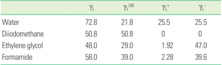
The surface energy analysis used the three-liquid method:
distilled water, formaldehyde (polar solvent), and diiodometh- ane (nonpolar solvent). Surface free energy and its compo- nents of each solvent employed are presented in Table 1. The contact angle for each solvent was measured.
Table 1. Surface free energy and its components of liquids em- ployed (mL/m2).
γL γLLW γL+ γL-
Water 72.8 21.8 25.5 25.5
Diiodomethane 50.8 50.8 0 0
Ethylene glycol 48.0 29.0 1.92 47.0
Formamide 58.0 39.0 2.28 39.6
γL: liquid-vapour interfacial tension, γLLW: Lifshitz-van der Waals component, γL+: liquid-vapour interfacial tension of electron donor, γL-: liquid-vapour interfacial tension of electron acceptor.
Osteoblast-enriched cell preparations were obtained from 21-day fetal calvaria of Sprague-Dawley rats by sequential collagenase digestion (Type II, Gibco BRL, Grand Island, NY, USA) as described previously [12]. The cells resulting from the third to fifth 15-minute digestions were pooled and cultured in BGJb media (Gibco BRL) supplemented with 10% heat-in- activated fetal bovine serum (FBS, Gibco BRL), 100 mg/mL penicillin (Gibco BRL), and 100 mg/mL streptomycin (Gibco BRL) at 37oC in a humidified atmosphere with 5% CO2-95% air.
Evaluation of cell adhesion
The cp-Ti and AHT were placed, under aseptic conditions, in the bottom of 12-well culture dishes, and then rinsed three times in 70% ethanol, exposed to ultraviolet light for 1 hour, and air dried in the cell culture hood.
The fetal rat calvarial cells were seeded at a density of 1×10⁴ cells/mL in the BGJb media. After a 3-day incubation period, the dishes were washed three times with phosphate buffered saline (PBS, Gibco BRL), and fixed with 2.5% glutaraldehyde (Sigma-Aldrich Co., St. Louis, MO, USA) in 100 mM cacodyl- ate buffer (Sigma-Aldrich Co.). The samples were dehydrated in increasing concentrations of ethanol (30%, 60%, 95%, and 100%), immersed in hexamethyldisilazane (Sigma-Aldrich Co.) for 15 minutes, air-dried, and immediately mounted on aluminum stubs and coated with carbon. SEM was then per- formed.
Evaluation of cell proliferation
Cell proliferation was assessed using a 3-(4,5-Dimethylthia- zol-2-yl)-2,5-diphenyltetrazolium bromide (MTT) assay using the fetal rat calvarial cells. Cells were cultured on each disc in 12-well plate at a density of 1×10⁴ cells/mL in the BGJb medi- um supplemented with 10% FBS. Following incubation, cell proliferation was assessed at 1, 3, and 5 days using a MTT as- say (CellTiter 96, AQueous One Solution, Promega, Madison, WI, USA). In these experiments, the amount of reduced foma- zan product is directly proportional to the number of viable cells. Fomazan accumulation was quantitated by absorbance at 490 nm using an enzyme-linked immunoabsorbent assay plate reader (VERSAmax, Dynamic Devices, Wilmington, DE, USA). The experiment was carried out in triplicate.
ALP activity
ALP activity was measured spectrophotometrically using the fetal rat calvarial cells. The cells were seeded onto 12 well dishes at a density of 1×10⁴ cell/mL in the BGJb medium con- taining 10% FBS, ascorbate 40 g/mL and 20 g/mL β-glycerol phosphate. After incubation for 7 days, the cells were washed with PBS, lysed in Triton 0.1% (Triton X-100, Promega) in PBS,
was mixed with 200 g of 10 mM p-nitrophenyl phosphate and 100 g of 1.5 M 2-amino-2-methyl-1-propanol buffer (Sig- ma-Aldrich Co.). The samples were incubated for 1 hour at 37oC. The ALP activity was measured from the absorbance reading at 405 nm with a spectrophotometer (SmartSpec, Bio- Rad Laboratories Inc., Hercules, CA, USA) and corrected for the cell number determined in parallel. All experiments were carried out in triplicate.
Statistical analysis
An analysis of variance for repeated measurements were performed to examine the data for surface roughness, surface energy, contact angle measurement, cell proliferation, and ALP activity with SPSS ver. 12.0 (SPSS Inc., Chicago, IL, USA).
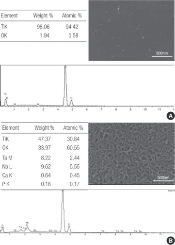
Figure 1. Scanning electron microscopy (SEM) and energy dispersed spectroscopy (EDS) analysis. (A) Commercially pure titanium (group cp-Ti) showed a uniform texture with porosity. EDS spectrum showed titanium and oxygen peak. (B) Alkali- and heat-treated titanium- 8tantalum-3niobium (group AHT) showed a flakelike morphology microprofile and dense structure. The proportion of oxygen in the alkali- and heat-treated titanium was higher than that in the com- mercially pure titanium. Calcium and phosphor peaks were detect- ed in EDS spectrum of group AHT but not in that of group cp-Ti.
A
B Element Weight % Atomic %
TiK 98.06 94.42
OK 1.94 5.58
Element Weight % Atomic %
TiK 47.37 30.84
OK 33.97 60.55
Ta M 8.22 2.44
Nb L 9.62 5.55
Ca K 0.64 0.45
P K 0.18 0.17
RESULTS
Surface characterization
Fig. 1 shows the SEM images and EDS spectra of samples.
spectrum showed titanium and oxygen peaks. Group AHT showed a flake-like morphology microprofile and dense struc- ture. The proportion of oxygen in group AHT was higher than that in group cp-Ti. Calcium and phosphor peaks were de- tected in the EDS spectrum of group AHT, but not in that of group cp-Ti.
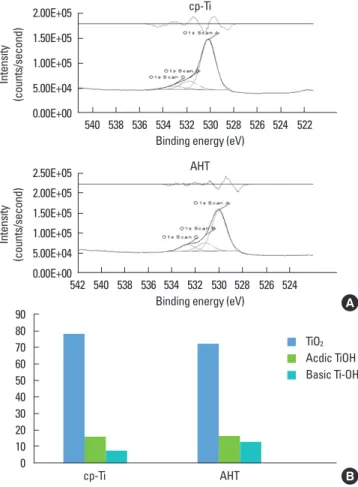
Fig. 2A shows the O1s XPS spectra for the surface of groups cp-Ti and AHT. The XPS spectra of O1s were classified into three Gaussian component peaks [13]. Titanium dioxide sur- faces have two hydroxide groups: an acidic hydroxide group (531.1 eV, TiOH) and a basic hydroxide group (532.4 eV, Ti-OH).
The TiOH group is acidic, whereas the Ti-OH group is basic and reacts readily with other anions. Fig. 2B shows the rela- tive ratios of peak areas for oxygen in surface oxide lattices (denoted by TiO2), oxygen in the hydroxyl residue of acidic titanium hydroxyl groups (bridging OH group, denoted by acidic TiOH) and oxygen in the hydroxyl residue of basic tita- nium hydroxyl groups (terminal OH group, denoted by basic Ti-OH). The alkali- and heat-treatment increased the amount of O in basic Ti-OH groups, meaning increased active OH groups on the surfaces.
Three-dimensional AFM images (5×5 m) showed nanoscale roughness on alkali- and heat-treated surfaces (Fig. 3). The surface topography of group AHT had spike shape than group cp-Ti. The mean surface roughness (Ra) measured by AFM was greater on group AHT than on group cp-Ti (19.1 nm and 0.6 nm, respectively).
Despite this difference in microroughness, the surface rough- ness (Ra) measured by the profilometer showed no significant
Table 2. Contact angle and surface energy.
cp-Ti AHT
Contact angle 59.00±5.27 32.02±3.98
Surface energy (dyne/cm) 46.92±4.04 64.68±2.73 Values are presented as mean±standard deviation.
cp-Ti: commercially pure titanium, AHT: alkali- and heat-treated titanium-8tantalum- 3niobium.
A
B 2.00E+05
1.50E+05 1.00E+05 5.00E+04 0.00E+00
2.50E+05 2.00E+05 1.50E+05 1.00E+05 5.00E+04 0.00E+00
540
542
536
536 538
540 538 534
534 532
532 530
530 528
528 526
526 524
524 522 Binding energy (eV)
Binding energy (eV) cp-Ti
AHT Intensity (counts/second)Intensity (counts/second)
90 80 70 60 50 40 30 20 10
0 cp-Ti AHT
TiO2
Acdic TiOH Basic Ti-OH
Figure 2. X-ray photoelectron spectroscopy analysis. (A) O1s spectra and (B) percentage of three oxygen species for commercially pure titanium (group cp-Ti) and alkali- and heat-treated titanium-8tanta- lum-3niobium (group AHT). Group AHT showed increased amount of hydroxyl groups on the surface layers. TiO2: titanium dioxide, TiOH: acidic titanium hydroxyl group, Ti-OH: basic titanium hy- droxyl group.
Figure 3. Atomic force microscopy (AFM) analysis of (A) commercially pure titanium (group cp-Ti) and (B) alkali- and heat-treated titanium-8tantalum-3niobi- um (group AHT). Three-dimensional AFM images (5×5 µm) showed nanoscale roughness on the group AHT surfaces.
The surface topography of group AHT had more spike shape than the group cp-Ti.
A B
difference (group cp-Ti, 0.31±0.07 µm; group AHT, 0.34±0.06 µm; P>0.05).
Table 2 presents the contact angle and surface energy group AHT showed a lower contact angle and higher surface ener- gy than group cp-Ti.
Cell responses
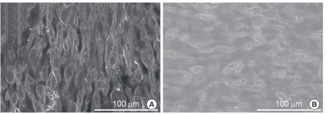
For each specimen, the cells were examined by SEM (Fig. 4).
The cells were spread extensively and totally flattened on group AHT surfaces. They were polygonal shapes with filopodial extensions, indicative of cell spreading. They did not have a regular orientation, and appeared scattered in all directions.
On group cp-Ti and group AHT surfaces, the cells spread po- lygonally and cell projections connecting the cells were visi- ble. No significant morphological difference was observed between cells on the cp-Ti, and alkali- and heat-treated sur-
faces. Thus, we can conclude that group AHT did not disturb the cell attachment.
Cell proliferation was measured by the MTT assay (Fig. 5).
On days 1, 3, and 5, group AHT showed a proliferation rate of 121%, 114%, and 106%, respectively, compared to group cp-Ti at the same respective points in time. The differences were statistically significant (P<0.05).
The cells on group AHT showed 20% higher ALP activity than on the group cp-Ti (Fig. 6). The differences were statisti- cally significant (P<0.01).
DISCUSSION
Since alkali and heat treatment of titanium surfaces was in- troduced, it has been reported that an apatite layer is formed on the alkali- and heat-treated surface and this apatite layer is osteoconductive [10,11]. However, the cellular response of osteoblasts on the surface of those implants has not been re- Figure 4. Cell adhesion examined by scanning electron microscopy (SEM). (A) The cells were spread extensively and totally flattened on the commercially pure titanium (group cp-Ti) surfaces. (B) The cells were spread extensively and totally flattened on the alkali- and heat-treated titanium-8tantalum-3niobium (group AHT). They were polygonal shapes with filopodial extensions, indicative of cell spreading. They did not have a regular orientation, and appeared scattered in all directions. The cells spread polygonally and cell projections connecting the cells were visible. No significant morphological difference was observed.
A B
Figure 6. Alkaline phosphatase (ALP) activity on the alkali- and heat- treated titanium-8tantalum-3niobium (group AHT) was the higher than on the commercially pure titanium (group cp-Ti). a)A statistically significant difference as compared with cp-Ti (P<0.05).
ALP activity (U/mg protein)
400 350 300 250 200 150 100 50
0 cp-Ti AHT
a)
Figure 5. Cell proliferation measured by the 3-(4,5-Dimethylthiazol- 2-yl)-2,5-diphenyltetrazolium bromide assay. On day 1, 3 and 5, the alkali- and heat-treated titanium-8tantalum-3niobium (group AHT) showed significantly greater cell proliferation compared with the commercial pure titanium (group cp-Ti). a)A statistically significant difference as compared with cp-Ti (P<0.05).
Optical density
1.2
1.0
0.8
0.6
0.4
0.2
0.0 Day 1 Day 3 Day 5
a)
a)
cp-Ti a)
AHT
assessed by SEM, cell proliferation assay, and ALP activity analysis.
In this study, primary osteoblasts were obtained from fetal rat calvaria. This is an excellent source of osteoblasts because cells from young animals proliferate rapidly. Cells from the third, fourth, and fifth digests were collected because these later digests provide a more pure culture, containing mostly cells that express an osteoblast-like phenotype [14].
Surface roughness can greatly affect the proliferation and protein synthesis of osteoblast cells that are cultured on a metal substrate during bone healing [15,16]. Many studies have demonstrated that roughness significantly influences cell responses. In this study, the surface roughness measured with a profilometer showed no significant difference be- tween group cp-Ti and AHT, although AFM and SEM analy- sis showed irregular surface morphology of group AHT. Na- noscale roughness shown in AFM images involves surface chemistry. Therefore, the enhanced cell response on alkali- and heat-treated surfaces might not be attributed to the sur- face roughness. Instead, it seemed that the difference in the microprofile and chemical properties contributed to an en- hanced cell response.
The finding that the surface roughness was not affected by the alkali- and heat-treatment is important. It has been re- ported that an implant with a rough surface might show more pronounced progression of peri-implantitis than that of a smooth surface [17], although rough surfaces allow enhanced osseointegration [18]. In this regard, an alkali- and heat-treat- ed implant might be more favorable for periimplant tissue health than an implant with a rough surface.
Alkali- and heat-treatment reduced the contact angle and increased the surface energy via alteration of surface chemi- cal composition. The lower contact angle and higher surface energy create a hydroxylated and hydrophilic surface, and promote the adhesion of relevant proteins [19]. These prop- erties can result in increased cell attachment and prolifera- tion. The hydrophilicity is associated with abundant hydroxyl groups. In this study, XPS results showed that alkaline treat- ment increased the number of OH groups.
In addition, a thin reactive layer of Ca-P (Ca-P layer) formed on alkali- and heat-treated surfaces. This result is consistent with the findings of other reports [4,10,11,20,21]. A Ca-P layer can form apatite in SBF and enhance the differentiation of osteoblasts. Several researchers have suggested that the for- mation of apatite should be associated with the formation of titanium oxide layer following alkali treatment and its densi- fication after heat treatment [20,22,23]. The apatite formed on alkali- and heat-treated surfaces would bind chemically to the apatite in the bone. In the present study, EDS analysis revealed
heat-treated surface. In addition, SEM analysis showed a dense structure of an alkali- and heat-treated surface. The Ti-OH groups formed on the surface are negatively charged and com- bine selectively with the positively charged Ca2+ ions in the fluid [24]. As a result, a positively charged surface combines with negatively charged phosphate ions to form an amor- phous calcium phosphate. This calcium phosphate spontane- ously transforms into the crystalline apatite.
The cell morphology was examined by SEM. The SEM eval- uations showed that the cells spread extensively and flattened on the group cp-Ti and AHT surfaces. The absence of signifi- cant morphological modification supports the cytocompati- bility of these metals. The lower contact angle, meaning high- er wettability, would promote cell spreading and attachment on the surface group AHT. Surface hydrophilicity is a factor that determines biocompatibility of biomaterials and is large- ly dependent on surface energy [25].
The cell proliferation on the group AHT surface was signif- icantly greater than on the group cp-Ti surface on days 1, 3, and 5. The lower contact angle and higher surface energy created a hydroxylated and hydrophilic surface, promoted the adhesion of relevant proteins, and increased cell attach- ment and proliferation [19].
In addition to increased cell proliferation, group AHT showed a higher level of ALP activity. The hydroxylated surface can form a calcium phosphate layer in SBF, which enhances the differentiation of osteoblasts [20]. The enhancement of ALP activity would contribute to the facilitation of osteoblastic differentiation.
Osteoblast cells undergo a temporal sequence with a change of phase during the development of their completely differ- entiated phenotype: proliferation, differentiation, and min- eralization [26]. Cells initially increase their number and pro- duce an extracellular matrix. The phase of differentiation fol- lows, characterized by the production of high levels of ALP, and modifications of the matrix that lead to the deposition of hydroxyapatite crystals. Cells grown on group AHT showed ALP levels significantly higher than on group cp-Ti (P<0.05).
ALP is an enzyme belonging to the group of membrane- bound glycoproteins. Although its physiological function re- mains unclear, ALP may play a key role in the formation and calcification of hard tissues [27]. Its expression and enzyme activity are frequently used as markers for osteoblast cells.
This finding is consistent with the results reported by Chosa et al. [28], and could be explained by the alkali- and heat-treat- ed surface increasing osteoblastogenesis and bone formation.
The results of this study have shown that group AHT had a better cell response than cp-Ti for bone remodeling. There- fore, group AHT might be a candidate for use in the prepara-
mal and human studies are needed before using this alloy in the clinical setting.
Osseointegration of dental implants depends on the cell responses around the implant. The changes in osteoblast proliferation, differentiation, and maturation are important events in bone remodeling. Group AHT formed an apatite surface layer that facilitated osteogenic differentiation. Since osteoblasts are central to bone remodeling, a biological re- sponse should be demonstrated to clarify the activity of group AHT in vitro.
In summary, these results show that alkali- and heat-treat- ment enhances cell proliferation and ALP activity, suggesting that group AHT may stimulate osteoblast differentiation and consequently facilitate bone remodeling.
CONFLICT OF INTEREST
No potential conflict of interest relevant to this article was reported.
ACKNOWLEDGEMENTS
This study was financially supported by research fund of Chonnam National University in 2010.
REFERENCES
1. Park JB, Lakes RS. Biomaterials: an introduction. 2nd ed.
New York: Kluwer Academic Publishers; 1992.
2. Cochran DL, Schenk RK, Lussi A, Higginbottom FL, Buser D. Bone response to unloaded and loaded titanium im- plants with a sandblasted and acid-etched surface: a his- tometric study in the canine mandible. J Biomed Mater Res 1998;40:1-11.
3. Li D, Ferguson SJ, Beutler T, Cochran DL, Sittig C, Hirt HP, et al. Biomechanical comparison of the sandblasted and acid-etched and the machined and acid-etched titanium surface for dental implants. J Biomed Mater Res 2002;60:
325-32.
4. Kokubo T, Miyaji F, Kim HM, Nakamura T. Spontaneous formation of bonelike apatite layer on chemically treated titanium metals. J Am Ceram Soc 1996;79:1127-9.
5. Shi S, Kirk M, Kahn AJ. The role of type I collagen in the regulation of the osteoblast phenotype. J Bone Miner Res 1996;11:1139-45.
6. Sisk MA, Lohmann CH, Cochran DL, Sylvia VL, Simpson JP, Dean DD, et al. Inhibition of cyclooxygenase by indo- methacin modulates osteoblast response to titanium sur- face roughness in a time-dependent manner. Clin Oral
7. de Groot K, Wolke JG, Jansen JA. Calcium phosphate coat- ings for medical implants. Proc Inst Mech Eng H 1998;
212:137-47.
8. Nishiguchi S, Nakamura T, Kobayashi M, Kim HM, Miyaji F, Kokubo T. The effect of heat treatment on bone-bond- ing ability of alkali-treated titanium. Biomaterials 1999;
20:491-500.
9. Nishiguchi S, Kato H, Neo M, Oka M, Kim HM, Kokubo T, et al. Alkali- and heat-treated porous titanium for ortho- pedic implants. J Biomed Mater Res 2001;54:198-208.
10. Nishiguchi S, Fujibayashi S, Kim HM, Kokubo T, Naka- mura T. Biology of alkali- and heat-treated titanium im- plants. J Biomed Mater Res A 2003;67:26-35.
11. Green J, Schotland S, Stauber DJ, Kleeman CR, Clemens TL. Cell-matrix interaction in bone: type I collagen modu- lates signal transduction in osteoblast-like cells. Am J Physi- ol 1995;268(5 Pt 1):C1090-103.
12. McCarthy TL, Centrella M, Canalis E. Further biochemical and molecular characterization of primary rat parietal bone cell cultures. J Bone Miner Res 1988;3:401-8.
13. Healy KE, Ducheyne P. Hydration and preferential molec- ular adsorption on titanium in vitro. Biomaterials 1992;13:
553-61.
14. Luben RA, Wong GL, Cohn DV. Biochemical characteriza- tion with parathormone and calcitonin of isolated bone cells: provisional identification of osteoclasts and osteo- blasts. Endocrinology 1976;99:526-34.
15. Lincks J, Boyan BD, Blanchard CR, Lohmann CH, Liu Y, Cochran DL, et al. Response of MG63 osteoblast-like cells to titanium and titanium alloy is dependent on surface roughness and composition. Biomaterials 1998;19:2219-32.
16. Kawahara H, Soeda Y, Niwa K, Takahashi M, Kawahara D, Araki N. In vitro study on bone formation and surface to- pography from the standpoint of biomechanics. J Mater Sci Mater Med 2004;15:1297-307.
17. Berglundh T, Gotfredsen K, Zitzmann NU, Lang NP, Lind- he J. Spontaneous progression of ligature induced peri- implantitis at implants with different surface roughness:
an experimental study in dogs. Clin Oral Implants Res 2007;18:655-61.
18. Abrahamsson I, Berglundh T, Linder E, Lang NP, Lindhe J.
Early bone formation adjacent to rough and turned en- dosseous implant surfaces. An experimental study in the dog. Clin Oral Implants Res 2004;15:381-92.
19. Pesskova V, Kubies D, Hulejova H, Himmlova L. The in- fluence of implant surface properties on cell adhesion and proliferation. J Mater Sci Mater Med 2007;18:465-73.
20. Sultana R, Kon M, Hirakata LM, Fujihara E, Asaoka K, Ichi- kawa T. Surface modification of titanium with hydrother-
470-9.
21. Jonasova L, Muller FA, Helebrant A, Strnad J, Greil P. Bio- mimetic apatite formation on chemically treated titani- um. Biomaterials 2004;25:1187-94.
22. Kim HM, Miyaji F, Kokubo T, Nishiguchi S, Nakamura T.
Graded surface structure of bioactive titanium prepared by chemical treatment. J Biomed Mater Res 1999;45:100-7.
23. Kim HM, Miyaji F, Kokubo T, Nakamura T. Effect of heat treatment on apatite-forming ability of Ti metal induced by alkali treatment. J Mater Sci Mater Med 1997;8:341-7.
24. Tamilselvi S, Raghavendran HB, Srinivasan P, Rajendran N. In vitro and in vivo studies of alkali- and heat-treated Ti-6Al-7Nb and Ti-5Al-2Nb-1Ta alloys for orthopedic im- plants. J Biomed Mater Res A 2009;90:380-6.
Cochran DL, et al. High surface energy enhances cell re- sponse to titanium substrate microstructure. J Biomed Mater Res A 2005;74:49-58.
26. Lian JB, Stein GS. The developmental stages of osteoblast growth and differentiation exhibit selective responses of genes to growth factors (TGF beta 1) and hormones (vita- min D and glucocorticoids). J Oral Implantol 1993;19:95-105.
27. Genge BR, Sauer GR, Wu LN, McLean FM, Wuthier RE.
Correlation between loss of alkaline phosphatase activity and accumulation of calcium during matrix vesicle-medi- ated mineralization. J Biol Chem 1988;263:18513-9.
28. Chosa N, Taira M, Saitoh S, Sato N, Araki Y. Characteriza- tion of apatite formed on alkaline-heat-treated Ti. J Dent Res 2004;83:465-9.