There are various causes of extensor tendon ruptures (ETRs) around the wrist, including distal radius fractures,1) carpal bone fractures or dislocation,2,3) Kienböck disease,4) rheumatoid arthritis (RA),5-7) and degenerative arthritis of the distal radioulnar joint (DRUJ).8-12) Ruptures of the extensor pollicis longus tendon in distal radius fractures, centrally located extensor tendons of fingers in Kienböck disease, and ulnar-sided extensor tendons in rheumatoid or degenerative arthritis of the DRUJ have frequently been
Computed Tomography-Based Morphologic Analysis of Osteoarthritis of the Distal Radioulnar
Joint Associated with Extensor Tendon Ruptures
Min-Gu Jang, MD, Youn Moo Heo, MD, Young Ki Min, MD, Tae Gyun Kim, MD, Byung Hak Oh, MD, Tae Hyeong Kim, MD
Department of Orthopedic Surgery, Konyang University College of Medicine, Daejeon, Korea
Background: Although the scallop sign is considered the most important risk factor for extensor tendon ruptures (ETRs) in pa- tients with osteoarthritis of the distal radioulnar joint (DRUJ), previous reports provide a limited understanding of the changes at DRUJ, as risk factors were examined in plain radiographs of the wrist. The aim of this study was to assess the changes of DRUJ using axial images of computed tomography (CT) in patients with DRUJ osteoarthritis and associated ETRs and to evaluate the re- lationship between the changes of DRUJ and ETRs.
Methods: Twelve patients with ETRs due to osteoarthritis of the DRUJ were enrolled. The changes of DRUJ were examined on axial images of CT and the following 8 parameters were measured: width of radius, anteroposterior (AP) length of radius, width of sigmoid notch (SN), AP length of SN, AP length of ulnar head, subluxation length of ulnar head, dorsal inclination of SN, and distance from Lister’s tubercle to SN. Radiological parameters of the DRUJ were measured in 60 control wrists without trauma or osteoarthritis, and the patient and control groups were statistically compared.
Results: Statistically significant differences were observed between the patient and control groups in all the radiological parame- ters except for the AP length of SN and AP length of ulnar head. The width of radius, AP length of radius, width of SN, subluxation length of ulnar head, and dorsal inclination of SN were greater and the distance from Lister’s tubercle to SN was smaller in the patient group than in the control group. The width of SN, dorsal inclination of SN, and distance from Lister’s tubercle to SN were statistically significant risk factors among the 8 parameters.
Conclusions: ETRs due to osteoarthritis of the DRUJ was related to the changes of DRUJ, especially the changes around SN of the distal radius. In addition to the existing risk factors, a decreased distance from Lister’s tubercle to SN and increased dorsal inclination of SN were identified as new risk factors. Axial images of CT were effective to evaluate degenerative changes at the DRUJ.
Keywords: Distal radioulnar joint, Extensor tendon, Osteoarthritis, Rupture, Computed tomography
Copyright © 2021 by The Korean Orthopaedic Association
This is an Open Access article distributed under the terms of the Creative Commons Attribution Non-Commercial License (http://creativecommons.org/licenses/by-nc/4.0) which permits unrestricted non-commercial use, distribution, and reproduction in any medium, provided the original work is properly cited.
Clinics in Orthopedic Surgery • pISSN 2005-291X eISSN 2005-4408 Received May 6, 2020; Accepted July 24, 2020
Correspondence to: Youn Moo Heo, MD
Department of Orthopedic Surgery, Konyang University Hospital, Konyang University College of Medicine, 158 Gwanjeodong-ro, Seo-gu, Daejeon 35365, Korea
Tel: +82-42-600-9125, Fax: +82-42-600-9090 E-mail: hurym1973@hanmail.net
reported.1-12) Progression of degenerative changes in the DRUJ induces dorsal subluxation of the ulnar head, struc- tural changes of the sigmoid notch (SN), or osteophyte formation around the joint, and as a result, this can lead to attritional ruptures of ulnar-sided extensor tendons of the fingers.8)
The ETRs caused by degenerative changes of the DRUJ were first reported by Vaughan-Jackson9) in 1948.
Since then, Freiberg and Weinstein7) have reported a characteristic radiological finding on the ulnar side of the distal radius in spontaneous ETRs in RA, which is known as the scallop sign. The scallop sign indicates scalloping of the ulnar aspect of the distal radius on the posteroante- rior (PA) radiograph of the wrist and is characterized by a slowly progressing sclerotic border on the SN of the radi- us.7) This finding is seen in cases of ETRs due to degenera- tive arthritis of the DRUJ. Ohshio et al.10) and Tada et al.11) reported severe degenerative changes at the DRUJ, positive ulnar variance, and dorsal subluxation of the ulnar head as risk factors of ETRs. Yamazaki et al.12) analyzed the radio- graphic morphology of DRUJs with ETRs and reported a severe degenerative change, radial shift of the ulnar head, and dorsal inclination of the SN as the risk factors. Howev- er, previous studies provide a limited understanding of the changes at the DRUJ as these changes were only examined on plain radiographs of the wrist.10-12) Furthermore, even the previous studies that examined DRUJs using com- puted tomography (CT) focused on dorsal subluxation of the ulnar head and instability in the DRUJ.13-15) The pur- pose of this study was to assess changes at the DRUJ using axial CT images of patients with degenerative arthritis of the DRUJ and ETRs and to investigate the relationship be- tween the changes and ETRs. We hypothesized that a CT- based morphologic analysis would reveal additional risk factors for osteoarthritic DRUJ with ETRs.
METHODS
Patients
Twelve patients who underwent surgical treatment of ETRs due to osteoarthritis of the DRUJ between April 2012 and August 2019 were enrolled. We conducted this study in compliance with the principles of the Declaration of Helsinki. The protocol of this study was reviewed and approved by the Institutional Review Board of Konyang University Hospital (IRB No. 2019-09-003). Written in- formed consents were obtained. There were 7 men and 5 women with a mean age of 70.8 years (range, 58–81 years).
Eight patients had ETRs in the right wrist and 4 in the left wrist. All patients complained of active extension loss and weakness of the involved fingers (Fig. 1). None of the pa- tients had a history of trauma, fracture, or inflammatory disease such as RA around the wrist. On physical exami- nation, dorsal protrusion of the ulnar head was confirmed in all the patients and swelling was observed around the 4th extensor compartment of the wrist in 5 patients. Se- vere osteoarthritis of the DRUJ was confirmed on plain radiographs of the affected wrist in all patients. Based on the physical and radiological findings, we diagnosed at- tritional ETRs in the wrist with degenerative changes of the DRUJ. We did not perform further examinations such as ultrasonography or magnetic resonance imaging for diagnosis, but performed CT to evaluate the progression of degenerative changes. To compare changes of the DRUJ, we reviewed the image reading of CT scans conducted from January 2017 to December 2018 and randomly se- lected CT scans of 60 patients without abnormal readings such as previous fractures or degenerative changes around the wrist. The control group consisted of 41 men and 19 women with a mean age of 46.8 years (range, 16–85 years).
All CT scans were obtained in the same posture (shoulder
A B C
R
Fig. 1. (A) A 75-year-old right-handed woman (patient no. 2) was unable to actively extend her 3rd, 4th, and 5th fingers at the metacarpophalangeal joints. (B) Plain radiograph showing severe osteoarthritic changes and a scallop sign at the distal radioulnar joint. (C) Intraoperative photograph showing ruptured 3rd, 4th, and 5th extensor digitorum communis and extensor digiti minimi tendons.
forward elevation and forearm pronation with the palm attached to the floor).
All patients were managed surgically and the state of extensor tendons was assessed intraoperatively (Table 1). Ruptures of the extensor digiti minimi (EDM) tendon and the 5th extensor digitorum communis (EDC) tendon were confirmed in all patients. Rupture of the 4th EDC tendon was found in 4 patients, the 3rd EDC tendon in 2 patients, and extensor indicis proprius tendon in 1 patient.
Two patients (patients no. 3 and 12) had intraoperatively identified additional tendon ruptures that had not been found in physical examination, which had no effect on the treatment process. The patients underwent distal ulnar re- section (Darrach procedure), stabilization of the resected distal ulna using distally based strip of the extensor carpi ulnaris tendon, advancement of the pronator quadratus muscle, and reconstruction of ruptured extensor tendons.
Assessment
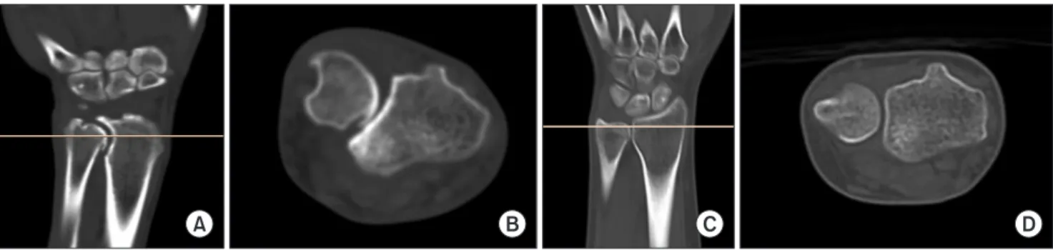
The changes at the DRUJ were assessed on axial images of CT. Axial images were selected from scout images of the deepest part of the SN in the coronal plane, which showed both SN and Lister’s tubercle (Fig. 2). For assessment of the changes at the DRUJ, the baseline was defined as the line connecting the volar cortex of the scaphoid facet and
the lunate facet of the distal radius (Fig. 3). A line parallel to the baseline was drawn from the volar edge of the SN (point A), and the point where the line from point A meets the radial cortical bone was designated as point B. Next, a line was drawn perpendicular to the baseline from the dor- sal edge of the SN (point C), and the point of contact of this line on the baseline was designated as point D. Point E is the point of intersection of lines AB and CD. A line parallel to the baseline was drawn from point C to Lister’s tubercle of the radius (point G) to the right, and the point where this line meets the ulnar cortical bone to the left was designated as point F. The straight line connecting points F and G was designated as line FG. A line perpendicular to line FG was drawn, denoting the greatest anteroposterior (AP) length of the ulnar head, and was designated as line HI. Point H and I are the posterior and anterior cortical bone of the ulnar head, respectively. The point of intersection of lines FG and HI was designated as point J, and the point where line FG meets the ulnar side of the cortical bone of Lister’s tubercle was designated as point K. The angle formed by a line joining points A and C (line AC) and line CE (∠ACE) was designated as the dorsal inclination of SN.
The following 8 parameters were measured using the points and lines described above: (1) width of radius (AB), (2) AP length of radius (CD), (3) width of SN (AE),
Table 1. Patient Summary
No. Age (yr)/sex Symptom onset
Extension limitation
(preoperative) Confirmed ruptured tendon
(intraoperative)
3rd 4th 5th EIP EDC (2nd) EDC (3rd) EDC (4th) EDC (5th) EDM
1 74/Male 3 wk ○ ○ ○
2 75/Male 4th/5th: 1 yr
3rd: 6 mo ○ ○ ○ ○ ○ ○ ○
3 77/Female 8 mo ○ ○ ○ ○ ○ ○
4 58/Male 4 day ○ ○ ○
5 81/Male 1 mo ○ ○ ○ ○ ○
6 64/Female 3 day ○ ○ ○
7 76/Male A few weeks ○ ○ ○
8 65/Female 2 wk ○ ○ ○
9 75/Female 5th: 6 yr
4th: 2 wk ○ ○ ○ ○ ○
10 63/Male 2 wk ○ ○ ○
11 70/Male 2 mo ○ ○ ○
12 67/Female 2–3 wk ○ ○ ○ ○ ○ ○
EIP: extensor indicis proprius, EDC: extensor digitorum communis, EDM: extensor digiti minimi.
(4) AP length of SN (CE), (5) AP length of ulnar head (HI), (6) subluxation length of ulnar head (HJ), (7) dorsal incli- nation of SN (∠ACE), and (8) distance from Lister’s tu- bercle to SN (CK). In addition, the following 4 ratios were calculated using the 8 parameters listed above: (a) width of SN/width of radius (AE/AB), (b) AP length of SN/AP length of radius (CE/CD)), (c) subluxation length of ulnar head/AP length of ulnar head (HJ/HI), and (d) distance from Lister’s tubercle to SN /width of radius (CK/AB).
Axial CT images were selected by an orthopedic hand specialist only (YMH) and the parameters were measured twice a week by the orthopedic hand specialist and one resident (YKM). We recorded the mean values of parameters. The measured parametric values of the pa- tient and control groups were compared using the Mann- Whitney test. And the importance among parameters was compared using multivariate regression analysis. The level of significance was set at p < 0.05.
RESULTS
In the patient group, the mean width of radius (AB) was 36.37 ± 4.10 mm (range, 29.12–44.58 mm), the mean AP length of radius (CD) was 23.59 ± 1.87 mm (range, 19.1–26.44 mm), the mean width of SN (AE) was 12.68 ± 2.91 mm (range, 13.69–19.88 mm), the mean AP length of SN (CE) was 16.17 ± 1.46 mm (range, 13.69–19.88 mm), the mean AP length of ulnar head (HI) was 17.50 ± 2.39 mm (range, 13.69–21.36 mm), the mean subluxation length of ulnar head (HJ) was 7.5 ± 2.98 mm (range, 2.19–11.49 mm), the mean dorsal inclination of SN (∠ACE) was 36.67° ± 9.24° (range, 16.1°–51.0°), and the mean distance from Lister’s tubercle to SN (CK) was 9.20 ± 1.75 mm (range, 4.88–13.57 mm). In the control group, the mean width of radius (AB) was 31.96 ± 2.95 mm (range, 26.87–38.67
A B C D
Fig. 2. Computed tomography (CT) images from the patient and control groups. (A) Coronal plane CT image showing the deepest sigmoid notch (SN) in the patient group. (B) Axial plane CT image corresponding to the scout image of the deepest SN shown in (A) was selected for assessment in the patient group. (C) Coronal plane CT image showing the deepest SN in the control group. (D) Axial plane CT image corresponding to the scout image of the deepest SN shown in (C) was selected for assessment in the control group.
Fig. 3. Measurement of the radiological parameters on an axial computed tomography (CT) image from the patient group. A: volar edge of the sigmoid notch (SN), B: point where a line parallel to the baseline drawn from point A meets the radial cortical bone, C: dorsal edge of SN, D:
point where a vertical line drawn from point C meets the baseline at right angles, E: point of intersection of (AB) and (CD), F: point where a line drawn parallel to the baseline from point C meets the ulnar cortical bone on the left, G: point where a line drawn parallel to the baseline from point C meets the radial cortical bone of Lister’s tubercle, H: point on the dorsal cortex of the ulna that is the start of a line drawn perpendicular to line FG and denoting the greatest anteroposterior (AP) length of the ulnar head, I: point on the posterior cortex of the ulna that is the end of a line perpendicular to (FG) and denoting the greatest AP length of the ulnar head, J: point of intersection of (FG) and (HI), K: point where (FG) meets the lateral border of Lister’s tubercle. (FB) is defined as width of radius, (CD) is AP length of radius, (AE) is width of SN, (CE) is AP length of SN, (HI) is AP length of ulnar head, (HJ) is subluxation length of ulnar head, (CK) is distance from the ulnar side of the cortical bone of Lister’s tubercle to the dorsal edge of SN, and ∠ACE is the angle formed by (AC) and (CE).
mm), the mean AP length of radius (CD) was 21.45 ± 1.70 mm (range, 18.57–26.44 mm), the mean width of SN (AE) was 4.61 ± 1.39 mm (range, 2.12–10.18 mm), the mean AP length of SN (CE) was 15.86 ± 1.46 mm (range, 12.89–18.77 mm), the mean AP length of ulnar head (HI) was 16.56 ± 1.49 mm (range, 13.78–19.98 mm), the mean subluxation length of ulnar head (HJ) was 4.04 ± 1.29 mm (range, 1.28–
6.66 mm), the mean dorsal inclination of SN (∠ACE) was 16.24° ± 4.54° (range, 6.8°–29.5°), and the mean distance from Lister’s tubercle to SN (CK) was 14.90 ± 2.45 mm (range, 10.27–20.88 mm).
Significant differences were observed between the patient and control groups in all the parameters except the AP length of SN (CE) and AP length of ulnar head (HI) (Table 2). The width of radius (AB), AP length of radius
(CD), width of SN (AE), subluxation length of ulnar head (HJ), and dorsal inclination of SN (∠ACE) were greater and the distance from Lister’s tubercle to SN (CK) was shorter in the patient group than in the control group (Fig.
4). The 4 ratios calculated using the 8 parameters were compared between the patient and control groups (Table 2). Statistically significant differences were observed in all the ratios between the patient and control groups. The width of SN/width of radius (AE/AB) and dorsal sublux- ation length of ulnar head/AP length of ulnar head (HJ/
HI) were greater and AP length of SN/AP length of radius (CE/CD) and distance from Lister’s tubercle to SN /width of radius (CK/AB) were shorter in the patient group than in the control group.
Table 2. Comparison of Parameters and Ratios between the Patient and Control Groups Based on Computed Tomography
Variable Patient (n = 12) Control (n = 60) p-value
Width of radius (mm) 36.37 ± 4.10 31.96 ± 2.95 0.002
AP length of radius (mm) 23.59 ± 1.87 21.45 ± 1.70 0.002
Width of SN (mm) 12.68 ± 2.91 4.61 ± 1.39 < 0.001
AP length of SN (mm) 16.17 ± 1.46 15.86 ± 1.46 0.626
AP length of UH (mm) 17.50 ± 2.39 16.56 ± 1.49 0.247
Subluxation length of UH (mm) 7.50 ± 2.98 4.04 ± 1.29 0.002
Dorsal inclination of SN (°) 37.67 ± 9.24 16.24 ± 4.54 < 0.001
Distance from Lister’s tubercle to SN (mm) 9.20 ± 1.75 14.90 ± 2.45 < 0.001
Width of SN/width of radius (%) 34.82 ± 7.93 14.37 ± 3.77 < 0.001
AP length of SN/AP length of radius (%) 68.77 ± 5.02 74.05 ± 5.20 0.011
Subluxation length of UH/AP length of UH (%) 42.02 ± 14.39 24.49 ± 7.78 0.001
Distance from Lister’s tubercle to SN/width of radius (%) 25.69 ± 6.20 46.56 ± 5.82 < 0.001 Values are presented as mean ± standard deviation.
AP: anteroposterior, SN: sigmoid notch, UH: ulnar head.
A B
Fig. 4. Comparison of computed tomo- graphy images of the distal radioulnar joint between the patient (A) and control (B) groups. The width of sigmoid notch (AE), dorsal subluxation of ulnar head (HJ), and dorsal inclination of sigmoid notch (∠ACE) were greater and the dis- tance from Lister’s tubercle to sigmoid notch (CK) was shorter in the patient group than in the control group.
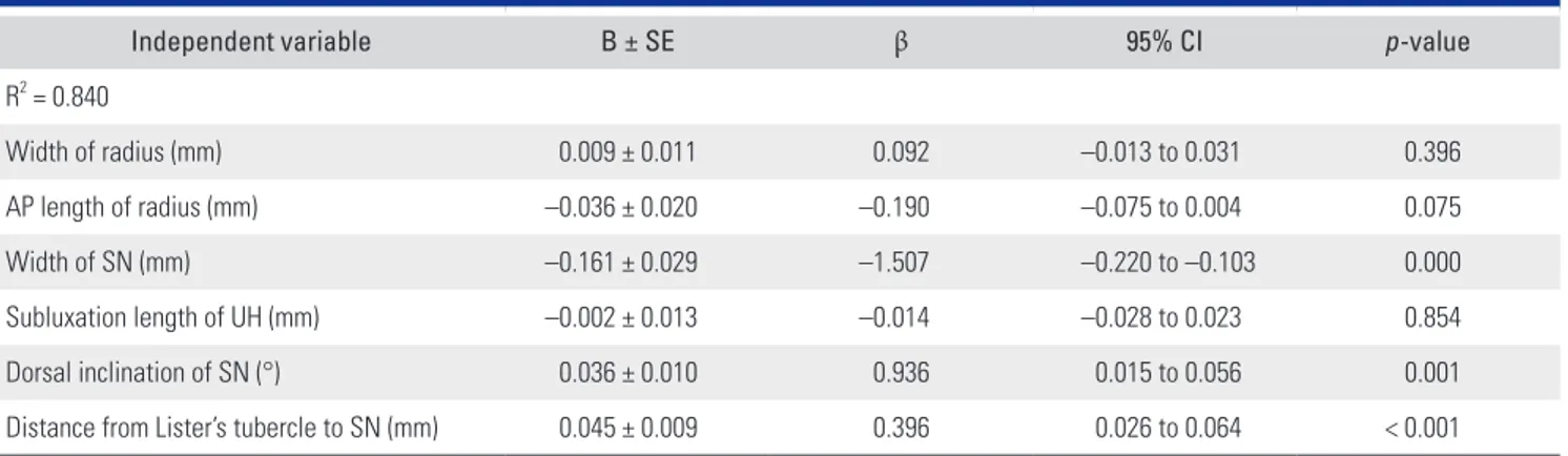
In the multivariate regression analysis, independent variables included the width of radius, AP length of radius, width of SN, subluxation of ulnar head, dorsal inclination of SN, and distance from Lister’s tubercle to SN; the AP length of SN and AP length of ulnar head without signifi- cant differences between the patient and control groups were excluded. The width of SN, dorsal inclination of SN, and distance from Lister’s tubercle to SN showed statistical significance as risk factors, whereas the width of radius, AP length of radius, and subluxation of ulnar head showed no statistical significance in patients with degenerative ar- thritis of the DRUJ and ETRs (Table 3).
DISCUSSION
In 1948, Vaughan-Jackson9) reported the first 2 cases of ETRs due to degenerative changes at the DRUJ. Ohshio et al.10) and Tada et al.11) observed severe degenerative chang- es at the DRUJ, positive ulnar variance, and dorsal sublux- ation of ulnar head in posteroanterior radiographs of the wrists with ETRs due to arthritis of the DRUJ. Yamazaki et al.12) analyzed PA radiographs of 41 wrists from 37 pa- tients with ETRs due to osteoarthritis of the DRUJ and compared with PA radiographs of 29 wrists without ETRs.
Unlike previous studies, they reported that there was no significant correlation between the ulnar head morphol- ogy, ulnar variance, and the incidence of ETRs. Further, they reported a scallop sign, radial shift of ulnar head, and dorsal inclination of SN as the radiological risk factors of ETRs. Dorsal subluxation of the ulnar head, which was reported as a radiological risk factor, should be evaluated on true lateral radiographs, but true lateral radiographs of all the patients could not be obtained in this study. Due to this limitation, the correlation between dorsal subluxation
of the ulnar head and ETRs could not be investigated. For this reason, CT images are more useful for the assessment of distal radioulnar congruency than radiographs.16,17) Even though some researchers compared axial CT images of wrists with subluxation of the ulnar head in healthy wrists, they focused on subluxation but did not study de- generative changes of DRUJ.7-9)
The current study analyzed axial CT images of the wrists in patients with osteoarthritis of the DRUJ and compared them with a control group. The results showed that the width of SN, dorsal subluxation of ulnar head, and dorsal inclination of SN were greater and the distance from Lister’s tubercle to SN was shorter in the patient group than in the control group. As the degenerative changes at the DRUJ progressed, the ulnar head was shift- ed to the radial side and displaced to the dorsal side.12) In addition, degeneration of the joint space was significantly more prominent on the dorsal proximal surface of the radius and the volar dorsal proximal surface of the ulna than in other areas.18) The changes of SN cause a decrease of the distance from the ulnar side of the cortical bone of Lister’s tubercle to the dorsal edge of the SN, and narrow- ing of the space where 3rd and 4th extensor compartments are located, resulting in attritional ETRs by the ulnar head.
In other words, radial shift and dorsal subluxation of the ulnar head means a greater shift of the dorsal edge of SN to the radial side than the volar edge of SN. That is, the de- generative changes are associated with the increased width of SN and dorsal inclination of SN and decreased distance from Lister’s tubercle to SN. And the increase in the sub- luxation length of ulnar head in the patient group was confirmed by dorsal displacement of the ulnar head. In the multivariate regression analysis, among independent parameters, the width of SN, dorsal inclination of SN, and
Table 3. Comparison of Importance among Significant Parameters by Multivariate Regression Analysis
Independent variable B ± SE β 95% CI p-value
R2 = 0.840
Width of radius (mm) 0.009 ± 0.011 0.092 –0.013 to 0.031 0.396
AP length of radius (mm) –0.036 ± 0.020 –0.190 –0.075 to 0.004 0.075
Width of SN (mm) –0.161 ± 0.029 –1.507 –0.220 to –0.103 0.000
Subluxation length of UH (mm) –0.002 ± 0.013 –0.014 –0.028 to 0.023 0.854
Dorsal inclination of SN (°) 0.036 ± 0.010 0.936 0.015 to 0.056 0.001
Distance from Lister’s tubercle to SN (mm) 0.045 ± 0.009 0.396 0.026 to 0.064 < 0.001 Values are presented as mean ± standard deviation.
SE: standard error, CI: confidence interval, AP: anteroposterior, SN: sigmoid notch, UH: ulnar head.
distance from Lister’s tubercle to SN showed statistical sig- nificance. These parameters reflected the changes around the SN of the distal radius, and these changes seemed to be associated with attritional ETR due to osteoarthritis of the DRUJ.
If a patient complains of extension limitation at the metacarpophalangeal (MCP) joint of ulnar side fingers, the physician should suspect an attritional ETR due to os- teoarthritis of the DRUJ. Extension of the 5th finger is per- formed by both EDM and 5th EDC tendons, and exten- sion is maintained by 5th EDC tendon if the EDM is only ruptured. Even if both tendons are ruptured, the end of a ruptured tendon still stays connected to the tendon sheath, which is attached to the extensor tendon of the adjacent finger or to the junctura tendinae. This may delay diag- nosis as some degree of MCP joint extension is still pos- sible. If diagnosis is delayed, attritional injury of extensor tendons progresses from ulnar side to radial side and may result in additional tendon ruptures. In our patients with long-term onset, additional tendon rupture that was not identified preoperatively was found intraoperatively and additional tendon reconstruction was required. Therefore, the surgery should be planned considering the possibility of additional ruptures. And the physician should explain complications, such as ETRs, which occur with degenera- tion to the patient, and recommend surgery in a timely manner before additional tendon rupture develops if inde- pendent 5th MCP extension is impossible.
Limitations of this study are the following. First, the changes of degenerative DRUJ were assessed in a small number of patients. Second, there was significant differ- ence in age between the patient group and control group because patients with osteoarthritis of the DRUJ were generally elderly. Last, the cutoff value for surgery was not determined because we did not make comparisons with patients without ETRs due to osteoarthritis of the DRUJ.
Henmi et al.19) compared healthy wrists with affected RA
wrists, which were divided into the ETRs group and non- rupture group. They set a cutoff value for subluxation of ulnar head to predict ETRs and determine the necessity of surgery. If more patients are enrolled in a later study, it may be possible to set a normal value for degenerative change and a cutoff value for the prediction of ETR and the determination of appropriate time for surgery. How- ever, there is a clinical limitation to a regular CT follow- up, which is only aimed at confirming the progression of arthritis in patients without tendon ruptures, because CT test has a risk of radiation exposure and most patients with osteoarthritis at the DRUJ have no functional limitation of the fingers.
In patients with degenerative changes at the DRUJ and associated ETRs, axial CT images are more useful to assess changes of the DRUJ than plain radiographs. The risk factors confirmed using axial CT images are as fol- lows: increased width of SN and dorsal subluxation of ulnar head, which were mentioned in previous studies, and decreased distance from Lister’s tubercle to SN and increased dorsal inclination of SN, which were first mea- sured and reported in this study. Changes around SN of the distal radius related to the width of SN, dorsal inclina- tion of SN, and distance from Lister’s tubercle to SN were also important. Radiological changes may be useful risk factors to help the physicians detect ETRs in patients with osteoarthritis of the DRUJ. However, as regular CT follow- up is not feasible for patients without tendon ruptures, we think the physical examination findings such as loss of independent extension of the 5th MCP is more important for early detection of attritional and progressive ETRs.
CONFLICT OF INTEREST
No potential conflict of interest relevant to this article was reported.
REFERENCES
1. Bunata RE. Impending rupture of the extensor pollicis lon- gus tendon after a minimally displaced Colles fracture: a case report. J Bone Joint Surg Am. 1983;65(3):401-2.
2. Stern PJ. Multiple flexor tendon ruptures following an old anterior dislocation of the lunate. A case report. J Bone Joint Surg Am. 1981;63(3):489-90.
3. Minami A, Ogino T, Hamada M. Rupture of extensor ten- dons associated with a palmar perilunar dislocation. J Hand Surg Am. 1989;14(5):843-7.
4. James JI. A case of rupture of flexor tendons secondary to Kienbock’s disease. J Bone Joint Surg Br. 1949;31(4):521-3.
5. Ryu J, Saito S, Honda T, Yamamoto K. Risk factors and pro- phylactic tenosynovectomy for extensor tendon ruptures in the rheumatoid hand. J Hand Surg Br. 1998;23(5):658-61.
6. Hsueh JH, Liu WC, Yang KC, Hsu KC, Lin CT, Chen LW.
Spontaneous extensor tendon rupture in the rheumatoid wrist: risk factors and preventive role of extended tenosyno- vectomy. Ann Plast Surg. 2016;76 Suppl 1:S41-7.
7. Freiberg RA, Weinstein A. The scallop sign and spontane- ous rupture of finger extensor tendons in rheumatoid ar- thritics. Clin Orthop Relat Res. 1972;83:128-30.
8. Chung DW, Kim KB, Lee YH. Extensor tendon rupture due to osteoarthritis of the distal radio-ulnar joints: a case re- port. J Korean Soc Surg Hand. 2001;6(2):215-8.
9. Vaughan-Jackson OJ. Rupture of extensor tendons by attri- tion at the inferior radio-ulnar joint: report of two cases. J Bone Joint Surg Br. 1948;30(3):528-30.
10. Ohshio I, Ogino T, Minami A, Kato H, Miyake A. Exten- sor tendon rupture due to osteoarthritis of the distal radio- ulnar joint. J Hand Surg Br. 1991;16(4):450-3.
11. Tada H, Hirayama T, Takemitsu Y. Extensor tendon rupture after osteoarthrosis of the wrist associated with nonrheuma- toid positive ulnar variance. Clin Orthop Relat Res. 1991;
(262):141-7.
12. Yamazaki H, Uchiyama S, Hata Y, Murakami N, Kato H. Ex- tensor tendon rupture associated with osteoarthritis of the distal radioulnar joint. J Hand Surg Eur Vol. 2008;33(4):469- 74.
13. Mino DE, Palmer AK, Levinsohn EM. The role of radiog- raphy and computerized tomography in the diagnosis of subluxation and dislocation of the distal radioulnar joint. J
Hand Surg Am. 1983;8(1):23-31.
14. Nakamura R, Horii E, Imaeda T, Nakao E. Criteria for di- agnosing distal radioulnar joint subluxation by computed tomography. Skeletal Radiol. 1996;25(7):649-53.
15. Naito K, Sugiyama Y, Aritomi K, et al. Assessment of dorsal instability of the ulnar head in the distal radioulnar joint:
comparison between normal wrist joints and cases of rup- tured extensor tendons. Eur J Orthop Surg Traumatol. 2016;
26(2):161-6.
16. Mino DE, Palmer AK, Levinsohn EM. Radiography and computerized tomography in the diagnosis of incongruity of the distal radio-ulnar joint: a prospective study. J Bone Joint Surg Am. 1985;67(2):247-52.
17. Nakamura R, Horii E, Imaeda T, Tsunoda K, Nakao E. Distal radioulnar joint subluxation and dislocation diagnosed by standard roentgenography. Skeletal Radiol. 1995;24(2):91-4.
18. Yoshida R, Beppu M, Ishii S, Hirata K. Anatomical study of the distal radioulnar joint: degenerative changes and mor- phological measurement. Hand Surg. 1999;4(2):109-15.
19. Henmi S, Yonenobu K, Akita S, Kuroda Y, Yoshida K. Diag- nosis of distal radioulnar joint subluxation in patients with rheumatoid wrist by computed tomography. Mod Rheuma- tol. 2007;17(4):279-82.