대한방사선의학회지 1994:31(2) :211- 216
뇌농양의 자기공명 영상 소견 I
박 만 수2 • 서 대 철 · 김 상 준3
목 적 :노|농앙의 특징적인 MR 소견을 알아보고자 하였다.
대상 및 방법 수술로서 확진된 9명의 뇌농앙 환자에서 MR 및 CT 영상을 후향적으로 분석해 보았다.
결 과:농앙의 모앙은 퉁글거나 (n =6), 다엽성 (n =2) 혹은 삼각형 (n = 1) 이었다. 전예에서 모든 병변이 피수질 경계부위에 위치하였고 안쪽 백질쪽으로 파급되는 앙상을 보여주었다. Gd-DTPA 조영 증강시 농약벽은 매끈한(smooth) 조영증강 (n =6) 불규칙하고 두꺼운 벽의 조영증강 (n=3) 을 보여 주었다. 특히 농앙벽의 일부분이 바깥쪽으로 아상돌출 (budike projection) 의 앙상을 4여|에서 보여주었 다. 농앙벽의 신호강도는 T1 강조영상에서 고신호강도 (n =5) 혹은 동신호강도 (n =4) 를 보여주었고 T2강조영상에서는 저신호강도 (n=6), 동신호강도 (n=2) 및 혼합된 저신호강도와 동신호강도
(n = 1) 를 보여주었다. 우|성병소 (n =2) 및 중등도의 주위부종 (n =9) 이 관찰되었고, 부비동염
(n =2), 뇌막염 (n=1), 뇌실염 (n =2) 및 경막하축농 (n =1)이 동반되었다.
결 론:뇌농앙은 MR 상 특징적인 농앙피막의 신호강도, 농앙벽의 일부분의 바깥쪽으로의 아상돌출 앙상, 농앙주위부종, 위성병소 및 뇌막염, 노|실염, 주변 부비동엽과 같은 동반된 소견이 특징적이었고 농양벽의 두께,모양등형태학적 소견은진단에 특이적이지 못했다.
서 료응 ‘-
뇌농양은 내과적 혹은 외과적 치료를 적절히 받지 못하 면 치명적인 질환으로서 조기진단 빛 치료가 매우 중요하 다. 전산화 단층촬영 (CT) 의 출현으로 현저한 사망율의 감소를 가져왔으며 (1), CT 및 자가공명영상 (MR) 에서 비교적 특정적 인 뇌농양의 소견들이 알려져 있다 (2-5). 그 러나 조영증강시 CT 및 MR에서의 뇌농양벽의 환상조영 증강은 뇌종양, 전이암 및 육아종에서도 유사하게 관찰되 어 감별진단이 용이하지 않을 경우가 많이 있다 (2, 6, 7).
저자들은 조영증강시 농양벽의 형태학적 소견, Tl 및 T2강조영상에서의 농양벽 의 신호강도를 분석하여 뇌농양 의 특정적인 MR 소견을 알아보고자 최근 3년간 본원에서 경험한 9명의 뇌농양 환자의 MR 소견을 후향적으로 분석 해보았다.
대상및방법
최근 3년간 본원에서 수술로서 확진된 9명의 뇌농양 환
1울산대학교 의과대학 방사선과학교실
2한립 대학교 의과대학 진 단방사선과학교실 3단국대학교 의과대학 방사선과학교실
이 논문은 1994년 3월 18일 접수하여 1994년 6월 2일에 채택되었음
자를 대상으로 후향적으로 MR 소견을 분석해 보았다.8 예에서는 CT가 같이 시행되었다. 원인균은 밝혀지지 않은 경우 (3예)가 제일 많았고 곰팡이, 포도상구균, 연쇄상구균 등 다양하였다 (Table 1).
연령은 34세에서 65세로 평균 46세였으며 남녀비는 5:4 였다. 사용한 기기는 1.5T 초전도 영상 (Signa ; General electric, Milwaukee, W I)를 사용하였고 외부병원의 MR 영상이 3예 포함되었다.6-7mm의 두께와 2-3mm의 간 격을 두고 영상을 얻었으며 전환자에서 Tl(600/20, TR/TE), T2(2500/80) 및 양자농도 강조 (2500/300) 축 상영상과 Tl 강조 시상영상, Gadolinium-DTPA(0.07- 0.
1 mmol/ kg) 주업 후 Tl강조영상을 얻었다.
농양의 위치,크기,모양및 농양내부와농양벽의 형태와 신호강도 그리고 뇌농양과 동반된 소견 (associated fin dings) 을 후향적으로 분석하였다.
결 과
농양의 모양은 원형 (n=6) , 다엽형 (n=2) 및 삼각형 (n=1) 이었으며 모두 피수질 경계부위에 위치하였고 전 예에서 단발성 (s이i tary) 이였다 (Fig.1-5). 농양의 위치는 전두엽 (n=3), 두정 엽 (n=2), 측두엽 (n=l) 및 소뇌 (n=
3) 에 위치하였다. 병변의 크기는 직경이 2.5-6cm이었고
고신호강도를 보여주었다. 9예 전부에서 Gadolini um-
DTPA 조영증강시 Tl 빛 T2강조영 상에서 보이 던 농양벽
보다더 두껍게 조영증강이 되었고CT 조영증강시 보이던 농양벽보다 더 두껍게 조영증강이 되었다. 농양벽은 비교 적 애끈한 형태 (n=6) (Fig. 2) 와 불규칙한 내외벽형태 (n
=6)(Fig. 3)을 보여주었q(Table 2). 9예중 4예에서 농
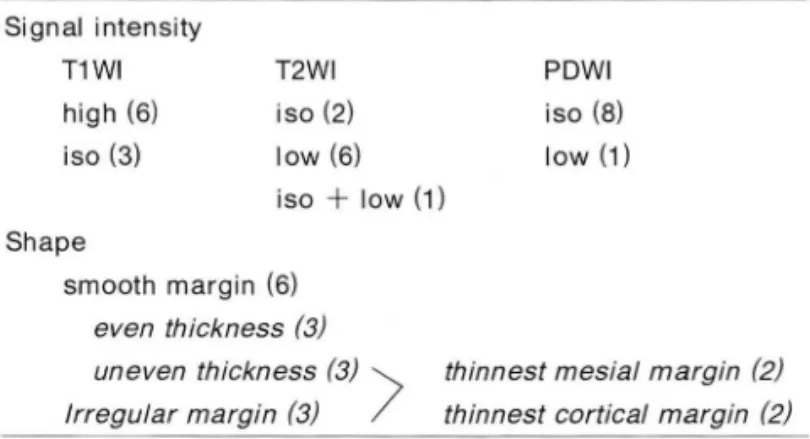
Table 2. Signal Intensity & Shape 01 Abscess Walls (n=9)
PDWI iso (8) low (1) T2WI
iso (2) low (6) iso
+
low (1) Signal intensityT1WI high (6) iso (3) 전예에서 병변 주위에 부종이 동빈되었는데 부종의 정도
는 병변 크기의 한배에서 두배정도였다. 농양벽의 신호강 도는 Tl강조영상에서 6예에서 백질보다 높은 고신호강도 를(Fig. 1), 3예에서 주위 백질과 동신호강도를(Fig. 2) 보 여주였다. 양지농도영상에서는 8예에서는 주위 회질과 통 신호강도를, 나머지 1 예에서는 저신호강도를 보여주었다.
T2강조영상에서는 6예에서 회질보다 저신호강도를(Fig.
1), 2예에서는 회질과 동신호강도를 보여주였고 (Fig. 2), 나머지 1예에서는 통신호강도와 저신호강도가 혼합되어 관찰되었다 (Table 2). 농양 내부의 신호강도는 전예에서 Tl 강조영상에서 뇌척수액 보다 높으나 회질보다 낮은 저 신호강도를 보여주었고 양자농도 및 T2강조영상에서는
211- 216 대 한방사선 의 학회 지 1994 : 31 (2)
thinnest mesial margin (2) thinnest cortical margin (2) Shape
smooth margin (6) even thickness (3) uneven thickness (3) \ Irregular margin (3) /
T1 WI : iso=isointense to white matter, high=hyperintense to white matter
T2WI, PDWI: iso=isointense to gray matter, low=hypointense to gray matter
1 1 1 1
f1
1 3 Table 1. Pathogens of Brain Abscess
Streptococcus Staphylococcus Aspergillus Peptostreptococcus
Peptostreptococcus
+
Fusobacterium Gram ( 十) cocciUnknown
Fig. 1. Abscess with high intensity cap- sule and satellite daughter lesions in a 37-year-old man. Pregadolinium T1-wei- ghted axial scan (a) demonstrates a left frontal lobe mass with a thin hyperin- tense rim (arrow) separating a markedly
hyp 이 ntense central cavity from relatively less hypointense edematous brain. On T2-weighted axial image (b), both abscess content and surrounding edema show high signal intensity, while the abscess wall appears hypointense to gray matter (arrows). On Gd-enhanced T1-weighted axial image (c), there is smooth rim en- hancement of abscess capsule. Note tw 。
satellite nodules (arrows) at the slightly upper level (d)
b
c
- 212
d a
양벽의 일부분이 바깥쪽으로 아상돌출 (budlike-projec- tion) 하는 양상을 보여주였다 (Fig. 4). 1 예에서는 농양내 부에 저신호강도와 고신호강도의 동심원성 고리 (concen tric ring) 양상을 보여주었다 (Fig. 4). 9예중 3예에서 균
일한 (uniform) 두께의 농양벽을 보여주었고 불균일한 두
께를 보인 6예중 2예에서는 뇌실쪽 벽이 피질쪽보다 앓았 고 2예에서는 반대로 피질쪽이 뇌설쪽보다 앓게 관찰되였 다 (Fig. 5). 뇌농양과 동반된 소견으로는 2예에서 농양주 위에 작은 2-3개의 위성 농양 (satellite nodule) 이 관찰되 었고(Fig. 1), 뇌막염 (n= 1), 뇌실염 (n=2), 부비동엽 (n=
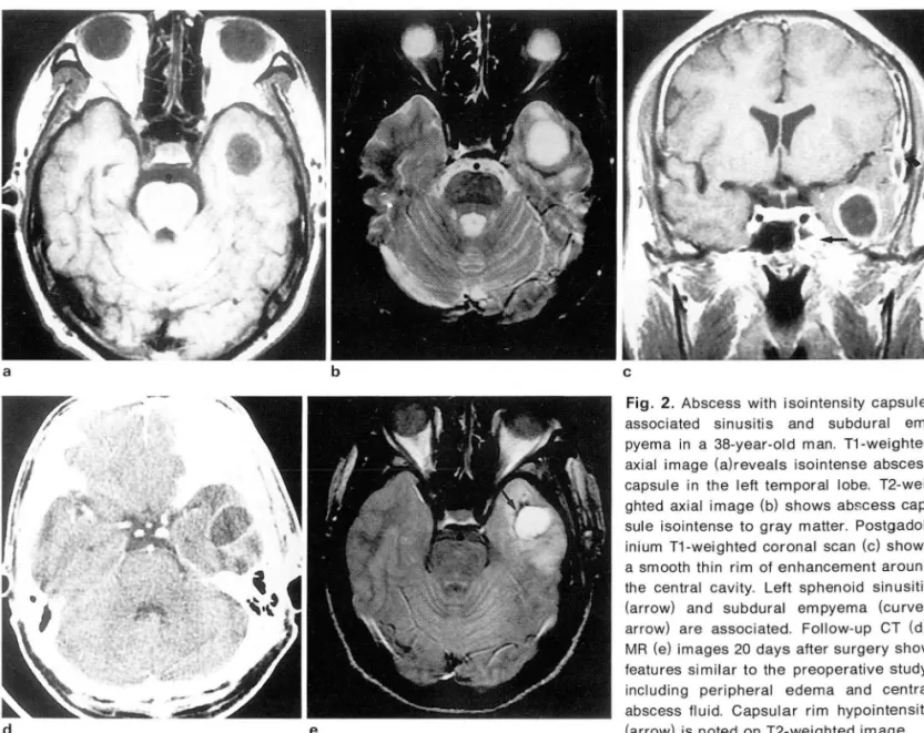
3) (Fig. 2, 4) 및 경막하 축농 (subdural empyema) (n = 1) (Fig.2) 이 동반되 었다.
고 찰
박만수 외 뇌농앙의 자기공명영상
터 2차적으로 혈행성으로 파급되는 예가 가장많다(1,
2) .
어른에서는 혈행성으로 파급되는 혐기성 박테리아 (anaer
obic nacteria) 흑은 혐기성과 호기성 (aerobic bacteria) 의 흔합균 주가 제일많고(1,
8
,9)
아이들에서는 포도상구 균 (staphylococci), 연쇄상구균( streptococci), 폐염균 (pneumococci) 이 가장 흔한 균주이다(10, 11). 혈행성으 로 생긴 뇌농양은 대부분 피수질 경계부위에 생기며 면역 결핍이 있는 환자를 제외하고는 주로 단발성으로 생기고,전두엽과 측두엽에 가장 호발한다 (2, 11). 저자들의 증례 에서는 전두엽,두정엽, 측두엽 및 소뇌에 다양하게 분포되 었고, 전예 모두에서 단발성을 보여주였다.
뇌농양은 2-3주에 걸쳐서 병리학적 변화를 보이는데 초기 4-5 일에는 급성뇌염기 (acute cere britis stage) 로 CT상 경계가 명확치 않은국소적인 저밀도음영이 나타나 고 조영증강은 되지 않거나 극소량의 조영증강을 보인다.
뇌농양은 과거 신경외과 수술이나 항생제 치료가 발전 MRI상
T1
강조영상에서 저신호강도,T2
강조영상에서 하기 전에는 이비학적 감염 (otorr hinologic infection)이 고신호강도를 보인다 (2, 3, 12). 1주말부터 만성뇌염기로 가장 흔한 원인을 차지하였으나 최근에는 두개 외부로 부 이행되는데 이 시기에는 괴사가 생겨 하나 혹은 두개의 큰a b
e
c
Fig. 2. Abscess with isointensity capsule, associated sinusitis and subdural em- pyema in a 38-year-old man. T1-weighted axial image (a)reveals isointense abscess capsule in the left temporal lobe. T2-wei- ghted axial image (b) shows abscess cap- sule isointense to gray matter. Postgadol- inium T1-weighted coronal scan (c) shows a smooth thin rim 01 enhancement around the central cavity. Left sphenoid sinusitis (arrow) and subdural empyema (curved arrow) are associated. Follow.up CT (d), MR (e) images 20 days after surgery show features similar to the preoperative study, including peripheral edema and central abscess Iluid. Capsular rim hypointensity (arrow) is noted on T2-weighted image
대 한밤사선의 학회 지 1994: 31(2) ’ 211- 216
괴사로 융화가 일어나고 괴사가 일어난 주위에 육아조직 이 형성된다. CT나 MRI상 두꺼운 환상 조영증강이 나타 나 조기 농양기와의 감별이 거의 불가능해진다 (2, 4 , 5). 2 주말부터 농양기가 시작되어 교질성 피막(C이lagen cap sule) 이 형성되는데 이는 섬유아세포(fibroblast) 에서 생 성되며 처음에는 앓고 불완전하나 몇주후 교원질 생성이 많아져 농양벽은 더 두꺼워진다. 농양벽이 성숙되어감에 따라 주변 부종의 양은 감소하고 벽 의 바깜쪽 gliotic reac- tion이 발달되어 농양벽은 세층을 이루게 되는데, 1. 내부
Fig. 3. A large abscess with thick irregular wall enhancement in a 34-year-old man. Postgadolinium T1-weighted coronal im-
의 육아조직층, 2. 교질층, 3. 외부의 교층(gliotic layer)이 다 (2). 이 시기의
CT
소견은 중심괴사부위는 비특이적 저 밀도음영이 나타나고 T1강조영상에서 뇌실질보다는 저신 호강도 뇌척수액보다는 고신호강도를 보여준다. Gd- DTPA 조영증강영상시 비교적 매끈하고 균일한 두께를 갖는 것이 특정이고 백질쪽벽이 피질쪽보다 앓은 것이 특 정으로 되어있다 (2). 저자들의 증례에서는 6예에서 애끈한 두께를 보여주었고 3예에서는 불규칙한 내외벽의 조영증 강을 보여주어 종양과의 형태학적 감별이 쉽지 않았고,9
예중 3예에서 균일한 두께의 농양벽을 보여주였고 불균일 한 두께를 보인 경우 백질쪽이 피질쪽보다 벽이 앓게 관
age shows a ring-enhancing mass with irregular thick wall in Fig. 5. A large abscess with smooth thin wall in a 34-year-old parietal lobe. At surgery, it was proved to be organizing ab- woman. On postgadolinium axial image, the abscess wall is
scess. thicker in the ventricular portion than in the cortical portion
a b c
Fig. 4. Abscess with budlike pr미 ection form the abscess caps비 e and concentric ring of variable signal in a 50 year-old woman. T1-weighted image (a) shows central abscess cavity material as hypointense relative to white matter, and hyperintense rim is seen at the margin(small arrows). T2-weighted image (b) shows concentric alternting zones of relative hypo- and hyperintensity (arrowheads
l.
Postgadolinium T1.weighted sagittal image reveal a characteristic budlike pr이ection from the abscess capsule (arrow). Associate ethmoid sinusitis is noted (curved arrow)- 214
찰된 경우가 2예, 오히려 피질쪽이 앓게 관찰된 경우가 2예 로 (Fig. 5) 뇌농양시 뇌실쪽의 벽이 피질쪽 벽보다 앓게 관찰된다는 문헌에 보고된 증례들과는 다소 차이가 있였 다. 그리고 농양벽의 일부분이 바깜쪽으로 아상돌출의 양 상을 9여l중 4예에서 보여 뇌농양에 비교적 특정적인 소견 으로 생각된다. 환자가 변역결핍이나 스테로이드치료를 받지않았다면 중등도의 부종이 뇌농양의 특정적인 소견으 로 부종의 양이 적거나 너무 많으면 농양의 가능성이 떨어 진다고 한다 (2-4,
12
,13).
저자들의 증례에서는 농양크기 의 1-2배 정도의 뇌부종의 양을 보여 문헌과 거의 같은 결과를얻었다.MRI 영상에서 펴막의 신호강도는 T1 강종영상에서 주 변 뇌실질과 통신호강도 혹은 고신호강도를 보이고 T2강 조영상에서 저신호강도가 특정적 인 소견이라고 한다 (2,
12).
저자들의 증례에서는 T1 강조영상에서 5예에서 고신호강도 4예에서 주위 백질과동신호강도를보여주었고 T2 강조영상에서는 6예에서 회질 보다 저신호강도를 2예에서 동산호강도를 보여주였다. MRI상 T2강조영상에서 피막 의 저신호강도는 몇가지 기전으로 설명되고 있다. 하나는 교원질의 존재에 기언한다는것이다.그러나이 설명은몇 가지 이유에서 옳지 않다. 첫째, 농양피막의 전신호강도는 교원질이 존재하지 않는 만성뇌염기에도 나타나고 둘째,
섬유조직은 전 pulse sequence에서 저신호강도를 보이는 데, 뇌농양피막은
T1
강조영상에서 동신호 흑은 고신호강 도를 보인다. 세째, 정상적인 섬유구조(fi brous structure) 는 저신호강도를 보이지만 병척인 섬유화는 신호강도가 다양해서 종종T2
강조영상에서 고신호강도를 보여준다 (12). 다른 가능성있는 기전은 출혈이다. 그러나 농양피막 에서 조직학적으로 출혈을 발견할 수 없었고(1 2) , 출혈에 의한신호강도는불균질하며 부분적이고불연속적인모양 을 보여 농양 피막에서 보이는 균일하고 연속적인 신호강 도와는 맞지않고, 출혈에의한 신호강도는 혈색소의 파괴 과정에 따라 신호강도의 변화를 보여 수개월 간 지속되는 피막의 신호강도와는 맞지 않다. 가장유력한 기전으로는 만성뇌염기나 농양기와 같이 염증이 활발할때 농양벽의 대식세포 (macrophage) 에서 생성된 산소유리기 (oxygen free radical) 의 susceptibility effect 에의한T2
감축에 의 한 것이라는 설이다 (2,3
,12).
뇌농양벽의 T2강조영상에 서의 저신호강도는 매우 특징적인 소견이나 아급성 및 만 성 혈종, 전이성암, 육아종 그리고 드물게 신경교종등에서 도 나타날수 있다(1 5-17). Haimes등은 환자의70%
정도 에서 농양내부에 저신호강도와 고신호강도의 동심원 고리 모양이 보이고 뇌농양의 비교적 특정적인 소견이라고 한 다(12). 저자들은 l 예에서 같은 소견을 관찰하였다 (Fig.4).
뇌농양벽의 T2강조영상에서의 저신호강도는 치료효과 판정에 도움을주는데 뇌농양의 환상벽은내과적 혹은외 과적 치료를 받아도 8개월까지 CT나 MRI에서 환상조영 층강이 지속되기도 한다(1 8). 그러나 환상조영증강이 남
박만수 외 뇌농앙의 자기공명영상
아있다 해도 내부에 괴사가 있고 계속해서 크기가 감소하 는 농양에 있어서는 치료가 실패한 것이 아니다 (5, 18). 농 양피막의 T2강조영상에서의 저신호강도는 대식세포의 활 동성의 지표가 되는 것으로 치료가 진행됨에 따라 대식세 포의 활동성은 계속해서 감소하므로 저신호강도의 감소 혹은 소실이 치료효과 판정에 보다 중요한 소견이다 (2,
12).
치료가 잘 진행되면 보통 4개월까지 농양벽의 저신호 강도는 소실된다 (2). Haimes등은 뇌농양의 외과적 처치 후 시기별로 MR로 추적검사를 시행하였는데 수술후 1 주 일 내에는 수술전과 비교하여 농양액, 주변부위의 부종, 농 양벽 저신호강도의 변화가 거의 없는 유사한 소견을 보여 주였고 그후부터 1년 사이의 추적검사에서는 부종의 크기,종괴효과 (mass effect) , 농양의 크기가 점차 줄어들었고 피막의 저신호강도가 점점 소실되는 소견을 관찰하였다
(1
2).
저자들은 l예에서 좌측 측두엽에 뇌농양에서 농을 배액 시킨후 20일후 추적
CT
및 MRI를 시행하였는데 농양의 크기, 환상조영정도, 내부 농양액의 변화가 거의 없었고 T2강조영상에서 피 막의 저신호강도를 보여주어 재 말된 뇌농양으로 생각하여 재수술을 시행하였는데 농 (pus) 은 발견할수 없었고 맑은 액체만을 발견할 수 있었다 (Fig. 2). 추적 MR검사 T2강조영상에서의 저신호강도는아마도수 술 후 생긴 출혈 흑은 뇌농양 배액 후남은농양벽의 교원 질 증가에 의한 변화로 생각되며 농양 수술후CT
및MR
추적검사로 치료효과 판정시 주의를 기울여야 할 것으로 생각된다.
결론적으로, 뇌농양의
MRI
소견은 특정적인 농양피막 의 신호강도, 농양벽 일부분의 바깥쪽으로의 아상돌출 (budlike projection), 위성병소, 중등도의 농양주위 부종 그리고 뇌막염, 뇌실염, 주변 부비동염, 경막하 축농(subdural empyema) 같은 동반된 소견이 비교적 특정적
인 소견이었고 뇌농양의 감별진단에 도움이 되리라고 생 각되며 농양벽의 두께, 모양등 형태학적 소견은 진단에 특 이척이지 못했다.
~~ I그 고 헌
1. Alvord CA, Shaw CM. Inlectious, allergic, and demyelination diseases 01 the nervous system. In: Newton TH, Pott DG, eds Radiology 01 the skull and brain: Anatomy and Pathology, Vol 3. St. Louis: CV Mosby, 1977; 3088-3172
2. Zimmerman RD, Weingerten K. Neuroimaging 01 cerebral ab- scess. Neuroimaging Clin North Am 1991 ; 1 : 1-162
3. Sze G, Zimmerman RD. The magnetic resonance imaging 01 inlections and inllammatory diseases. Radiol Clin North Am 1988 ; 26 : 839-859
4. Enzmann DR, Britt RH, Placone R. Staging 01 human brain abscess by Computed tomography. Radiology 1983; 146 703-708
5. Whelan MA, Hilal SK. Computed tomography as a guide in the diagnosis and lollow-up 01 brain abscess. Radiology 1980; 135: 667-670
대 한 방사 선 의 학회 지 1994: 31(2): 211- 216
6. Kaulmann OM, Leeds NE. Computed tomography in the diag- 13. Kim SM, Chang KH, Han MH, Kim SJ, Cha SH. Brain abscess nosis 01 intracranial abscess. Neurology 1977; 27: 1069-1073 : MR imaging leatures. J of Korean Radiol Soc 1992; 28(4) 7. New PFJ, Oais KR, Ballantine Jr HT. Computed tomography 513-518
in cerebral abscess. Radiology 1976; 121 : 641-646 14. Oestian S, Heier La, Zimmerman RO, et al : Meningeal fibrosis 8. Lukes SA, Norman O. Computed tomography in acute following chronic ventricular shunting. AJNR 1989; 1 0: 1021
disseminated encephalitis. Ann Neuro/1983;13:567-572 15. Gomori JM, Grossman RI, Goldberg HI, et al. Intracranial 9. Samson OS, Clark K. A current review of brain abscess. Am hematoma: imaging by high lield MR Radiology 1985; 157
J Med 1973; 54: 201-21 0 87-93
10. Zimmerman RO, Bilaniuk LT, Sze G. Intracranial inlection. In: 16. Zimmerman RO, Heier LA, Snow RB, et al. MR imaging Brant-Zawadzki M, Norman 0, eds. Magnetic resonance features of acute intracranial hemorrhage studied at 0.5T with imaging of the central nervous system. New York: Raven emphasis on sequential intensity changes on multiple pulse
press, 1987; 235-257 sequences. AJNR 1988; 9: 47-57
11. Parker JC Jr, Oyer MC. Nuerologic inlections due to bacteria, 17. Gupta RK, Jena A, Sharma a, et al. MR imaging 01 intra- fungi, and parasites. In: Ooris RL, Robertson OM, eds. Text- cranial tuberculomas. J Comput Assist Tomogr 1988; 12(2) book 01 neuropathology. Baltimore :Williams and Wilkins, 280-285
1985;632-703 18. Oobikin JF, Healton EB, Oickinson PCT, et al. Nonspecilicity
12. Haimes AB, Zimmerman RO, Morgello S, et al. MR imaging 01 ring-enhancement in “medically cured" brain abscesses of brain abscess. AJNR 1989;10:279-2919 Neurology 1984;34:139-144
Journal of the Korean Radiological Society, 1994: 31(2): 211- 216
MR Findings of 8rain Abscess
Man SOO Park. M.D.
l,
Dae Chul Suh. M.D .• Sang Joon Kim. M.D.2Department o( Diagnostic Radiology
,
Asan Medical Center,
University o( Ulsan College o( Medicine 1Department o( Radiology, Hallym University College o( Medicine2Department o( Radiology, Dankook University College o( Medicine
Purpose: To analyze the imaging features 01 brain abscess
Materials and Methods: MR studies 01 nine patients with surgically verified brain abscess were retrospectively reviewed.
Results: The shape of abscesses were round(n=6), multilobulated(n=2) or triangular(n=1). AII lesions were located in corticomed 비 lary junction and extended into white matter. On gadolinium-DTPA enhanced images, smooth rim-like(n=6) or irregular thick enhancement(n=3) of abscess wall was noted. Budlike projection from the abscess caps비 e was found in 4 cases. The signal intensity 01 abscess caps비 e was either hyperintense (n=5) or isointense(n=4) relative to white matter on T1-weighted images, and hypointense(n=6), isointense (n=2) or mixed hypo and isointense(n=1) on T2-weighted images. Satellite nodules were lound in 2 cases. PNS inflammation(n=2), meningitis(n=1), ventriculitis(=2) and subdural empyema(n=1) were associated
Conclusion : The MR features 01 brain abscess included characteristic intensity of abscess caps 비 e, budlike pr이 ection from the abscess wall, moderate amount of peripheral edema, satellite nodules, and associated men- ingitis, ventriculitis or PNS infection. The morphology of abscess wall was not specific lor the diagnosis of brain abscess
Index Words: Brain. abscess Brain. MR
Address reprint requests to: Man 800 Park, M.D., Department of Diagnostic Radiology Asan Medical Center, University of Ulsan College of Medicine. Tel. 82-361-52-9970 Fax.82-361-55-6244