ORIGINAL ARTICLE
한국의 화농성 간농양 환자들의 예후 인자: 단일기관 후향 연구
손세훈, 김국현, 박재현, 김태년
영남대학교 의과대학 내과학교실 소화기내과
Predictors of Mortality in Korean Patients with Pyogenic Liver Abscess: A Single Center, Retrospective Study
Se Hoon Sohn, Kook Hyun Kim, Jae Hyun Park, and Tae Nyeun Kim
Division of Gastroenterology and Hepatology, Department of Internal Medicine, Yeungnam University College of Medicine, Daegu, Korea
Background/Aims: The mortality rate of pyogenic liver abscess (PLA) has decreased dramatically, but it remains a potentially life threatening disease. Most cases are cryptogenic or occur in elderly men with underlying biliary tract disease. Although several studies have addressed the characteristics and etiology of PLA, research on factors affecting PLA-associated mortality is lacking. This study intended to identify the clinical and radiological features, pathogens, complications, and predictors of mortality in Korean PLA patients.
Methods: The medical records of 231 PLA patients diagnosed at Yeungnam University Medical Center between January 2010 and January 2014 were analyzed. A diagnosis of PLA was made based on imaging studies and blood and abscess cultures.
The clinical, radiological, and laboratory findings of patients were analyzed.
Results: The mean patient age was 64.0±12.9 years and the male to female ratio was 1.5:1. Klebsiella pneumoniae was the predominant organism isolated from hepatic abscesses (69.9%) and blood (74.2%). The most common complication was pleural effusion (35.8%) and most common co-infection was cholangitis (8.2%). The overall mortality rate of PLA was 6.9%
(16/231), and was significantly higher in patients with a history of liver abscess (OR 5.970, 95% CI 1.207-29.529; p=0.028), bilirubinemia (>2 mg/dL) (OR 9.541, 95% CI 2.382-38.216; p=0.001), thrombocytopenia (<140×103/L) (OR 4.396, 95%
CI 1.130-17.106; p=0.033), or anemia (<12 g/dL) (OR 13.277, 95% CI 1.476-119.423; p=0.021).
Conclusions: The prognosis of PLA appears to be dependent on underlying pathologies and severity of condition. More aggressive treatment should be considered if a poor prognosis is expected. (Korean J Gastroenterol 2016;67:238-244)
Key Words: Pyogenic liver abscess; Risk factors; Klebsiella pneumoniae
Received March 26, 2016. Revised April 25, 2016. Accepted April 26, 2016.
CC This is an open access article distributed under the terms of the Creative Commons Attribution Non-Commercial License (http://creativecommons.org/licenses/
by-nc/4.0) which permits unrestricted non-commercial use, distribution, and reproduction in any medium, provided the original work is properly cited.
Copyright © 2016. Korean Society of Gastroenterology.
교신저자: 김국현, 42415, 대구시 남구 현충로 170, 영남대학교 의과대학 내과학교실 소화기내과
Correspondence to: Kook Hyun Kim, Division of Gastroenterology and Hepatology, Department of Internal Medicine, Yeungnam University College of Medicine, 170 Hyeonchung-ro, Nam-gu, Daegu 42415, Korea. Tel: +82-53-620-3576, Fax: +82-53-654-8386, E-mail: Email: [email protected]
Financial support: None. Conflict of interest: None.
INTRODUCTION
Pyogenic liver abscess (PLA) is an uncommon disease, with an incidence ranging from 0.008% to 0.022% among hospitalized patients.1,2
However, it remains a life threatening disease and ac- counts for 48% of visceral and 13% of intra-abdominal
abscesses.3 Early diagnosis is difficult, despite diagnostic developments such as ultrasonography (US) and CT, as pre- sentation is often non-specific and diagnosis requires a high degree of clinical suspicion. Over the past decades, the treat- ment paradigm for PLA has shifted. In particular, clinical ap- praisal and surgical drainage have been increasingly re- placed by accurate imaging and percutaneous drainage.
Image-guided percutaneous drainage offers a low-risk ap- proach, even in critically ill patients, and achieves effective drainage without the need for general anesthesia or surgery.4-6 PLA risk factors include diabetes, an underlying hepatobiliary or pancreatic disease, and liver transplan- tation.7-9 Biliary disease accounts for 21-31% of PLAs and ex- trahepatic biliary obstruction can lead to ascending chol- angitis and PLA. The risk of PLA is higher in patients with dia- betes or metastatic cancer,1 but half of all PLA cases are idiopathic. PLA is an important clinical problem with a sig- nificant mortality rate, and in the 1990s, its reported mortal- ity rates ranged from 8% to 31%.5,10-12 However, little in- formation is available regarding risks for PLA-associated mortality. This study was undertaken to identify clinical and radiological features, pathogens, complications, and pre- dictors of mortality in Korean patients with PLA.
SUBJECTS AND METHODS
1. Patients
Two hundred and thirty one consecutive patients with bac- terial liver abscess admitted to Yeungnam University Medical Center in Korea between January 2010 and January 2014 were the subjects of this retrospective study. Patients with the following characteristics were included: (1) clinically sus- picious symptoms of liver abscess, including fever, chills, and abdominal pain; (2) US or CT findings compatible with liver abscess. Patients with the following characteristics were ex- cluded: (1) age <18 years, (2) those without blood or pus cul- ture data, (3) those with an amebic or eosinophilic liver abscess. Basic clinical data, underlying diseases, clinical manifestations, laboratory results, and radiologic imaging findings were obtained. The final study outcome was death.
The study was approved by the Institutional Review Board of Yeungnam University Medical Center.
2. Culture method
Blood cultures were performed on all patients within 24 hours of admission. Two sets of blood samples were collected for culture sensitivity from different venipuncture sites and inoculated into aerobic and anaerobic culture bottles.
Abscess aspiration was performed under CT or US guidance and a pus sample was sent for gram staining and culture. PLA was defined as a hepatic lesion demonstrated by US and/or
CT in a patient with compatible clinical symptoms and signs including features of sepsis, right upper quadrant pain, and abnormal liver function, plus one or more of the following: (a) a positive culture for lesion aspirate, (b) a positive blood cul- ture, or (c) clinical response to antibiotic treatment. All cases were treated on an individual basis after identifying the pre- dominant pathogen present by microbiological evaluation.
3. Statistical analysis
Statistical analysis used PASW Statistics version 18.0 for Windows (IBM Co., Armonk, NY, USA). Quantitative variables are expressed as means±SD. Student’s t-test was used to an- alyze continuous variables, and Fisher’s exact or Pearson’s chi-square tests were used to compare categorical variables.
The outcome was the hospital mortality rate. Univariate anal- ysis was performed using Pearson’s chi-square or Fisher’s exact test and analysis of variance. Variables significantly as- sociated with hospital mortality on univariate analysis were subjected to logistic regression analysis to identify the in- dependent risk factors. Null hypotheses of no difference were rejected if p-values were less than 0.05, or, equivalently, if the 95% CIs of risk point estimates excluded 1.
RESULTS
1. Baseline characteristics
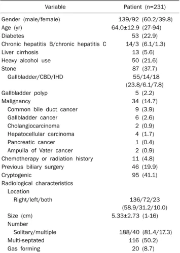
The baseline characteristics of the 231 study subjects are provided in Table 1. The mean patient age was 64.0±12.9 years (range, 27-94 years) and the male to female ratio was 1.51:1. The mean hospital stay was 17.51±12.61 days (range, 1-132 days). Clinically, 190 (82.3%) patients had fever and 35 (15.2%) patients had low blood pressure (<90/60 mmHg) upon admission due to PLA. Of the 231 patients, 53 (22.9%) patients had diabetes, 17 (7.4%) patients had chronic hep- atitis, 87 patients (37.7%) had a gallbladder or biliary tract stone, and 34 (14.7%) patients had a malignancy.
The radiologic characteristics of liver abscesses are sum- marized in Table 1. Abscesses were located in the right lobe in 136 (58.9%) cases, the left lobe in 72 (31.2%) cases, and in both lobes in 23 (10.0%) cases. Abscesses ranged from 1 to >16 cm in diameter (mean 5.33± 2.73 cm). One hundred and eighty-eight (81.4%) patients had a single abscess and 40 (17.3%) patients had multiple abscesses. Abscesses were multi-septated in 116 (50.2%) patients and gas for-
Table 3. Co-infections and Complications of Pyogenic Liver Abscess
Variable Patient (n=231)
Co-infections
Cholangitis 19 (8.2)
Intra-abdominal abscess 17 (7.4)
Cholecystitis 16 (6.9)
Pneumonia 13 (5.6)
Urinary tract infection 6 (2.6)
Endophthalmitis 4 (1.7)
Othersa 4 (1.7)
Complications
Pleural effusion 83 (35.9)
Acute renal failure 2 (0.9)
Portal vein thrombosis 2 (0.9) Gastrointestinal bleeding 1 (0.4) Values are presented as n (%).
aInclude 1 empyema, 1 osteomyelitis, 1 infective endocarditis, 1 pyogenic arthritis.
Table 2. Micro-organisms Isolated by Site
Micro-organism Total (n=124) Blood (n=31) Abscess (n=93) Both (n=26)
Klebsiella pneumoniae 88 (71.0) 23 (74.2) 65 (69.9) 19 (73.1)
Escherichia coli 13 (10.5) 3 (9.7) 10 (10.8) 3 (11.5)
Enterococcus spp. 4 (3.2) 1 (3.2) 3 (3.2) 1 (3.8)
Streptococci spp. 4 (3.2) 2 (6.5) 2 (2.2) 2 (7.7)
Pseudomonas spp. 3 (2.4) 1 (3.2) 2 (2.2) 1 (3.8)
Staphylococci spp. 1 (0.8) 0 1 (1.1) 0
Etc.a 11 (8.9) 1 (3.2) 10 (10.8) 0
Values are presented as n (%).
aInclude 2 Acinetobacter baumannii, 2 Enterobacter cloacae, 2 Aeromonas hydrophila, 1 Citrobacter braakii, 1 Citrobacter freundii, 1 Actinomycosis, 1 Salmonella typhi, 1 Klebsiella oxytoca.
Table 4. Causes of Death in Patients with Pyogenic Liver Abscess
Variable Patient (n=16)
Sepsis 11 (68.8)
Cancer progression 2 (12.5)
Empyema 2 (12.5)
Asphyxia 1 (6.3)
Values are presented as n (%).
Table 1. Patient Baseline Characteristics
Variable Patient (n=231)
Gender (male/female) 139/92 (60.2/39.8)
Age (yr) 64.0±12.9 (27-94)
Diabetes 53 (22.9)
Chronic hepatitis B/chronic hepatitis C 14/3 (6.1/1.3)
Liver cirrhosis 13 (5.6)
Heavy alcohol use 50 (21.6)
Stone 87 (37.7)
Gallbladder/CBD/IHD 55/14/18
(23.8/6.1/7.8)
Gallbladder polyp 5 (2.2)
Malignancy 34 (14.7)
Common bile duct cancer 9 (3.9)
Gallbladder cancer 6 (2.6)
Cholangiocarcinoma 2 (0.9)
Hepatocellular carcinoma 4 (1.7)
Pancreatic cancer 1 (0.4)
Ampulla of Vater cancer 2 (0.9)
Chemotherapy or radiation history 11 (4.8) Previous biliary surgery 46 (19.9)
Cryptogenic 95 (41.1)
Radiological characteristics Location
Right/left/both 136/72/23
(58.9/31.2/10.0)
Size (cm) 5.33±2.73 (1-16)
Number
Solitary/multiple 188/40 (81.4/17.3)
Multi-septated 116 (50.2)
Gas forming 20 (8.7)
Values are presented as n (%) or mean±SD (range).
CBD, common bile duct; IHD, intrahepatic duct.
mation was observed in 20 (8.7%) patients.
2. Blood and abscess cultures and abscess-associated complications
Klebsiella pneumoniae was the predominant organism as determined by abscess aspiration (69.9%) anhd blood culture
(74.2%) (Table 2). Abscess-associated complications are documented in Table 3. The most common complication was pleural effusion (35.9%) and most common co-infection was cholangitis (8.2%), followed by cholecystitis (6.9%) and pneu- monia (5.6%).
3. Risk factors related with mortality
The overall mortality rate was 6.9% (16/231) and the main causes of death were sepsis (68.8%; n=11) and empyema
Table 5. Significant Mortality Risk Factors
Variable Univariate Multivariate
OR (95% CI) p-value OR (95% CI) p-value
Liver cirrhosis 7.630 (2.051-28.383) 0.008
Cancer history 5.719 (2.002–16.334) 0.002
Biliary operation history 4.658 (1.645-13.188) 0.005
Recurrence 3.882 (1.129-13.352) 0.045 5.970 (1.207-29.529) 0.028
Pleural effusion 6.085 (1.895-19.540) 0.001
Hypoalbuminemia (<3.5 g/dL) 1.778 (1.58-2) 0.001
Bilirubinemia (>2 mg/dL) 7.574 (2.351-24.397) 0.001 9.541 (2.382-38.216) 0.001
Thrombocytopenia (<140×103/L) 5.952 (1.855-19.23) 0.001 4.396 (1.130-17.106) 0.033
Anemia (<12 g/dL) 25 (3.28–200) 0.001 13.277 (1.476-119.423) 0.021
Gas forming abscess 8.614 (2.733-27.151) 0.001
Diabetes 1.166 (0.346-3.659) 0.776
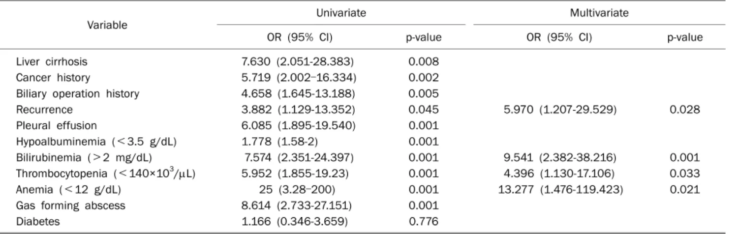
(12.5%; n=2) (Table 4). Risk factors found to be significantly associated with mortality by univariate analysis were liver cir- rhosis (p=0.008, OR 7.630), underlying malignancy (p=
0.002, OR 5.719), a previous biliary operation (p=0.005, OR 4.658), a gas forming abscess (p=0.001, OR 8.614), a history of abscess recurrence (p=0.045, OR 3.882), pleural effusion (p=0.001, OR 6.085), hypoalbuminemia (p=0.001, OR 1.778), bilirubinemia (p=0.0001, OR 7.574), thrombocytope- nia (p=0.001, OR 5.952), and anemia (p=0.0001, OR 25) (Table 5). Diabetes was not found to be related with mortality (p=0.776, OR 1.166).
Multivariate logistic regression analysis showed that ab- scess recurrence (OR=5.970, 95% CI 1.207-29.529, p=
0.028), bilirubinemia (>2 mg/dL, OR 9.541, 95% CI 2.382-38.216; p=0.001), thrombocytopenia (<140×103/L, OR 4.396, 95% CI 1.130-17.106; p=0.033), and anemia (<12 g/dL, OR 13.277, 95% CI 1.476-119.423; p=0.021) were independent risk factors for mortality (Table 5).
Regarding treatment modalities, 67 patients underwent antibiotic treatment alone, 143 patients received percuta- neous abscess drainage (PCD) insertion, and 21 patients re- ceived aspiration alone. Eight patients were surgically treat- ed, including one case of enucleation due to endophthalmitis.
DISCUSSION
Recent reports estimated the annual incidence of PLA at 2.3 cases per 100,000 of the population and found that most PLA patients are elderly (mean age, 64 years),7,13 which is about 10 years older than subjects mentioned in previous reports.14,15 Older research reported that Escherichia coli
was the most important pathogen in the etiology of PLA, but during the last two decades, K. pneumoniae is observed to be the major causative pathogen.16 Many studies were con- ducted at Asian institutions, in which a large proportion of PLA cases (43% to 66%) were attributed to Klebsiella spe- cies, typically in association with diabetes or of cryptogenic origin.6,15,17,18 On the other hand, in Western studies E. coli re- mains the most common causative organism.19,20 In the present study, K. pneumoniae predominated (71.0%), fol- lowed by E. coli (10.5%), consistent with previous inves- tigations conducted in Asia.15,21,22 In addition, outcomes of E.
coli PLA were inferior to those of K. pneumoniae PLA, al- though this may have been due to inherent demographic and clinical differences. Chan et al.7 found that mortality rate of E. coli PLA tended to be higher than that of K. pneumoniae PLA (p=0.066). In the present study, patients with E. coli PLA were older than K. pneumoniae PLA patients, while gender ra- tios were similar. Rates of cancer, biliary operation, and ERCP were significantly higher in E. coli PLA patients. In addition, there were 8 (8.8%) cases of extended-spectrum beta-lacta- mase (ESBL)-positive K. pneumoniae and 4 (30.8%) cases of ESBL-positive E. coli, but no association was observed be- tween these factors and mortality (Table 6).
The cause of liver abscess was cryptogenic in 41.1% pa- tients, comparable to previous reports,10,22,23 followed by bili- ary origin (37.7%), diabetes (22.9%), and hepatobiliary malig- nancy (10.4%). Choledocholithiasis, benign and malignant tumors, and postoperative strictures are also associated with PLA.1,5,10,24,25 In fact, any source of intra-abdominal ab- scess, such as acute diverticulitis, appendicitis, inflam- matory bowel disease, and perforated hollow viscus, can
Table 6. Baseline Characteristics, Co-morbidities and Treatment Outcomes of Klebsiella pneumoniae and Escherichia coli Pyogenic Liver Abscess
K. pneumonia (n=91) E. coli (n=13) p-value
Male 52 (57.1) 7 (53.8) 0.822
Age (yr) 64.29±12.21 72.00±10.44 0.033
Co-morbidity
Diabetes 24 (26.4) 3 (23.1) 1.000
CRF 2 (2.2) 0 (0.0) 1.000
Heavy alcohol use 19 (20.9) 1 (7.7) 0.454
GB stone 22 (24.2) 3 (23.1) 1.000
Biliary stone 12 (13.2) 4 (30.8) 0.113
Biliary operation 13 (14.3) 8 (61.5) 0.001
ERCP History 15 (16.5) 7 (53.8) 0.006
Cancer related abscess 11 (12.1) 5 (38.5) 0.028
Positive of ESBLs 8 (8.8) 4 (30.8) 0.042
Image study
Abscess size (cm) 5.73±2.56 5.14±3.00 0.448
Gas forming abscess 10 (11.0) 2 (15.4) 0.644
Solitary abscess 74 (81.3) 8 (61.5) 0.241
Abscess location 0.137
Right 60 (65.9) 5 (38.5)
Left 22 (24.2) 5 (38.5)
Both or junction 9 (9.9) 3 (23.1)
Treatment outcomes
Median length of hospital stay (day) 19.02±15.03 14.76±10.45 0.327
PCD insertion 74 (81.3) 10 (76.9) 0.712
Operation 0 (0) 1 (7.7) 0.125
Mortality 5 (5.5) 3 (23.1) 0.060
Values are presented as n (%) or mean±SD.
CRF, chronic renal failure; GB, gallbladder; ESBLs, extended-spectrum beta-lactamases; PCD, percutaneous abscess drainage.
cause PLA via portal pyemia.1 PLA by hematogenous in- fection via the hepatic artery is hypothesized to originate from hepatic seeding of bacteria, in cases of systemic in- fection, such as endocarditis or urinary sepsis. Huang et al.11 reported that malignant biliary obstruction had become the leading cause of liver abscess. In PLA, a solitary lesion is more common in the right lobe,26 which can be explained by the larger size of the right lobe and its receipt of most mesen- teric-portal blood flow.11 Diabetes is a risk factor for PLA, and the main pathogen is K. pneumoniae in diabetes related PLA.
In the present study, liver abscesses were observed in right lobe in 60 patients (65.9%), 10 patients (8.7%) exhibited a gas forming pattern, and 45.0% were related to diabetes.
Gas-forming liver abscesses are uncommon but occur in 7-24% of PLA cases and bring a high risk of mortality.27 In the present study, although gas formation was found to predict mortality by univariate analysis, this relationship was not found to be significant by multivariate analysis.
Significant advances and changes have been made in the management of PLA over the past 15 years. Traditionally, pa-
tients with PLA were treated by surgical drainage and anti- biotic therapy, but today percutaneous aspiration/drainage under US or CT guidance is the procedure of choice.5,6,14 In the present study, 67 (29.0%) of the 231 patients were treat- ed using antibiotics alone, 143 (61.9%) patients by PCD in- sertion, and eight (3.4%) patients surgically. However, these different modalities did not influence mortality significantly.
We administered third generation cephalosporin (ceftriaxone) and metronidazole empirically until the identity of the organ- ism and its sensitivity were determined, and continued anti- biotic therapy until abscess cavity obliteration was observed by CT and there were no signs of infection. When clinical signs of infection resolved completely in the presence of a small ab- scess, as determined by CT, patients were discharged on oral antibiotics.
PLA often leads to complications such as pleural effusion, acute renal failure, multiple organ failure, abdominal ab- scess, and portal or hepatic vein thrombosis.28 In the present study, there were 83 (35.9%) cases of pleural effusion and 4 (1.7%) cases of endophthalmitis. Septic endophthalmitis
due to K. peumoniae is sometimes devastating. In the pres- ent study, the single affected patient ultimately underwent enucleation.29
Over the past few decades, mortality associated with PLA has decreased gradually. Prior to 1980, case series mortal- ities were consistently greater than 50%. Improvements in mortality rates in the 1980s and 1990s were attributed to highly effective broad spectrum antibiotics and the advent of cross sectional CT and US imaging. Mortality rates during and after the 1990s was between 4-10%.6,14,15,17 However, an as- sociation between mortality and malignancy was recently highlighted by Mezhir et al.,30 who reported a mortality rate as high as 29%. In the present study, the overall mortality rate was 6.9%, but the mortality rate associated with malignancy was almost twice as high (12.5%).
The high mortality rate of PLA highlights the importance of prognostic factors, prompting research into risk factors.
Although no consensus has been reached, marked leukocy- tosis, hypoalbuminemia, hyperbilirubinemia, altered renal function, septic shock, higher Acute Physiologic Assessment and Chronic Health Evaluation II (APACHE II) score, and pleural effusion are significantly associated with mortality.10,12,22,31,32
Six independent risk factors predict severe complications in K. pneumonia-related PLA: thrombocytopenia (<100,000/
mm3), alkaline phosphatase >300 U/L, gas formation, an APACHE III score of >40, the use of cefazolin (rather than ex- tended-spectrum cephalosporin), and delayed drainage.33 Co-morbid malignancy is considered a grave factor, probably because of the presence of malnutrition and immuno- suppression.10,11,34 In the present study, the risk factors asso- ciated with PLA mortality as determined by multiple re- gression were recurred abscess, jaundice (>2 mg/dL), thrombocytopenia (<140×103/L), and anemia (<12 g/dL), which largely concurs with previous studies.10,11,34 Recurred abscess has not been commonly mentioned as a prognostic factor, but in the present study it is associated with underlying malignancy and biliary disease, underlining the need for care in the approach in PLA patients with such co-morbidities.
The main limitation of the present study is its retro- spective, single center design. Furthermore, the true preva- lence of PLA may have been under-estimated because we col- lected cases with a definitive diagnosis to reduce con- founding. Nevertheless, our study has several strengths.
First, it adds to factors already identified associated with
mortality in PLA, aiding treatment decision-making in high risk cases. Second, we evaluated underlying diseases and complications and explored relations between underlying diseases, complications, and disease outcomes.
Summarizing, the study shows that a history of liver abscess, bilirubinemia (>2 mg/dL), thrombocytopenia (<140×103/
L), and anemia (<12 g/dL) are independently associated with mortality in patients with PLA, and suggests clinicians pay special attention to patients with these factors.
REFERENCES
1. Branum GD, Tyson GS, Branum MA, Meyers WC. Hepatic abscess. Changes in etiology, diagnosis, and management. Ann Surg 1990;212:655-662.
2. Pitt HA, Zuidema GD. Factors influencing mortality in the treat- ment of pyogenic hepatic abscess. Surg Gynecol Obstet 1975;140:228-234.
3. Altemeier WA, Culbertson WR, Fullen WD, Shook CD. Intra-ab- dominal abscesses. Am J Surg 1973;125:70-79.
4. Johannsen EC, Sifri CD, Madoff LC. Pyogenic liver abscesses.
Infect Dis Clin North Am 2000;14:547-563.
5. Rintoul R, O'Riordain MG, Laurenson IF, Crosbie JL, Allan PL, Garden OJ. Changing management of pyogenic liver abscess. Br J Surg 1996;83:1215-1218.
6. Wong WM, Wong BC, Hui CK, et al. Pyogenic liver abscess: retro- spective analysis of 80 cases over a 10-year period. J Gastroen- terol Hepatol 2002;17:1001-1007.
7. Chan KS, Chen CM, Cheng KC, Hou CC, Lin HJ, Yu WL. Pyogenic liver abscess: a retrospective analysis of 107 patients during a 3-year period. Jpn J Infect Dis 2005;58:366-368.
8. Mohsen AH, Green ST, Read RC, McKendrick MW. Liver abscess in adults: ten years experience in a UK centre. QJM 2002;95:
797-802.
9. Thomsen RW, Jepsen P, Sørensen HT. Diabetes mellitus and pyo- genic liver abscess: risk and prognosis. Clin Infect Dis 2007;
44:1194-1201.
10. Chu KM, Fan ST, Lai EC, Lo CM, Wong J. Pyogenic liver abscess.
An audit of experience over the past decade. Arch Surg 1996;
131:148-152.
11. Huang CJ, Pitt HA, Lipsett PA, et al. Pyogenic hepatic abscess.
Changing trends over 42 years. Ann Surg 1996;223:600-607;
discussion 607-609.
12. Lee KT, Sheen PC, Chen JS, Ker CG. Pyogenic liver abscess: multi- variate analysis of risk factors. World J Surg 1991;15:372-376;
discussion 376-377.
13. Petri A, Höhn J, Hódi Z, Wolfárd A, Balogh A. Pyogenic liver ab- scess: 20 years' experience. Comparison of results of treatment in two periods. Langenbecks Arch Surg 2002;387:27-31.
14. Barakate MS, Stephen MS, Waugh RC, et al. Pyogenic liver ab- scess: a review of 10 years' experience in management. Aust N Z J Surg 1999;69:205-209.
15. Lee KT, Wong SR, Sheen PC. Pyogenic liver abscess: an audit of 10 years' experience and analysis of risk factors. Dig Surg 2001;18:459-465; discussion 465-466.
16. Shon AS, Bajwa RP, Russo TA. Hypervirulent (hypermucoviscous) Klebsiella pneumoniae: a new and dangerous breed. Virulence 2013;4:107-118.
17. Tan EY, Lee CW, Look Chee Meng M. Spontaneous pneumo- peritoneum resulting from the rupture of a gas-forming pyogenic liver abscess. ANZ J Surg 2005;75:251-252.
18. Foo NP, Chen KT, Lin HJ, Guo HR. Characteristics of pyogenic liver abscess patients with and without diabetes mellitus. Am J Gastroenterol 2010;105:328-335.
19. Cerwenka H, Bacher H, Werkgartner G, et al. Treatment of pa- tients with pyogenic liver abscess. Chemotherapy 2005;51:366- 369.
20. Alvarez Pérez JA, González JJ, Baldonedo RF, et al. Clinical course, treatment, and multivariate analysis of risk factors for pyogenic liver abscess. Am J Surg 2001;181:177-186.
21. Seeto RK, Rockey DC. Pyogenic liver abscess. Changes in etiol- ogy, management, and outcome. Medicine (Baltimore) 1996;
75:99-113.
22. Tazawa J, Sakai Y, Maekawa S, et al. Solitary and multiple pyo- genic liver abscesses: characteristics of the patients and effi- cacy of percutaneous drainage. Am J Gastroenterol 1997;92:
271-274.
23. Chou FF, Sheen-Chen SM, Chen YS, Lee TY. The comparison of clinical course and results of treatment between gas-forming and non-gas-forming pyogenic liver abscess. Arch Surg 1995;
130:401-405; discussion 406.
24. Gyorffy EJ, Frey CF, Silva J Jr, McGahan J. Pyogenic liver abscess.
Diagnostic and therapeutic strategies. Ann Surg 1987;206:
699-705.
25. Kandel G, Marcon NE. Pyogenic liver abscess: new concepts of an old disease. Am J Gastroenterol 1984;79:65-71.
26. Park JH, Lee TH, Kim ST, et al. Clinical features of pyogenic liver abscess according to age group. Korean J Gastroenterol 2010;56:90-96.
27. Huang CY, Chou WK, Lin MS, Tsai KC, Sun JT. Gas-forming pyo- genic liver abscess. QJM 2009;102:885-886.
28. Pang TC, Fung T, Samra J, Hugh TJ, Smith RC. Pyogenic liver ab- scess: an audit of 10 years' experience. World J Gastroenterol 2011;17:1622-1630.
29. Liu YC, Cheng DL, Lin CL. Klebsiella pneumoniae liver abscess associated with septic endophthalmitis. Arch Intern Med 1986;146:1913-1916.
30. Mezhir JJ, Fong Y, Jacks LM, et al. Current management of pyo- genic liver abscess: surgery is now second-line treatment. J Am Coll Surg 2010;210:975-983.
31. Chou FF, Sheen-Chen SM, Chen YS, Chen MC, Chen FC, Tai DI.
Prognostic factors for pyogenic abscess of the liver. J Am Coll Surg 1994;179:727-732.
32. Mischinger HJ, Hauser H, Rabl H, et al. Pyogenic liver abscess:
studies of therapy and analysis of risk factors. World J Surg 1994;18:852-857; discussion 858.
33. Lin JC, Yeh KM, Chang FY. The distant metastasis of pyogenic liv- er abscess caused by Klebsiella pneumoniae serotype K2 and the underlying disease of diabetes mellitus should be carefully interpreted. Clin Infect Dis 2007;45:1531-1532; author reply 1532-1533.
34. Yeh TS, Jan YY, Jeng LB, Chen TC, Hwang TL, Chen MF. Hepatocel- lular carcinoma presenting as pyogenic liver abscess: character- istics, diagnosis, and management. Clin Infect Dis 1998;26:
1224-1226.