© 2017 The Korean Ophthalmological Society
This is an Open Access article distributed under the terms of the Creative Commons Attribution Non-Commercial License (http://creativecommons.org/licenses /by-nc/3.0/) which permits unrestricted non-commercial use, distribution, and reproduction in any medium, provided the original work is properly cited.
Original Article
Oral Administration of Cilostazol Increases Ocular Blood Flow in Patients with Diabetic Retinopathy
Duck Jin Hwang1,2, Joo Young Shin1, Hyeong Gon Yu1
1Department of Ophthalmology, Seoul National University Hospital, Seoul National University College of Medicine, Seoul, Korea
2HanGil Eye Hospital, Incheon, Korea
Purpose: To investigate the effect of cilostazol on ocular hemodynamics and to determine whether the adminis- tration of cilostazol increases the ocular blood flow in patients with diabetic retinopathy.
Methods: This prospective observational study investigated the effect of orally administered cilostazol on dia- betic retinopathy. Before and after administration for 1 week, pulsatile ocular blood flow (POBF) and retrobul- bar hemodynamics were measured using a POBF analyzer and transcranial Doppler imaging, respectively.
Visual acuity, intraocular pressure, and blood pressure were also evaluated before and after treatment.
Results: Twenty-five eyes of 25 patients were included in this study. POBF increased significantly (16.8 ± 4.6 µL/sec vs. 19.6 ± 6.2 µL/sec, p < 0.001) after administration of cilostazol, while no significant change was iden- tified in visual acuity, intraocular pressure, and blood pressure. Mean flow velocity in the ophthalmic artery as measured with transcranial Doppler imaging also increased significantly after medication (23.5 ± 5.6 cm/sec vs. 26.0 ± 6.9 cm/sec, p = 0.001). The change in POBF directly correlated with the change in mean flow veloc- ity (r = 0.419, p = 0.007).
Conclusions: Cilostazol was effective in increasing ocular blood flow in patients with diabetic retinopathy, possi- bly by modulating retrobulbar circulation.
Key Words: Blood flow velocity, Cilostazol, Diabetic retinopathy, Pulsatile flow
Medications that modulate blood flow are widely used in the treatment of cerebrovascular and cardiovascular dis- eases. These systemic medications may also affect ocular blood flow, as retinal circulation is similar to cerebral cir-
culation [1-4]. Cilostazol, an antiplatelet agent, has been used for the prevention of ischemic stroke in Japan and other Asian countries [5]. Cilostazol acts as a direct and in- direct antiplatelet agent by inhibiting platelet activation and improving overall vascular endothelial function [5,6].
This antiplatelet effect is mediated by cyclic guanosine monophosphate, which inhibits phosphodiesterase-3 and also induces vasodilation [7,8]. Cilostazol also reportedly enhances cerebral blood flow in patients with chronic cere- bral infarction. However, few studies have examined the
Received: January 6, 2016 Accepted: February 17, 2016
Corresponding Author: Hyeong Gon Yu, MD, PhD. Department of Oph- thalmology, Seoul National University College of Medicine, #103 Dae- hak-ro, Jongno-gu, Seoul 03080, Korea. Tel: 82-2-2072-2438, Fax: 82-2- 741-3187, E-mail: [email protected]
effects of systemic medications on ocular blood flow.
The pulsatile ocular blood flow (POBF) analyzer (Para- digm Medical Instruments, Salt Lake City, UT, USA) (Fig.
1A) is a device that noninvasively measures ocular blood flow by measuring changes in intraocular pressure (IOP) caused by pulsatile rhythmic filling of the intraocular ves- sels with a pneumatic applanation tonometer [9-11]. The device’s design is based on the assumption that venous outflow from the eye is non-pulsatile. Moreover, ocular ri- gidity, which is used to derive changes in ocular volume from changes in IOP, is assumed to be the same in all sub- jects. The calculation of POBF is automatically derived from the beat-to-beat IOP variation as measured using the five pulses that are closest to each other. POBF is calculat- ed on a “per-minute” or “per-second basis” as beat-to-beat variation in IOP, which provides an indirect assessment of total ocular blood flow.
Changes in ocular blood flow have been known to cor- relate with the development and progression of diabetic
retinopathy, leading to macular edema and retinal neovas- cularization [12-14]. Although the exact nature of ocular blood flow abnormalities in diabetic retinopathy has not yet been established, the effect of pathogenic mechanisms such as vascular and rheological abnormalities and long- term hyperglycemia on the microvasculature seem to alter the blood flow in patients with diabetic retinopathy [15-19].
In addition, ocular ischemia caused by this dysregulated blood flow induces the expression of cytokines such as vascular endothelial growth factor, which also may affect retinal blood flow [20-22]. Thus, there is a possibility that eyes with diabetic retinopathy may potentially benefit from medications altering ocular blood flow. While previ- ous studies have mainly focused on retinal blood flow in di- abetic retinopathy, interest in investigating the hemodynam- ics of the entire ocular circulation has been prompted by the development of the POBF analyzer, which is a clinical- ly feasible technique for the measurement of POBF [9-11].
It has been suggested that the pulsatile component of ocu- lar blood flow is a reliable parameter for the evaluation of the choroidal circulation.
Therefore, in this study, we investigated the effect of ci- lostazol on ocular hemodynamics and evaluated whether the administration of cilostazol increases ocular blood flow in patients with diabetic retinopathy.
Materials and Methods
Participants
Patients over 30 years of age with type 2 diabetes, dia- betic retinopathy, and a visual acuity of 20 / 50 or better were enrolled in this prospective observational study. All participants were seen at Seoul National University Hospi- tal from October 2011 to March 2012. The baseline POBF analyses were performed in conjunction with a transcrani- al Doppler (TCD) examination and blood pressure (BP) measurements. Ophthalmic evaluations of best-corrected visual acuity (BCVA), IOP, and Cirrus HD optical coher- ence tomography (OCT; Carl Zeiss Meditec, Dublin, CA, USA) measurements were performed. One week after the administration of cilostazol (Pletal; Otsuka Pharmaceuti- cal, Tokyo, Japan), all examinations were repeated. All POBF examinations were performed by a single trained examiner (DJH) to reduce inter-examiner bias. The central Fig. 1. (A) The pulsatile ocular blood flow analyzer (Paradigm
Medical Instruments, Salt Lake City, UT, USA), (B) the transcra- nial Doppler image using a TCD150M device (Spencer Technolo- gies, Seattle, WA, USA). This case shows a peak systolic velocity (PEAK), mean flow velocity (MEAN), end diastolic velocity (DIAS) and pulsatility index (PI) of the left ophthalmic artery at 1 week after the administration of cilostazol.
A
B
foveal thickness (CFT, the mean retinal thickness of the central 1 mm circle), the macular cube volume, and the macular cube average thickness were measured using a Cirrus HD OCT unit. When both eyes were eligible for the study, each eye was individually evaluated; however, a computer algorithm randomly selected only one eye for use in the statistical analysis. The type of diabetic retinop- athy was also classified into two subgroups: a non-prolifer- ative diabetic retinopathy (NPDR) group and a panretinal photocoagulation (PRP)-treated proliferative diabetic reti- nopathy (PDR) group.
Upon enrollment, the clinical history of each patient was thoroughly evaluated. After screening, patients were ad- ministered cilostazol twice daily at a dose of 100 mg for 1 week (200 mg per day). Patients who were taking aspirin at the time of enrollment in this study were asked to stop aspirin treatment for 2 weeks before beginning the ci- lostazol regimen. The 200-mg cilostazol dose was contin- ued for 1 week if no complication or severe event was ob- served. The administration of any other anti-platelet drugs that could strongly influence the effect of cilostazol was prohibited during the study period. However, pre-existing prescription drugs for chronic diseases such as hyperten- sion and diabetic mellitus were continued throughout the study period.
Patients with the following conditions were excluded from the study: a history of retinal surgery; ocular diseas- es, excluding diabetic retinopathy; new retinal vessels ob- served with fluorescein angiography; macular edema with a CFT exceeding 300 μm; severe hypertension (defined as having a systolic BP greater than 180 mmHg or a diastolic BP greater than 110 mmHg); hypersensitivity to the ci- lostazol treatment; pregnancy or the possibility of preg- nancy; uncontrolled diabetes (hemoglobin A1c >8%); angi- na pectoris, myocardial infarction, or heart failure.
POBF measurement
A POBF analyzer was used to assess blood flow accord- ing to methods previously described [9-11]. This noninva- sive technique assessing ocular blood flow is based on the IOP pulse. A modified applanation prism with distensible film at the contact surface is used to measure blood veloci- ty. A pneumotonometer is used to create a waveform rep- resenting the ocular pulse. The amplitude and pulse rate (PR) of this waveform are then used to calculate POBF.
The POBF analyzer has been found to be reliable and re- producible in measuring ocular pulse amplitude (PA), pulse volume (PV), PR, and POBF [9,10,23,24].
Retrobulbar vessel (ophthalmic artery) blood velocity The TCD studies were performed using a TCD150M de- vice (Spencer Technologies, Seattle, WA, USA) (Fig. 1B), which calculates a power M-mode Doppler image and pro- vides a single-gate spectrogram. TCD has shown good re- producibility and repeatability in the assessment of the ophthalmic artery [25]. The peak systolic velocity (PSV), mean flow velocity (MFV), end diastolic velocity (EDV), and pulsatility index (PI) of the ophthalmic artery were evaluated at baseline and 1 week after the administration of cilostazol. The ophthalmic artery can be situated above or below the optic nerve in the posterior orbit and passes forward into the nasal orbit in a horizontal plane slightly superior to that of the optic nerve. These vessels were ex- amined at a point approximately 25 mm behind the globe in the nasal orbit. At this point, the vessels lie in a straight position that facilitates accurate measurements. The PI was calculated using the following formula (PSV – EDV) / (MFV), which was used to characterize the peripheral vas- cular resistance.
Statistical analyses
Statistical analyses were performed using a commercial- ly available software package PASW ver. 18.0 (SPSS Inc., Chicago, IL, USA). Changes between values measured at baseline and those measured 1 week after the administra- tion of cilostazol were evaluated using the paired t-test and Wilcoxon signed-rank test for normally distributed data and data that were not normally distributed, respectively.
Significant differences in the POBF analyzer and TCD values between the subgroups (NPDR group vs. PRP-treat- ed PDR group) were evaluated using the Mann-Whitney test for nonparametric data. Analysis of covariance was performed to adjust for age, gender, and the duration of di- abetes mellitus in the subgroup analysis. Spearman’s non- parametric regression analysis was used to determine the correlation between POBF and TCD at the ophthalmic ar- tery. A p-value less than 0.05 was considered to be statisti- cally significant.
Ethics statement
The study was approved by the institutional review board of Seoul National University Hospital (no. 0620113380) and followed the guidelines of Good Clinical Practice. Written informed consent for participation was obtained from all patients after they had been given a thorough explanation of this study. The study was carried out in accordance with the tenets of the Declaration of Helsinki.
Results
We enrolled 25 eyes from 25 patients with diabetic reti- nopathy. The mean age was 62.5 ± 12.0 years and 16 pa- tients (64%) were male. The mean duration of diabetes mellitus was 14.6 ± 7.0 years. The PDR group (18.4 ± 6.4 years) had a longer duration of diabetes mellitus than the NPDR group (11.6 ± 6.0 years) (p = 0.001). Cilostazol was well tolerated by all of the patients, and no complications or severe adverse events were observed during the 1 week of administration. There were no significant changes in vi-
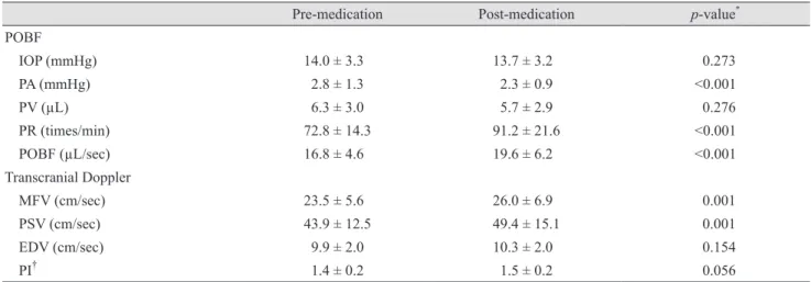
Table 1. Changes in POBF analyzer and transcranial Doppler parameters after cilostazol administration
Pre-medication Post-medication p-value*
POBF
IOP (mmHg) 14.0 ± 3.3 13.7 ± 3.2 0.273
PA (mmHg) 2.8 ± 1.3 2.3 ± 0.9 <0.001
PV (µL) 6.3 ± 3.0 5.7 ± 2.9 0.276
PR (times/min) 72.8 ± 14.3 91.2 ± 21.6 <0.001
POBF (µL/sec) 16.8 ± 4.6 19.6 ± 6.2 <0.001
Transcranial Doppler
MFV (cm/sec) 23.5 ± 5.6 26.0 ± 6.9 0.001
PSV (cm/sec) 43.9 ± 12.5 49.4 ± 15.1 0.001
EDV (cm/sec) 9.9 ± 2.0 10.3 ± 2.0 0.154
PI† 1.4 ± 0.2 1.5 ± 0.2 0.056
Values are presented as mean ± standard deviation.
POBF = pulsatile ocular blood flow; IOP = intraocular pressure; PA = pulse amplitude; PV = pulse volume; PR = pulse rate; MFV = mean flow velocity in the ophthalmic artery; PSV = peak systolic velocity in the ophthalmic artery; EDV = end diastolic velocity in the oph- thalmic artery; PI = pulsatility index.
*Wilcoxon signed-rank test; †(PSV – EDV) / (MFV).
µL/sec
POBF p < 0.001
Pre-medication Post-medication 30
20
10
0
cm/sec
MFV of OA p = 0.001
Pre-medication Post-medication 40
30 20 10 0
Fig. 2. One week after cilostazol administration, (A) pulsatile ocular blood flow (POBF) had increased by 17.1% (p < 0.001) and (B) mean flow velocity (MFV) of ophthalmic artery (OA) had increased by 11.7% (p = 0.001). Values are means with error bars representing stan- dard deviation.
A B
sual acuity, IOP, or BP after medication. Among the OCT parameters, there were no significant changes in the CFT (255.9 ± 26.4 μm vs. 254.0 ± 27.4 μm, p = 0.111), macular cube volume (10.32 ± 1.0 mm3 vs. 10.3 ± 1.0 mm3, p = 0.845), or macular cube average thickness (286.7 ± 29.0 μm vs. 287.3 ± 28.2 μm, p = 0.111), respectively.
POBF analysis
The PA, PR, and POBF measurements exhibited signifi- cant changes after cilostazol administration, as shown in Table 1. At 1 week after cilostazol administration, PA had decreased by 17.9% (2.8 ± 1.3 mmHg vs. 2.3 ± 0.9 mmHg, p < 0.001), and PR had increased by 25.3% (72.8 ± 14.3 times/min vs. 91.2 ± 21.6 times/min, p < 0.001). Mean POBF was 16.8 ± 4.6 µL/sec at baseline and increased by 17.1% to 19.6 ± 6.2 µL/sec after 1 week of cilostazol medi-
cation (p < 0.001) (Fig. 2A). PV remained relatively con- stant throughout the study period (p > 0.05).
Retrobulbar vessel (ophthalmic artery) blood velocities Table 1 also shows the change in the TCD parameters induced by cilostazol. MFV was 23.5 ± 5.6 cm/sec at base- line and then increased significantly by 11.7% to 26.0 ± 6.9 cm/sec with medication administration (p = 0.001) (Fig.
2B). PSV also increased by 12.8% (43.9 ± 12.5 cm/sec vs.
49.5 ± 15.1 cm/sec, p = 0.001). EDV and PI remained rela- tively constant throughout the study.
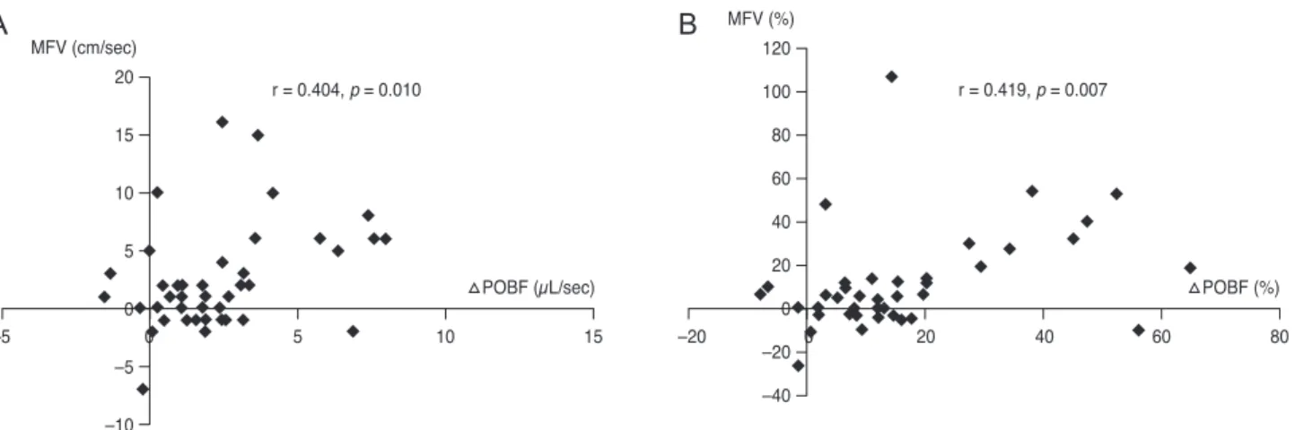
Correlation between the POBF and TCD parameters A moderate correlation between changes in POBF and TCD was observed. The changes in the POBF were found
Table 2. Comparison of POBF and MFV in the NPDR and PDR groups
NPDR (n = 12) PDR (n = 13) p-value* p-value†
Pre-POBF 17.5 ± 4.6 15.9 ± 4.4 0.346 0.326
Post-POBF 20.2 ± 7.2 18.7 ± 4.6 0.868 0.272
△POBF 2.8 ± 3.8 2.8 ± 2.6 0.431 0.728
Pre-MFV 22.9 ± 5.9 24.2 ± 5.4 0.352 0.243
Post-MFV 24.5 ± 6.5 27.6 ± 7.1 0.177 0.144
△MFV 1.6 ± 4.4 3.4 ± 4.6 0.211 0.675
Values are presented as mean ± standard deviation.
POBF = pulsatile ocular blood flow; MFV = mean flow velocity in the ophthalmic artery; NPDR = non-proliferative diabetic retinopathy;
PDR = proliferative diabetic retinopathy; Pre-POBF = POBF at baseline; Post-POBF = POBF after medication; ∆POBF = post-POBF – pre-POBF; Pre-MFV = MFV in the ophthalmic artery at baseline; Post-MFV = MFV in the ophthalmic artery after medication; ∆MFV = post-MFV – pre-MFV.
*Mann-Whitney U-test; †Age, gender, and duration of diabetes mellitus were adjusted by analysis of covariance.
20 15 10 5 0 –5 –10 –5
MFV (cm/sec)
r = 0.404, p = 0.010
△POBF (µL/sec) △POBF (%)
0 5 10 15
120 100 80 60 40 20 0 –20 –40 –20
MFV (%)
r = 0.419, p = 0.007
0 20 40 60 80
Fig. 3. Spearman’s regression analysis showed that the change in pulsatile ocular blood flow (POBF) was found to correlate with the change in mean flow velocity (MFV) of the ophthalmic artery: (A) absolute change: r = 0.404, p = 0.010; (B) percentage change: r = 0.419, p = 0.007.
A B
to correlate with the changes in MFV (absolute change: r = 0.404, p = 0.010; percentage change: r = 0.419, p = 0.007, respectively), as shown in Fig. 3A and 3B, respectively.
The changes in the POBF also correlated with the changes in PSV and EDV (r = 0.393, p = 0.012 and r = 0.337, p = 0.034, respectively).
Comparison of the NPDR and PDR groups
After dividing the 25 patients into two groups (a NPDR group and PRP-treated PDR group), subgroup analyses were performed for the POBF and TCD parameters. There was no significant difference in any of the POBF or TCD parameters between the two groups. A summary of the re- sults can be seen in Table 2. The analysis of covariance re- vealed no significant inter-group differences after adjust- ment for age, sex, and duration of diabetes mellitus (p >
0.05). Neither age nor gender was found to be a significant predictor of changes in POBF or MFV (p = 0.275 and p = 0.114, respectively).
Discussion
In this study, ocular blood flow as assessed by the POBF analyzer increased with oral administration of cilostazol in patients with diabetic retinopathy patients. This increase in ocular blood flow was parallel to the increase in retrobul- bar vessel blood flow velocity as measured with TCD. The same trend of increased ocular blood flow was observed irrespective of the degree of diabetic retinopathy.
The average increase in POBF observed in this study was 17.1%. This change in ocular blood flow was significant even after considering the previously proven test/retest variability [9,10,23,24]. Furthermore, to minimize the in- terobserver variability, all measurements were performed by a single experienced operator, which is very important in measuring POBF [26]. POBF values have a wide normal range and low discriminating power [27], so intra-individ- ual comparisons with repeated examinations, as performed in this study, are more useful than inter-individual com- parisons. POBF analysis also showed that cilostazol ad- ministration significantly decreased PA and increased PR, which have been reported to be negatively correlated with each other in healthy subjects [28], and PV did not change significantly. These trends may explain the increase in
POBF, which is affected by PA, PR, and PV [9-11].
However, the outcomes of the present study should be discussed with an awareness of the associated instrumen- tal limitations. Currently, there is no established gold stan- dard for the assessment of ocular blood flow, and therefore validation of any given method is limited to comparison with other methods. Although the measurement of ocular blood flow with the POBF analyzer has empirical validity, it is necessary to consider the accuracy of our results. This is important because the measurement of blood flow from the ocular pulse is based on specific assumptions [11], hence no single method may provide a complete descrip- tion of ocular blood flow. We therefore performed TCD to also evaluate retrobulbar vessel velocity, and found that the mean velocity in the ophthalmic artery had increased by 11.7 %. These changes in the TCD parameters of retrob- ulbar hemodynamics suggest that cilostazol treatment may modulate the velocity of blood in the retrobulbar vessels.
Changes in POBF were found to moderately correlate with changes in MFV by TCD, although ocular blood flow ac- counts for only a small portion of blood flow through the ophthalmic artery [29]. Changes in POBF also correlated with changes in PSV and EDV as well as MFV. Despite the fact that the POBF and TCD measure different phe- nomena, the simultaneous increase in POBF and retrobul- bar blood flow, as measured by the TCD, raises the possi- bility that the increase in POBF may reflect an actual increase in ocular blood flow, which may be affected by the increase in retrobulbar blood flow.
The exact mechanism of cilostazol in increasing ocular blood flow remains to be clarified. Although cilostazol re- portedly enhances cerebral blood flow in cases of chronic cerebral infarction, its effect on ocular blood flow has not been investigated, and its ability to increase ocular blood flow remains unknown, especially in the diabetic state. To the best of our knowledge, this is the first study designed to elucidate the effect of systemic medication on ocular blood flow in patients with diabetic retinopathy. It may be speculated that the increased ocular blood flow observed in this study was due to the vasodilatory effect of ci- lostazol, as cilostazol is known to act as a platelet inhibitor and vasodilator. The PI calculated from the parameters measured by TCD, which reflects vascular resistance, showed no significant change. The blood flow velocity was observed to increase, thereby increasing the retrobulbar blood flow. Cilostazol may affect the autoregulatory func-
tion of ocular blood flow and cause the vascular resistance to remain constant despite increased cerebral blood flow, thereby allowing increased ocular blood flow.
No significant change was identified in the IOP, BP, BCVA, and OCT thickness parameters, such as the CFT and the macular cube volume at 1 week after the adminis- tration of cilostazol. With the increased ocular blood flow induced by cilostazol, autoregulatory mechanisms may have decreased ocular vascular resistance to maintain a constant IOP. Additionally, cilostazol may have a direct ef- fect on inducing vasodilation in the ocular vasculature.
However, the exact mechanism and the effect on retinal blood flow remain unclear. Spectral domain-OCT parame- ters do not represent retinal blood flow, and there are no reliable methods currently available to assess retinal blood flow. The challenge of evaluating the effects of cilostazol on retinal blood flow and its long-term clinical outcomes remains to be solved in future studies.
Previous studies on cilostazol reported a lower mean POBF in the PRP-treated PDR group as compared to the NPDR group. However, our results showed no significant difference in POBF values between the NPDR group and PDR groups. In addition, the POBF of the NPDR and PDR groups were higher than those reported in previous studies for patients with diabetic retinopathy. Perrott et al. [30] re- ported a mean value of 15.9 µL/sec for the NPDR group, which was lower than the values reported in this study.
Savage et al. [19] reported mean values of 15.7 µL/sec for moderate to severe NPDR and 10.3 µL/sec for PRP-treated PDR, both of which were also lower than the values re- ported in this study (16.8 µL/sec for NPDR and 19.6 µL/sec for PDR). This may be due to variability in the conditions of the patients enrolled in these studies, as most related studies have reported a small sample size, and only pa- tients with relatively good vision were enrolled in our study. In addition, the interpretation and comparison of the POBF values may be limited by differences in patient age, sex, and the classification of diabetic retinopathy.
The clinical significance of this increase in ocular blood flow with the administration of cilostazol in patients with diabetic retinopathy remains to be evaluated. Studies de- scribing the role of altered ocular blood flow in patients with diabetic retinopathy report conflicting results. In- creased ocular blood flow does not directly correlate with either improved visual acuity or delayed progression of di- abetic retinopathy. Our understanding of the role of ocular
blood flow in the pathophysiology of diabetic retinopathy is in its infancy, and attempts to pharmacologically modify the progression of diabetic retinopathy are also in their primitive stages. To the best of our knowledge, our study is the first to evaluate the effects of systemic medication on ocular blood flow in diabetic retinopathy. The results of this study suggest that cilostazol may have potential value in increasing ocular blood flow in patients with diabetic retinopathy, although the long-term benefits need to be elucidated. With regard to safety, none of the patients in this study showed any complications or severe adverse events during medication administration. Moreover, in a recent report [31], the addition of cilostazol to a regimen of aspirin therapy did not increase the bleeding tendency when compared with a regimen consisting of aspirin alone.
Cilostazol treatment could therefore safely be added to an ongoing antiplatelet regimen without increasing the risk of bleeding in patients with diabetic retinopathy.
There are some limitations to this study that should be considered when interpreting the results. Most important- ly, there was no control group of normal non-diabetic sub- jects. This is a major limitation in an interventional drug study. Second, the sample size was small, with a relatively brief study period. A large-scale long-term comparative study with a control group will be necessary to confirm these findings.
In this study, cilostazol increased ocular blood flow by more than 10% without any significant side effects in pa- tients with diabetic retinopathy. This demonstrates its po- tential as a tool for increasing blood flow in diabetic reti- nopathy, although its long-term clinical benefit remains to be clarified. Further studies are required to investigate whether increased ocular blood flow due to cilostazol can slow the progression of diabetic retinopathy or improve vi- sual outcome.
Conflict of Interest
No potential conflict of interest relevant to this article was reported.
Acknowledgements
The authors disclosed receipt of the following financial support for the research: Otsuka Pharmaceutical provided financial support for this investigator-initiated research.
The sponsor or funding organization had no role in the de- sign or conduct of this research.
Statistical analyses were aided by the consultation of Medical Research Collaborating Center of Seoul National University College of Medicine and Seoul National Uni- versity Hospital.
References
1. Patton N, Aslam T, Macgillivray T, et al. Retinal vascular image analysis as a potential screening tool for cerebrovas- cular disease: a rationale based on homology between cere- bral and retinal microvasculatures. J Anat 2005;206:319-48.
2. Delaey C, Van De Voorde J. Regulatory mechanisms in the retinal and choroidal circulation. Ophthalmic Res 2000;32:249- 56.
3. Li G, Shih YY, Kiel JW, et al. MRI study of cerebral, reti- nal and choroidal blood flow responses to acute hyperten- sion. Exp Eye Res 2013;112:118-24.
4. Yoshida Y, Sugiyama T, Utsunomiya K, et al. A pilot study for the effects of donepezil therapy on cerebral and optic nerve head blood flow, visual field defect in normal-tension glaucoma. J Ocul Pharmacol Ther 2010;26:187-92.
5. Uchiyama S, Demaerschalk BM, Goto S, et al. Stroke pre- vention by cilostazol in patients with atherothrombosis:
meta-analysis of placebo-controlled randomized trials. J Stroke Cerebrovasc Dis 2009;18:482-90.
6. Goto S. Cilostazol: potential mechanism of action for anti- thrombotic effects accompanied by a low rate of bleeding.
Atheroscler Suppl 2005;6:3-11.
7. Tanaka T, Ishikawa T, Hagiwara M, et al. Effects of ci- lostazol, a selective cAMP phosphodiesterase inhibitor on the contraction of vascular smooth muscle. Pharmacology 1988;36:313-20.
8. Schror K. The pharmacology of cilostazol. Diabetes Obes Metab 2002;4 Suppl 2:S14-9.
9. Langham ME, Farrell RA, O’Brien V, et al. Blood flow in the human eye. Acta Ophthalmol Suppl 1989;191:9-13.
10. Silver DM, Farrell RA, Langham ME, et al. Estimation of pulsatile ocular blood flow from intraocular pressure. Acta
Ophthalmol Suppl 1989;191:25-9.
11. Krakau CE. Calculation of the pulsatile ocular blood flow.
Invest Ophthalmol Vis Sci 1992;33:2754-6.
12. Sakata K, Funatsu H, Harino S, et al. Relationship between macular microcirculation and progression of diabetic mac- ular edema. Ophthalmology 2006;113:1385-91.
13. Mendivil A, Cuartero V, Mendivil MP. Ocular blood flow velocities in patients with proliferative diabetic retinopathy and healthy volunteers: a prospective study. Br J Ophthal- mol 1995;79:413-6.
14. Cuypers MH, Kasanardjo JS, Polak BC. Retinal blood flow changes in diabetic retinopathy measured with the Heidel- berg scanning laser Doppler flowmeter. Graefes Arch Clin Exp Ophthalmol 2000;238:935-41.
15. Aiello LP, Gardner TW, King GL, et al. Diabetic retinopa- thy. Diabetes Care 1998;21:143-56.
16. Bursell SE, Clermont AC, Kinsley BT, et al. Retinal blood flow changes in patients with insulin-dependent diabetes mellitus and no diabetic retinopathy. Invest Ophthalmol Vis Sci 1996;37:886-97.
17. Feke GT, Tagawa H, Yoshida A, et al. Retinal circulatory changes related to retinopathy progression in insulin-de- pendent diabetes mellitus. Ophthalmology 1985;92:1517-22.
18. Geyer O, Neudorfer M, Snir T, et al. Pulsatile ocular blood flow in diabetic retinopathy. Acta Ophthalmol Scand 1999;77:522-5.
19. Savage HI, Hendrix JW, Peterson DC, et al. Differences in pulsatile ocular blood flow among three classifications of dia- betic retinopathy. Invest Ophthalmol Vis Sci 2004;45:4504-9.
20. Campochiaro PA; C99-PKC412-003 Study Group. Reduc- tion of diabetic macular edema by oral administration of the kinase inhibitor PKC412. Invest Ophthalmol Vis Sci 2004;45:922-31.
21. Aiello LP, Cahill MT, Cavallerano JD. Growth factors and protein kinase C inhibitors as novel therapies for the medical management diabetic retinopathy. Eye (Lond) 2004;18:117-25.
22. Yokota T, Ma RC, Park JY, et al. Role of protein kinase C on the expression of platelet-derived growth factor and en- dothelin-1 in the retina of diabetic rats and cultured retinal capillary pericytes. Diabetes 2003;52:838-45.
23. Butt Z, Oʼbrien C. Reproducibility of pulsatile ocular blood flow measurements. J Glaucoma 1995;4:214-8.
24. Morgan A, Hosking S. Ocular blood flow tonometer repro- ducibility: the effect of operator experience and mode of application. Ophthalmic Physiol Opt 2001;21:401-6.
25. Baxter GM, Williamson TH. Color Doppler imaging of the
eye: normal ranges, reproducibility, and observer variation.
J Ultrasound Med 1995;14:91-6.
26. Schmetterer L, Dallinger S, Findl O, et al. Noninvasive in- vestigations of the normal ocular circulation in humans.
Invest Ophthalmol Vis Sci 1998;39:1210-20.
27. Aydin A, Wollstein G, Price LL, Schuman JS. Evaluating pulsatile ocular blood flow analysis in normal and treated glaucomatous eyes. Am J Ophthalmol 2003;136:448-53.
28. Gekkieva M, Orgul S, Gherghel D, et al. The influence of sex difference in measurements with the Langham ocular blood flow system. Jpn J Ophthalmol 2001;45:528-32.
29. Hayreh SS. The ophthalmic artery: III. branches. Br J Oph- thalmol 1962;46:212-47.
30. Perrott RL, Drasdo N, Owens DR, North RV. Can pulsatile ocular blood flow distinguish between patients with and without diabetic retinopathy? Clin Exp Optom 2007;90:445- 50.
31. Kim SH, Chang HW, Choi TH, et al. Cilostazol effectively reduces the decrease of flow volume in a thrombotic anas- tomosis model in a rat: a novel application of ultrasonogra- phy for evaluation. Ann Plast Surg 2010;64:482-6.