www.ogscience.org 534
Case Report
Obstet Gynecol Sci 2020;63(4):534-537 https://doi.org/10.5468/ogs.19232 pISSN 2287-8572 · eISSN 2287-8580
Introduction
Histiocytosis is a complex and heterogeneous group of disor- ders of unknown etiology. Moreover, differentiating the types of histiocytosis can be quite challenging and requires a com- bination of clinical, radiological, and immunohistochemical features [1]. Erdheim-Chester disease (ECD) is characterized by the accumulation of foamy macrophages, chronic inflam- mation, fibrosis, and organ failure [2]. ECD tumors contain lipid-laden macrophages that are CD68-positive and CD1a-/
S100-negative [3]. ECD primarily affects the bones and may involve virtually every organ and tissue with protean clinical manifestations, ranging from asymptomatic bone involve- ment to multisystemic, life-threatening forms [2].
We describe a unique case of multi-retroperitoneal organ involvement, mainly the ureter, kidney, rectum, bladder, and uterus, with no skeletal bone involvement.
Case report
A 73-year-old woman visited our hospital with vaginal spot-
ting for a month. She had also been experiencing abdominal pain along with pus-like urine. Transvaginal ultrasonography revealed a 5-cm mass on the anterior uterus. The mass was suspected to have invaded the bladder and rectum. Labo- ratory studies revealed an elevated white blood cell count of 12.5 103/μL and a C-reactive protein level of 23.17 mg/
dL. However, other tumor markers (CA125, CA19-9, carci- noembryonic antigen) were within the normal range. Mag- netic resonance imaging also revealed a 5.2×5.8×6.8-cm3
Retroperitoneal Erdheim-Chester disease without
skeletal bone involvement mimicking uterine sarcoma with multiple organ involvement
Hae Min Kim, MD
1,2, Gun Oh Chong, MD, PhD
1,2, Min Ju Kim, MD
1,2, Ji Young Park, MD, PhD
3, Yoon Hee Lee, MD
1,2Department of Obstetrics and Gynecology, 1School of Medicine, Kyungpook National University, 2Kyungpook National University Chilgok Hospital;
3Department of Pathology, School of Medicine, Kyungpook National University, Daegu, Korea
Erdheim-Chester disease (ECD) is a rare type of non-Langerhans cell histiocytosis and is characterized by the diffuse histiocytic infiltration of multiple organs. Retroperitoneal ECD, especially with uterine involvement, is extremely rare.
We report about a 73-year-old woman who presented with vaginal spotting for a month and experienced abdominal pain along with pus-like urine. Computed tomography revealed an irregular mass-like lesion in the uterus, possibly a uterine sarcoma, invading the ureter, rectosigmoid, and bladder. A tissue biopsy of the retroperitoneal mass revealed typical morphological and immunohistochemical features of ECD. However, clinical features, especially long bone involvement, did not coincide with ECD, and BRAF V600E gene mutation was not detected. We made a diagnosis of atypical retroperitoneal ECD mimicking uterine sarcoma with multiple organ involvement.
Keywords: Erdheim-Chester disease; Uterus; Ureter
Received: 2019.12.09. Accepted: 2020.02.27 Corresponding author: Gun Oh Chong, MD, PhD
Department of Obstetrics and Gynecology, Kyungpook National University Chilgok Hospital, 807 Hoguk-ro, Buk-gu, Daegu 41404, Korea
E-mail: gochong@knu.ac.kr
https://orcid.org/0000-0003-4887-4017
Articles published in Obstet Gynecol Sci are open-access, distributed under the terms of the Creative Commons Attribution Non-Commercial License (http://creativecommons.
org/licenses/by-nc/3.0/) which permits unrestricted non-commercial use, distribution, and reproduction in any medium, provided the original work is properly cited.
Copyright © 2020 Korean Society of Obstetrics and Gynecology
www.ogscience.org 535 Hae Min Kim, et al. Retroperitoneal histiocytosis
irregular mass lesion in the uterus that invaded the right pelvic sidewall, bladder, rectosigmoid colon, and right ureter (Fig. 1A and B).
A computed tomography urography scan of her abdomen also revealed a lobulated soft tissue mass in the uterus that invaded the right mid ureter, severe hydronephrosis, and diffuse wall thickening at the rectosigmoid junction of the colon (Fig. 1C).
Explorative laparotomy was performed based on the sus- picion of a uterine sarcoma that invaded the right ureter, rectum, and bladder. A lobulated uterine mass approximately
7 cm in size was agglomerated with the rectum and bladder and invaded the right ureter and pelvic wall. The right ureter was filled with pus-like fluid. Total abdominal hysterectomy, bilateral salpingo-oophorectomy, low anterior resection and small bowel resection, appendectomy, right nephrectomy, and partial cystectomy were performed. Frozen sections of the uterus, kidney, and rectum showed diffuse histiocytic in- filtration. The final histopathology showed diffuse histiocytic infiltration within the entire myometrium, rectum, right ure- ter, right kidney, and bladder. Tumor cells had large amounts of cytoplasm with a foamy appearance; however, there were
Fig. 1. (A) Magnetic resonance image showing a lobulated irregular mass (white arrow) at the left side of the uterus (U: uterus). (B) Mag- netic resonance image showing bladder invasion (white arrow) and rectosigmoid invasion (yellow arrow). (C) Computed tomographic im- age showing severe hydronephrosis and hydroureter (white arrow).
A B C
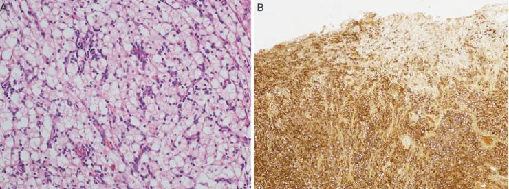
Fig. 2. (A) Image of histopathologic features of the uterus demonstrating clear and foamy histiocytes (hematoxylin and eosin stain,
×200). (B) Immunohistochemical staining image demonstrating the strong positive reactivity of histiocytic markers (CD68, ×40).
A B
www.ogscience.org 536
Vol. 63, No. 4, 2020
no atypia or mitosis (Fig. 2A). For differential diagnosis of histiocytic proliferation in the female genital tract, further diagnostic workup including immunohistochemistry (CD68, S100, and cytokeratin) and molecular pathology (BRAF V600E gene) was performed. Immunohistochemical staining was positive for CD68 (Fig. 2B) and negative for S100 and cytokeratin. BRAF V600E gene mutation was absent in real- time PCR. The bone scan revealed no uptake in long bones.
The patient was regularly followed up at 3-month intervals with no medical therapy, and she remained without evidence of recurrence 8 months after surgery.
Discussion
ECD is an extremely rare type of non-Langerhans cell his- tiocytosis involving multiple organs including the bones, central nervous system, heart, lung, retroperitoneum, and skin. Skeletal involvement is the most frequent manifesta- tion, with 96% of patients displaying the characteristic ra- diological finding of bilateral symmetric long bone sclerosis [4]. However, ECD has heterogeneous and diverse clinical manifestations. Atypical presentations can be challenging for the diagnosis of ECD. ECD with no skeletal involvement is extremely rare, and only a few cases have been reported [5].
Our patient had no skeletal involvement.
Histologically, there is proliferation of foamy lipid-laden histiocytes surrounded by Touton giant cells. Due to the lack of cytological atypia in the cells, the neoplastic nature of the proliferation is often missed. Immunohistochemical staining for CD68, CD163, and FACTOR XIII is positive, with CD1a, Langerin, and S100 being negative. The BRAF V600E muta- tion is present in more than 50% of ECD cases, with other mutations such as N/KRAS and PIK3CA described in BRAF- negative ECD [6].
In a prospective investigation of 60 adults with ECD con- ducted at the National Institutes of Health (NIH) clinical cen- ter, one-third of patients had a history of renal impairment before their NIH visit [7]. A total of 65% of patients exhibited encasement of the kidney and involvement of the retroperi- toneal space, causing renal artery stenosis in 54% of cases and ureteropelvic junction obstruction in 51% of cases [7].
However, a study investigating abdominal involvement dur- ing ECD reported that there was no uterine involvement [8].
ECD has no standard therapy, although consensus guide-
lines for clinical management were recently reported [9].
Empiric treatments include anti-inflammatory, immunosup- pressive, and chemotherapeutic agents, and recent reports of BRAF V600E and MAPK pathway gene mutations in ECD have suggested treatment with BRAK and MEK inhibitors [7].
Our patient had multiple retroperitoneal organ involvement without skeletal bone involvement. Tissue biopsy and immu- nohistochemical studies suggested ECD diagnosis. However, the BRAF V600E gene mutation was not found. To the best of our knowledge, uterine involvement in ECD has not been reported. Differential diagnosis of ECD in the female genital tract includes a variety of infectious, inflammatory, and neo- plastic disorders. Xanthogranulomatous inflammation involv- ing the uterus is typically centered in the endometrium and involves a combination of neutrophils, plasma cells, lympho- cytes, and hemosiderin-laden histiocytes [10]. Unlike ECD, uterine Rosai-Dorfman disease involves lymphocytophagocy- tosis or emperipolesis [11].
Our patient presented atypical ECD with pathologic and immunohistochemical characteristics that coincided with ECD and clinical characteristics such as skeletal bone involve- ment and BRAF V600E gene mutation that did not coincide with ECD. To the best of our knowledge, this is the first case report of retroperitoneal ECD with mainly uterine involve- ment. To correctly diagnose atypical ECD, communication among clinicians, radiologists, and pathologists is vital.
Conflict of interest
No potential conflict of interest relevant to this article was reported.
Ethical approval
The study was performed in accordance with the principles of the Declaration of Helsinki. Written informed consents were obtained.
Patient consent
The patients provided written informed consent for the pub- lication and the use of their images.
www.ogscience.org 537 Hae Min Kim, et al. Retroperitoneal histiocytosis
References
1. Favara BE, Feller AC, Pauli M, Jaffe ES, Weiss LM, Arico M, et al. Contemporary classification of histiocytic disor- ders. Med Pediatr Oncol 1997;29:157-66.
2. Campochiaro C, Tomelleri A, Cavalli G, Berti A, Dagna L.
Erdheim-Chester disease. Eur J Intern Med 2015;26:223- 9.
3. Haroche J, Arnaud L, Amoura Z. Erdheim-Chester dis- ease. Curr Opin Rheumatol 2012;24:53-9.
4. Mazor RD, Manevich-Mazor M, Shoenfeld Y. Erdheim- Chester disease: a comprehensive review of the litera- ture. Orphanet J Rare Dis 2013;8:137.
5. Salama H, Kojan S, Abdulrahman S, Azzumeea F, Alhe- jazi A. Erdheim-Chester disease with no skeletal bone involvement and massive weight loss. Case Rep Hematol 2017;2017:3862052.
6. Emile JF, Diamond EL, Hélias-Rodzewicz Z, Cohen-Au- bart F, Charlotte F, Hyman DM, et al. Recurrent RAS and PIK3CA mutations in Erdheim-Chester disease. Blood
2014;124:3016-9.
7. Estrada-Veras JI, O’Brien KJ, Boyd LC, Dave RH, Durham B, Xi L, et al. The clinical spectrum of Erdheim-Chester disease: an observational cohort study. Blood Adv 2017;1:357-66.
8. Nikpanah M, Kim L, Mirmomen SM, Symons R, Papa- georgiou I, Gahl WA, et al. Abdominal involvement in Erdheim-Chester disease (ECD): MRI and CT imaging findings and their association with BRAFV600E mutation.
Eur Radiol 2018;28:3751-9.
9. Diamond EL, Dagna L, Hyman DM, Cavalli G, Janku F, Estrada-Veras J, et al. Consensus guidelines for the di- agnosis and clinical management of Erdheim-Chester disease. Blood 2014;124:483-92.
10. Blanco C, Fernández F, Buelta L, Garijo F, Val-Bernal JF, Sánchez S. Xanthomatous endometritis. Appl Pathol 1989;7:273-6.
11. Pan LY, Offman SL, Warnke RA, Longacre TA. Uterine Rosai-Dorfman disease (sinus histiocytosis with massive lymphadenopathy). Int J Gynecol Pathol 2014;33:432-6.