Original Articles Korean Circulation J 1998;;;;28((((7): ): ): 1077-1083 ):
Clinical Features and Molecular Diagnosis of CATCH-22 Syndrome
Jung Yun Choi, MD 1,2 , Jeong-Wook Seo, MD 1,3 , Myoung Hee Kim, PhD 4 , Eul Kyung Kim, MD 3 , Jung-Sun Kim, MD 3 , Ho Sung Kim, MD 2 ,
Chong Heon Lee, DDS 3 , Hyangsuk Hur, PhD 4 , Eun Jung Bae, MD 5 , Chung IL Noh, MD 1,2 and Yong Soo Yun, MD 1,2
1
Heart Research Institute, Medical Research Center, Seoul National University, Seoul,
2Departments of Pediatrics and
3Pathology, Seoul National University College of Medicine, Seoul,
4Korea Research Institute of Bioscience and Biotechnology, KIST, Taejon,
5Department of Pediatrics, Sejong General Hospital, Buchon, Kyunggi-do, Korea
CATCH-22 증후군의 임상 소견 및 분자유전학적 진단
서울대학교 의학연구원 심장연구소,
1서울대학교 의과대학 소아과학교실
2및 병리학교실,
3한국과학기술연구원 생명공학연구소,
4부천세종병원 소아과
5최정연
1,2
·서정욱1,3
·김명희4
·김을경3
·김정선3
·김호성2
이종헌3
·허향숙4
·배은정5
·노정일1,2
·윤용수1,2
국 문 초 록
연구배경
:CATCH-22 증후군은 염색체 22q11위치의 결손과 특정 심기형, 특이한 얼굴 모양, 흉선과 부갑 상선의 형성부전 등을 일으키는 질환이다. 본 연구는 분자유전학적 방법으로 확인된 CATCH-22 증후군 12
예의 임상 소견을 요약하고 진단 방법의 문제점을 검토하였다.
방 법:임상적으로 CATCH-22 증후군이 의
심되고 세포유전학적 검사와 fluorescent in-situ hybridization(FISH) 검사에서 염색체 22q11위치의 결손 이 확인된 12예의 환자에 대하여 환자의 얼굴 모양을 7가지 관점에서 분석하였고, 심장 기형과 동반 기형을 조사하였으며, Southern blot을 시행하여 유전자 결손을 확인하였다.
결 과:12명의 환자는 외견상 특이한 얼굴 모양이었으나 7가지 기준으로 분석하여 4가지 이상의 소견을 나타낸 환자가 7명이었고, 1~3가지 만족 한 경우가 5명이었다. 8명의 심장 기형은 Fallot 4징이었고 그중 7명은 폐동맥 폐쇄가 동반되었다. 다른 4명 의 환자의 진단은 각각 총동맥간, 폐동맥 협착, 동맥관 개존증, 심방중격 결손이었다. Southern blot 검사는 8 명에서 시행되었는데 6명에서 양성이었다.
결 론:CATCH-22 증후군은 Fallot 4징을 비롯한 주요 심기형 을 동반하는 유전적 심장질환이며, 분자 유전학적 진단을 위해서는 FISH 검사와 Southern blot 분석이 필요 하다. ( (( (Korean Circulation J 1998;28( (( (7) )) ):1077-1083) )) )
중심 단어
:CATCH-22 증후군・누두동맥간 안면기형 증후군・DiGeorge 증후군・22q11번 염색체・선성 심기형.
논문접수일:1998년 6월 22일/심사완료일:1998년 7월 21일
Corresponding author:Jeong-Wook Seo, MD. 28 Yongon-Dong, Chongno-Gu, Seoul 110-799, Korea
Department of Pathology, Seoul National University, College of Medicine, Seoul, KoreaTEL:(02) 740-8268・FAX:(02) 765-5600 E-mail:[email protected]
Introduction
CATCH-22 is an acronym of cardiac defect, abn- ormal face, thymic hypoplasia, cleft palate, and hypo- calcemia associated with microdeletion of chromosome 22q11.
1)This syndrome encompasses heterogeneous groups of patients with DiGeorge syndrome (DGS),
2)velocardiofacial syndrome (VCFS),
3)and conotruncal anomaly face syndrome (CTAFS).
4)This syndrome is currently considered to be the second most com- mon cause of congenital heart disease, preceded only by Down’s syndrome.
5)In a survey in the northern England, deletion of 22q11 was estimated to account for 5% of all congenital heart defects and showed a minimum prev- alence of 1 in 4000 births.
5)The prevalence of this syndrome in Korea and the type of associated cardiac lesions are not known, but in view of the relative inci- dence of congenital heart disease, it is estimated that th- ere are at least ten thousand cases.
The important aspect of this syndrome is that the cardiac lesion is less severe and the mental retardation is milder than those in Down’s syndrome.
5)Number of patients with 22q11 deletion, therefore, is expected as being far more than that of Down’s syndrome in the clinical practice. Moreover, phenotypic and genotypic features are so variable and variability between mono- zygotic twins has been reported.
6)Several genes are being studied but no gene have been proven specific for this syndrome.
7-10)The fluorescent in situ hybridization (FISH) is currently understood as the most specific me- thod to diagnose this genetic defect.
11)12)We performed FISH study for metaphase chromosomes using a D22S75 probe. Twelve patients were collected to reveal genetic features of CATCH-22 syndrome. We report the clinical and genetic profile of those Korean cases of CATCH-22 syndrome. Eight cases among them were additionally studied by Sout-hern blot analysis of genomic DNA using DGCR680 and pDH-1 probes.
Materials and Methods
Patients
A total of twelve cases of congenital heart disease
with abnormal face were proven to have a deletion at the chromosome 22q11 by FISH analysis during the one-year period ending June 1997 (Table 1). Clinical diagnosis was performed at Seoul National University Children’s Hospital (eight cases), Sejong General Hospital (four cases). Facial features were analyzed according to seven independent items (Table 1). Faces with positive result on four or more items were interpreted as abnormal face, faces with two or three positive items being equivocal. Cardiac diagnosis was confirmed by echocardiography, angiocardiography and/or surgery.
Fluorescent in situ hybridization for metaphase chromosome
Metaphase chromosome slides were prepared from culture of peripheral lymphocytes by a standard met- hod which included exposure to a mitotic arrestant (colcemid), treatment with a hypotonic solution (0.075M KCl), fixation (3:1 mixture of methanol and glacial acetic acid) and dehydration (70%, 80%, 90% ethanol). DNA on slides was denatured in 70%
formamide/2X saline sodium citrate (SSC) for 2
minutes at 70℃, immediately dehydrated through a
cold (-20℃) ethanol series, and air dried. The hybr-
idization mixture containing the digoxigenin-labeled
D22S75 DGCR probe with D22S39 chromosome 22
control probe (Oncor, U.S.A.) was placed on denat-
ured chromosome slides for in situ hybridization. After
overnight hybridization at 37℃ in a moist chamber,
slides were washed once for 5 minutes in 2X SCC at
72℃, and once for 2 minutes in 1X phosphate buffered
distilled water (PBD) at room temperature ; they
were incubated with anti-digoxigenin fluorescein in
1X PBD/5% BSA at 37℃ for 60 minutes and rinsed
three times in 1X PBD (2 minutes each time). For
amplification, slides were treated with rabbit anti-
sheep antibodies at 37℃ for 30 minutes and washed
in three changes of 1X PBD at room temperature (2
minutes each time). They were then incubated with
FITC-rabbit antibody at 37℃ for 30 minutes, and then
washed three times in 1X PBD. Slides were stained
with propium iodide and mounted with fluorescent
mounting media (DA-KO, U.S.A.). For observation of
fluorescence signals in the chromosome, a fluoresc- ence microscope equipped with appropriate fluore- scence filter sets was used.
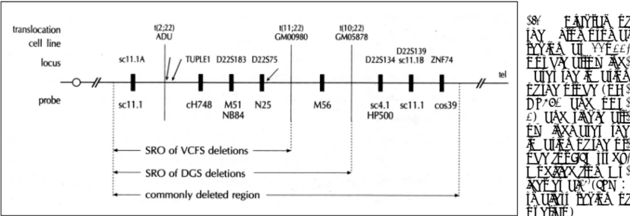
Production of probes for Southern hybridization
We used a mixture of two DNA probes, DGCR680 and pDH-1.
13)14)DGCR680 was obtained from human genomic DNA using two primers based on ADU breakpoint, where a DiGeorge syndrome patient had balanced translocation.
15)The other probe pDH1 was obtained by screening from a human liver cDNA library using DGCR680 as a probe. These together covered 1300 base pairs within the deleted sequence of chromosome 22
16)(Fig. 1). Details of our probes are described elsewhere.
13)14)Southern blot analysis
Genomic DNA extracted from peripheral blood of patients was digested with restriction enzyme HindIII.
After Southern hybridization, the test probe signal at 18 kilobases and control signal at 16 kilobases were measured by densitometer.
13)The values representing locus copy number were obtained, standardized from quantitative analysis of the hybridization signals obtained with the 18kb fragment and compared to a low copy repeat intensity. It was decided that a value less than 1.50 indicated deletion.
13)14)Results
Of the twelve patients, eight cases were male and four cases were female;their ages ranged from 3
month to 16 years (Table 1). Although all of twelve patients were initially suspected for CATCH-22 syndr- ome from the facial features obsertved by pediatric cardiologists, only seven cases were determined to have abnormal facial morphology according to our criteria using seven items (Table 1). A cleft palate was present in a patient and a high arched palate in six.
Nine patients showed delayed development. Two cases had umbilical hernia. The main cardiac lesion of eight patients were tetralogy of Fallot (TOF) and seven of them had pulmonary atresia. Two cases had other ano- malies in the ventricular outflow tract, being common arterial trunk or pulmonary stenosis. Two cases had a patent arterial duct or atrial septal defect(ASD) (Table 1).
All of twelve patients had positive result on FISH study. Eight cases among twelve patients were studied with Southern blot analysis. Six cases among eight patients were positive for Southern blot analysis.
There were six patients with positive FISH and positive Southern analysis. Five of them had typical facial features but one had an equivocal face. The cardiac lesion in every case was TOF and pulmonary atresia.
Both of two cases with positive FISH and negative Southern analyses had equivocal face and the cardiac lesions were TOF or ASD.
Four cases were studied only by FISH method. The cardiac diagnoses were TOF, pulmonary stenosis, common arterial trunk and isolated patent arterial duct (PDA). The facial morphology of two cases had ty- pical facial features.
Fig. 1. Diagram of
the chromosomal region at 22q11.Double arrow ind- cates the location of the probe (DG- CR680 and pDH- 1) and single arr- ow indicates the location of the pr- obe for FISH study.
Modified from Mu- lder et al.16)(SRO:
smallest region of overlap)
Discussion
DiGeorge syndrome was the first to be recognized as a group of defects currently understood as CATCH- 22 syndrome
2)and the association of cardiac anomalies in this immunologic defect was studied in 1972.
17)The hypocalcemic features, such as seizure, and cellular immune deficiency are seen early in life, but they become less apparent with age, probably due to secondary compensations. Frequent cardiac lesions are interrupted aortic arch (IAA), CAT, TOF with PA, TOF, and isolated VSD.
5)Velocardiofacial syndrome (VCFS) or Shprintzen syndrome, with its characteri- stic facial features comprising a prominent nose, broad nasal root, narrow palpebralfissures and retrognathia were reported.
3)18)19)Typical cases show a high inci- dence of craniofacial anomalies;cleft palate (98%), pharyngeal hypotonia (90%), retrognathia (80%) and malar flatness (70%);the incidence of congenital heart disease has been reported as 82%
20);Driscoll et al. and Shprintzen et al. reported that VSD and TOF were the common cardiac lesions.
18)21)The Japanese
group led by Takao
22)was the first to reco-gnize the major contribution of the phenotype to the patient population with the ventricular outflow tract defects.
The prevalence of CTAFS, based on the facial features in a series of TOF, was 12.8%, but this figure was 48%
in the subgroup of TOF associated with PA and major aortopulmonary collateral arteries.
22)Among clinical cases of CTAFS, the most common cardiac defect was TOF (92%);half of these cases were ass- ociated with the PA and the systemic pulmonary collateral arteries.
23)Within chromosome region 22q11, reported frequ- encies of association with deletion are 89% in DGS, 81% in VCFS, and 84% in CTAFS.
21)23)But it is also possibile that non-deletion patients have undete-ctable smaller deletions or point mutations within critical genes in this region.
24)Deletions were also observed in 20-30% of such unselected, nonsyndromic patients with CAT, IAA, and TOF.
25)Among patients requiring surgery for congenital heart disease, 5% to 10% may have this single genetic abnormality.
26)Parental deletions are found in approximately 25% of patients with CATCH-22, though variable expression within a Table 1. Facial features, cardiac diagnosis and molecular findings of patients with CATCH-22 syndrome. Clinical
decision of being“abnormal” was made when there are four or more positive findings. Clinical decision of being“equivocal” was made when there are one to three positive findings on their face. Every case showed positive reaction for the fluorescent in-situ hybridization study.Age/ Facial features* Palate Develop- Ca/P** Others Clinical Cardiac Aortic Southern No. Sex 1 2 3 4 5 6 7 ment decision lesions*** arch blot
1 3m/M -++++++ High Normal 8.6/6.2 Abnormal TOF, PA Rt 1.12
2 21m/M +-+++++ High Delayed Abnormal TOF Lt
3 15m/M ?- ?++++ Normal Delayed Umb. hernia Abnormal PS Rt
4 3m/F +---+-- High Normal Equivocal CAT Lt
5 9m/M +----+- High Delayed Equivocal PDA Lt
6 31m/M ? ?++++? High Delayed 9.0/4.9 Umb. hernia Abnormal TOF, PA, PDA Lt 1.00 7 30m/F ? ? ? ? ?+? Normal Delayed 8.9/6.6 Equivocal TOF, PA Rt 0.43 8 7m/F --+--+- Normal Delayed Equivocal TOF, PA 2.32
9 2yr/F ? ? ?+++- Normal Delayed Equivocal ASD 1.94
10 6m/M --++++- High Delayed Abnormal TOF, PA 0.49
11 16yr/M +++--++ Cleft Delayed 8.3/7.1 Abnormal TOF, PA Lt 0.78 12 3yr/M ?+++?+- Normal Normal 9.1/4.9 Abnormal TOF, PA Rt 1.21
*1, long face;2, flat malar area;3, depressed nose and narrow alae;4, retarded mandible;5, small A- shaped mouth;6, hypertelorism;7, bloating eyelid
**Ca/P, serum level of calcium and phosphorus
***TOF, Tetralogy of Falllot;PA, pulmonary atresia;ASD, atrial septal defect;PS, pulmonary stenosis;DORV, double outlet right ventricle;CAT, common arterial trunk;PDA, patent ductus arteriosus
family is well documented.
1)6)27)Among 40 parents of children with congenital heart disease, one of 14 fathers and five of 26 mothers had CTAFS and in one father and four mothers there was deletion.
23)Our study aims to reveal the clinical significance of this genetic defect in the pediatric cardiological pra- ctice in Korea and to assess the significance of diff- erent genetic studies in the diagnosis of this syndrome.
This study is the first report on the systematic study on this syndrome in Korea.
As is shown in Table 1, facial features of individual cases were so variable that no single criteria could be used as an indicator of this syndrome. But we could define an abnormal face when one had abnormalities on four or more items and equivocal when one had abnormalities on two or three items. The incidence of this disease, therefore, varies when we define this disease through the clinical examination fo the facial features only. We could divide facial features into abn- ormal, equivocal or normal faces. But this classification system should be a simple screening tool for patients with congenital heart disease.
In view of the variability of phenotypic features observed in association with 22q11 deletions, we cannot precisely predict outcome on the basis of mo- lecular studies at this time. The observed variability may reflect the amount of deleted genes in that critical region, or may be dependent upon genetic background, in utero environment, or parental origin of the dele- tion.
28)Correlation between genotype and phenotype will require a detailed molecular analysis of the deleted region to determine which region or genes specify individual features of the phenotyppe. Further molecular studiesinvolving the functional characteri-stics of probes D22S75, DGCR680 and pDH-1 should also be perfor- med. Other tools for the molecular diagnoses include RFLP (Restriction Fragment Length Polymorphism), DNA dosage analysis in addition to the FISH or Sou- thern blot analysis. But one of important aspect of the study would be those for their family members.
We conclude that CATCH-22 syndrome has variable facial, cardiac and genetic features, and the combined use of probes is recommended for a more accurate
diagnosis.
Summary
Background: : : :
CATCH-22 syndrome is a common genetic disorder with features of cardiac defect, abnormal face, thymic hypoplasia, cleft palate, and hypocalcemia, along with microdeletion at chromosome 22. This study is to report twelve Korean patients with CAT-CH-22 syndr- ome diagnosed by the fluorescent in situ hybridization (FISH) method.
Method: : : :
Clinical features were analyzed according to the FISH result and the Southern blot analysis using new probes DGCR680 and pDH-1 was performed to correlate with the clinical findings and FISH results.
Twelve patients were studied by FISH met-hod and eight of them were studied by Southern blot anal- ysis.
Results: : : :
Seven patients had typical facial features for CAT- CH-22 syndrome, but five patients had equivocal face, although they were originally suspected to have the conotruncal face. The main cardiac lesion of eight patients were tetralogy of Fallot (TOF) and seven of them had pulmonary atresia. Two cases had other ano- malies in the ventricular outflow tract, being common arterial trunk or pulmonary stenosis. Two cases had a patent arterial duct or atrial septal defect (ASD). All of twelve patients had positive result on FISH study.
Among eight patients with positive FISH study, six cases were positive for Southern blot analysis.
Conclusion: : : :
We conclude that CATCH-22 syndrome has variable facial, cardiac and genetic features, and the combined use of probes is recommended for a more accurate diagnosis.
KEY WORDS:CATCH-22 syndrome・Conotruncal
anomaly face syndrome・DiGeorge syndrome・Chro-
mosome 22q11・Congenital heart disease.
■ Acknowledgment
The authors are indebted to Drs. Hye Soon Kim, Hong Ryang Kil, Soon Sung Park, Seong Ho Kim, Se Jung Sohn, Mee Hye Oh, Shi Joon Yoo and Heung Jae Lee who actively particip- ated in our collection of clinical cases. Drs. Dong Soo Kim and Soo Kyung Choi helped our cytogenetic study. We are also grateful to Ms. Sung Hee Hong and GiJin Kim for their valued technical assistance with cytogenetic study.
이 논문은 1996년도 과학기술처 의과학 연구비(96-B00- 02-001-004)의 지원으로 이루어졌음.
REFERENCES