서의 ERK1/2와 p38 MAPK
가톨릭대학교 의과대학 내과학 교실, 가톨릭대학교 의과대학 성모병원 호흡기 질환 연구실1
김용현, 윤형규, 김치홍, 안중현, 권순석, 김영균, 김관형, 문화식, 박성학, 송정섭, 조경숙1
Muc5ac Gene Expression Induced by Cigarette Smoke is Mediated Via a Pathway Involving ERK1/2 and p38 MAPK
Yong Hyun Kim, M.D., Hyoung Kyu Yoon, M.D., Chi Hong Kim, M.D., Joong Hyun Ahn, M.D., Soon Seog Kwon, M.D., Young Kyoon Kim, M.D., Kwan Hyoung Kim, M.D., Hwa Sik Moon, M.D., Sung Hak Park, M.D., Jeong Sup Song, M.D., Kyung Sook Cho1
Division of Pulmonology, Department of Internal Medicine and 1Institute of Respiratory Disease, St. Mary’s Hospital, College of Medicine, the Catholic University of Korea, Seoul, Korea
Object : Cigarette smoking is a major cause of mucus hypersecretion, which is a pathophysiological feature of many inflammatory airway diseases. Mucins, which are an important part of the airway mucus, are synthesized from the Muc gene in airway epithelial cells. However, the signaling pathways for cigarette smoke-induced mucin synthesis are unknown. The aim of this study was to determine the signal pathway for smoking induced Muc5ac gene expression.
Methods : A549 cells were cultured and transiently transfected with the Muc5ac promoter fragment. These cells were stimulated with 5% cigarette smoke extract (CSE) alone or with CSE after a pretreatment with various signal transduction pathway inhibitors (AG1478, PD98059 and SB203580). The Muc5ac promoter activity was examined using the luciferase reporter system, and the level of phosphorylated EGFR, ERK1/2, p38 MAPK and JNK were all examined using Western blot analysis. Muc5ac mRNA expression was also examined using reverse transcriptase polymerase chain reactions (RT-PCR).
Results : 1. The peak level of luciferase activity of the Muc5ac promoter was observed at 5% concentration and after 3 hours of incubation with the CSE. The level of EGFR phosphorylation and the luciferase activity of the transfected cells caused by the CSE were significantly suppressed by AG1478 or PD98059 (P<0.01).
2. CSE phosphorylated ERK1/2 or p38 MAPK but not JNK. The Muc5ac mRNA expression level was increased by the CSE but that was suppressed by PD98059 or AG1478.
3. The CSE-induced phosphorylation of ERK1/2 was blocked by PD98059 and that of p38 MAPK was blocked by either PD98059 or SB203580. Either PD98059 or SB203580 suppressed the luciferase activity of the transfected cells (P<0.0001).
Conclusion : The Muc5ac mRNA expression level was increased by the CSE. The increased CSE-induced transcriptional activity was mediated via EGF receptor activation, which led to ERK1/2 and p38 MAPK phosphorylation.
(Tuberc Respir Dis 2005; 58: 590-599)
Key words : Cigarette smoke, MUC5AC, MAPK, Signal transduction
Address for correspondence : Jeong Sup Song, M.D.
150-713 Division of Pulmonology, Department of Internal Medicine and Institute of Respiratory Disease, St. Mary’s Hospital, College of Medicine, the Catholic University of Korea, Seoul, Korea Phone : 02-3779-1146 Fax : 02-780-3132 E-mail : jssong@catholic.ac.kr
Received : Feb. 21. 2005 Accepted : May. 19. 2005
서 론
흡연은 오래 전부터 만성폐쇄성폐질환 (Chronic ob
structive lung disease)과 죽상경화증의 주요 위험 인 자 1-4로 잘 알려져 있다. 또 흡연은 만성폐쇄성폐질환 환자에서뿐만 아니라 건강한 사람에서도 기도 내 점 액분비를 증가시킨다. 특히 만성폐쇄성폐질환의 주요 임상 양상인 점액의 과다 분비는 이 질환의 중요한 병 태생리학적 소견이며 과도하게 분비된 점액은 하부기 도의 폐쇄를 일으켜 호흡곤란과 나아가 호흡부전을 야기하는 중요한 원인이 된다5. 기도 점액은 기도상피 를 덮고 있으며 수분의 증발을 막고 병균이나 화학적 자극 물질들로부터 상피를 보호하는 역할을 하는데,
이는 대기도 상피에 존재하는 배상세포와 점막 하 분 비선에 존재하는 점액세포와 장액세포에서 분비된 다6-8. 점액은 95%의 물과 소량의 지방, 단백질 및 특 정 유전자에 의해 만들어지는 당 단백질 등으로 구성 되어 있으며6 어떤 이유에 의해 과다 분비가 일어나면 문제를 일으킨다. 점액의 과다 분비는 기도 내 배상세 포의 증식 및 화생 (metaplasia)때문이다9. 점액의 과 다 분비를 일으키는 원인은 매우 다양한데 이 중 한 가지가 흡연이다.
담배연기 내에는 대략 4000 여 가지의 다양한 물질 들이 기체 혹은 결정의 형태로 존재하며 nitrogen oxide, hydrogen peroxide, hydrogen cyanide, acrolein 등의 다양한 산화제를 포함하고 있다10. 이들 산화제는 만 성폐쇄성폐질환, 기관지확장증, 낭성섬유증과 같은 기 도 내 염증질환의 병태생리에 중요한 역할을 한다고 알려져 있다. 기도내의 점액분비를 유발하고 조절시 키는 기전은 아직까지 정확하게 알려져 있지 않지만 점액의 주성분인 점액 당단백질을 합성하는데 관여하 는 기도상피세포내의 점액 유전자, 즉 Muc 유전자가 있음이 알려져 있다. 현재까지 인체에서는 15종류의 Muc 유전자가 존재한다11. 그 중에서도 Muc5ac 유전 자에 의해 합성되는 당단백은 정상인과 기관지 천식 환자에서 기도 분비액의 주된 성분으로 알려져 있고12 기도 내 배상세포 증식 및 화생과 가장 관련성이 있다 고 보고 되었다13.
최근 이 Muc 유전자의 발현 과정에 대한 여러 연 구에서 유전자를 활성화시키는 세포 내 신호전달 과정에 표피성장인자 (epidermal growth factor) 수 용체가 관여함이 밝혀졌다14,15. 하지만, 표피성장인 자 수용체 활성화 이후의 세포 내 신호전달 과정 에 대해서는 확실하게 규명이 되어 있지 않고 일부 의 연구들에서 이 신호전달 과정에 mitogen-activated protein kinase (MAPK)가 관련되어 있음을 최근 보고하였다16-18. 저자들은 흡연에의 노출이 점액 유 전자의 발현을 활성화시키고 이 유전자의 발현에 어떠한 MAPK가 신호전달 과정에 관여하는지 알아 보고자 기도상피세포의 특성을 가지고 있는 폐선암 세포주인 A549세포를 담배연기 추출물에 노출시켜 실험하였다.
방 법 1. 세포배양
사람의 폐선암 세포주인A549 세포 (KCLB, 한국세 포주은행, Korea)를 10% 우 태아혈청 (Glbco-BRL, California, USA), penicillin (Glbco-BRL) 100 U/mL, streptomycin (Glbco-BRL) 100 ㎍/mL, HEPES 640 (Sigma, St. Louis, USA) 25 mmol 배지에서 37℃, 5% CO2 의 배양기에서 배양하였다.
2. 담배연기 추출 용액 (cigarette smoke extract solution)
25 mL의 RPMI1640 medium (10% 우 태아혈청, HEPES 50 mmol without penicillin and streptomy
cin)내로 one puff/min의 비율로 5분 동안 담배연기를 모았다. pH 7.4가 되도록 적정을 하고, 0.22 ㎛ filter unit (Millipore, Bedford)을 사용하여 여과를 하였다.
이때 만들어진 담배연기 추출용액은 사용하기 30분전 에 만들어 썼다. 또한 이 용액은 동일한 배지로 5%
혹은 10%로 희석하여 세포에 처리하였다.
3. Transient transfection과 세포 자극
A549세포를 6-well 조직배양 용기 (Nunc, Roskilde, Denmark)에 1×10⁵ 세포/mL의 양으로 2mL씩 접종 후 48시간 동안 배양하여, 용기의 80% 정도 채워 자랐을 때 Muc5ac의 transfection 실험을 실시하였다. Trans
fection을 수행할 PGL3-Muc5ac-3752pro luciferase reporter plasmid와 PGL3-basic vector는 실험을 수 행하기 전에 각각 200 ng/㎕로 정량화하였고, 대조군 으로 사용할 β-galactosidase의 양은 100 ng/㎕로 정 량화 하였다. Transfection전에 세포는 혈청이 없는 배지로 세척을 하고 각 용기 당 800㎕의 혈청이 없는 배지를 넣어 미리 적응을 시켰다. 한 튜브에는 혈청이 없는 배지 300㎕, PGL3-Muc5ac-3752pro luciferase reporter plasmid 5㎕, plus reagent (Gibco-BRL) 5㎕, β-galactosidase 3㎕를 혼합하여 준비하고 다른 한 튜
브에는 혈청이 없는 배지 300㎕, LIPOFECTAMINETM reagent (Gibco-BRL) 4㎕를 넣은 후 각각의 튜브를 상온에서 15분간 반응시킨 후, 두 개의 튜브를 잘 섞 어서 상온에서 15분간 반응을 시켜 복합체를 형성하 도록 하였다. 준비된 복합체를 미리 적응시킨 배양 용 기에 200㎕씩 골고루 넣은 후, 20% FBS를 1 mL을 배 양 용기에 첨가하여 24시간 동안 배양하였다. Trans
fection한 세포 주를 혈청이 없는 배지에 24시간 배양 한 후, 5% 담배연기 추출 용액과 mitogen-activated protein kinase kinase (MAPKK)의 억제제인 PD98059 40 mmol (Sigma), epidermal growth factor receptor (EGFR) kinase의 억제제인 AG1478 (Calbiochem, Bad Soden, Germany) 50 ㎛ol, p38 MAPK 억제제인 SB203580 (Calbiochem) 50 ㎛ol을 넣어 30분간 전 처 리한 후, 다시 5% 담배연기 추출용액으로 3시간 자극 하였다. phosphate buffer solution (PBS)으로 2번 세 척하고 reporter lysis buffer (Promega, Madison, USA) 를 넣고 상온에서 20 분간 반응시켜 12,000 rpm에서 15 초간 원심 분리한 후, 세포 단백질을 얻었다.
4. Luciferase receptor assay
얻어진 세포 단백질을 20㎕으로 정량 한 후 lucife
rase 활성화 정도를 측정하기 위해서 세포 추출 단백 질에 100㎕의 luciferase assay substrate (Promega) 를 넣어 반응시킨 후, luminometer (Turner Designs, Turku, Finland)를 이용하여 활성화 정도를 측정하였 다. 또한 각각의 활성도는 β-galactosidase를 이용하 여 보정하였다.
5. 역전사효소 연쇄중합반응 (Reverse transcription polymerase chain reaction)
A549 세포가 배양 용기의 80% 정도 채워 자랐을 때, transient transfection 실험과 동일한 조건으로 Tri- ZolTM reagent (Invitrogen, California, USA) 500 ㎕ 로 상온에서 5분간 반응시키고 제조자가 제시한 방법 으로 RNA를 추출하였다. 추출한 RNA는 spectropho
tometer (Bio-RAD, California, USA)를 이용해 1 ㎍/㎕
로 정량화 하였고, 여기에서 1 ㎕를 채취하여 역전사 효소 (SUPERSCRIPT Ⅲ, Invitrogen) 0.5㎕로 BioNeer Accupower PCR premix (BioNeer, Seoul, Korea)를 이용하여 역전사효소 연쇄중합반응을 수행하였다. 활 성화 정도는 GAPDH를 사용하여 보정하였으며 사용 한 Muc5ac 및 GAPDH의 primer 염기서열은 다음과 같다.
Muc5ac ; (F) 5’ –TCCGGCCTCATCTTCTCC–3’
(R) 5’ –ACTTGGGCACTGGTGCTG–3’
GAPDH ; (F) 5’ –ACCACAGTCCATGCCATCAC–3’
(R) 5’ –TCCACCACCCTGTTGCTGTA–3’
6. Western blot
A549 세포를 위에서 언급한 방법으로 처리하여 배 양하고 Bradford assay (Bio-RAD) 방법으로 정량하 여 세포 단백질을 준비하였다. 준비된 세포 단백질 30
㎍을 10% SDS-polyacrylamide gel에서 120V에서 약 1시간 30분간 전기영동을 하였다. 이어 PVDF mem
brane (Bio-RAD)으로 4℃, 250mA에서 1시간 30분간 전기영동 하여 단백질을 전이시키고 차단 용액으로 차단시켰다. Phosphorylated EGFR (P-EGFR/EGFR), phosphorylated extracellular signal-related kinase (P-ERK/ERK), phosphorylated p38 MAPK (P-p38 MAPK/p38 MAPK), phosphorylated c-Jun N-ter
minal kinase (P-JNK/JNK)에 대한 일차항체 (rabbit anti-human, Santacruz, California & Cell signaling, Beverly, MA, USA)로 4℃에서 밤새 반응시키고 세척 후 이차항체 (anti-rabbit IgG HRP-linked antibody 또는 anti-mouse IgG HRP-linked antibody, Cell si
gnaling)로 1시간 30분 동안 반응시켰다. 세척 후, ECLTM reagent (Amersham-Pharmacia Biotech, Seoul, Korea) 를 이용하여 제조자가 제시한 방법으로 반응시킨 후, X-ray 필름에 감광시키고 자동 현상기를 이용하여 현 상하였다.
7. 통계 방법
통계적 검정은 SPSS 프로그램을 사용하여 one-way
Figure 2. Effect of incubation time on the CSE-induced Muc5ac transcriptional activity. Transcriptional activity of Muc5ac promoter was the highest when cells were exposed to 5% CSE for three hours (PGL3-basic:
PGL3-basic vector without Muc5ac promoter region,
* P<0.01).
Figure 1. Effect of cigarette smoke extract solution (CSE) concentration on the transcriptional activity of Muc5ac promoter. MUC5AC reporter plasmid was transfected into A549 cells and transfected cells were treated with CSE for one hour. The transcriptional activity of Muc5ac promoter was the highest when the transfected cells were exposed to 5% CSE.
Luciferase activity was expressed as a relative value to a PGL3-basic value (PGL3-basic: PGL3-basic vector without Muc5ac promoter region, CTL: control/Muc5ac promoter transfected, CSE: cigarette smoke extract solution, * P<0.01).
Figure 3. Western blot analysis of epidermal growth factor receptor (EGFR) and phosphorylated epidermal growth factor receptor (P-EGFR). Western blot analy
sis was performed under conditions as described in methods. A549 cells were incubated with 5% CSE for three hours. EGFR phosphorylation was increased by CSE and decreased by EGFR tyrosine kinase inhibitor, AG1478 or mitogen-activated protein kinase kinase inhibitor, PD98059 pretreatment (M: marker, CTL: control/
MUC5AC promoter transfected, 5% CSE: 5% cigarette smoke extract solution)
ANOVA와 independent sample T-test를 사용하였으 며 P 값이 0.05 이하인 경우를 통계적 유의성이 있는 것으로 하였다.
결 과
1. 담배연기 추출물 자극에 의한 Muc5ac 유전자 전사 활성도의 증가 및 표피성장인자 수용체의 인산화
담배연기 추출물의 농도를 달리하여 A549 세포를 자극하였을 때 5% 담배연기 추출용액일 때 가장 높은 luciferase 활성도를 보여 이 농도에서 Muc5ac pro
moter의 전사가 가장 증가됨을 알 수 있어 이후 세포 자극 시 5% 담배연기 추출 용액을 사용하였다 (그림 1). 자극시간에 따른 활성도는 일반적으로 자극시간이 길수록 활성도가 더 증가하는 경향을 보였으며 3시간
자극 시 가장 높은 활성도를 보였다 (그림 2). Western blot을 이용하여 표피성장인자 수용체의 인산화, 즉 활성화된 수용체를 본 결과 대조군에 비해 담배연기 추출물 자극에 의해 수용체의 인산화가 증가 되었고 이는 수용체 인산화를 억제하는 AG1478에 의해 억제 되었다 (그림 3).
PGL3-basic + - - - -
MUC5AC - + + + +
5% CSE - - + + +
PD98059 - - - + -
AG1478 - - - - +
Figure 4. The effect of cell signal transduction inhibi
tors on luciferase activity. Mitogen-activated protein kinase kinase inhibitor, PD98059 and EGFR tyrosine kinase inhibitor, AG1478 suppressed the CSE-induced luciferase activity (PGL3-basic: PGL3-basic vector with
out Muc5ac promoter region, MUC5AC: Muc5ac pro
moter transfected, 5% CSE: 5% cigarette smoke extract solution, * P<0.01).
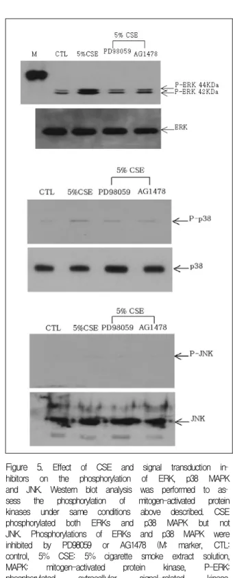
Figure 5. Effect of CSE and signal transduction in
hibitors on the phosphorylation of ERK, p38 MAPK and JNK. Western blot analysis was performed to as
sess the phosphorylation of mitogen-activated protein kinases under same conditions above described. CSE phosphorylated both ERKs and p38 MAPK but not JNK. Phosphorylations of ERKs and p38 MAPK were inhibited by PD98059 or AG1478 (M: marker, CTL:
control, 5% CSE: 5% cigarette smoke extract solution, MAPK: mitogen-activated protein kinase, P-ERK:
phosphorylated extracellular signal-related kinase, P-JNK: phosphorylated c-Jun N-terminal kinase).
2. 세포 내 신호전달 경로로써 MAPK와 Muc5ac 유전자의 전사 발현
Muc5ac 유전자의 전사과정에서 세포 내 신호를 전 달하는데 표피성장인자 수용체가 관여하며 그 하위 신호전달 과정에 MAPK, 특히 ERK1/2가 관계 있음 이 알려져 있어 저자들은 표피성장인자 수용체의 tyr
osine kinase 억제제인 AG1478, MAPK kinase 억제 제인 PD98059를 사용하여 Muc5ac 유전자의 전사가 억제되는지 luciferase 활성도를 통하여 관찰하였다.
담배연기 추출물 자극 시 증가된 Muc5ac 유전자의 전사는 이들 억제제에 의하여 모두 억제되었다 (그림 4).
Western blot을 이용하여 3가지 MAPK의 활성화된 형태, 즉 인산화 된 kinase 활성도를 본 결과 담배연기 추출물의 자극에서 인산화 된 ERK1/2와 p38 MAPK 의 활성화는 관찰되었으나 JNK의 활성화는 볼 수 없 었다. 또 표피성장인자 수용체의 인산화를 억제하는 AG1478에 의해 ERK 1/2와 p38 MAPK 활성도는 예 상대로 억제되었고 MAPK kinase 억제제인 PD98059 처리시 ERK 1/2와 p38 MAPK 활성도 역시 감소하였 다 (그림 5). 이를 다시 확인하기 위하여 역전사효소 중
합연쇄반응을 이용하여 Muc5ac mRNA 발현을 관찰 해 보니 담배연기 추출물 자극 시 Muc5ac mRNA 발 현이 증가되었고 AG1478과 PD98059에 의해 Muc5ac mRNA 발현은 억제되었다 (그림 6).
Figure 7. Western blot analysis of ERK and p38 MAPK. PD98059 inhibited phosphorylation of both ERKs and p38 MAPK. SB203580 also inhibited p38 MAPK phosphorylation as much as to the level of control and CSE treatment group (CTL: control, CSE:
5% cigarette smoke extract solution).
Figure 6. Reverse transcription polymerase chain re
action (RT-PCR) analysis of Muc5ac mRNA expression in A549 cells. PCR reaction was run on a 1% agarose gel and the band density was expressed as a relative value using densitometry measurement. CSE increased the Muc5ac mRNA expression. Pretreatment of PD98059 or AG1478 suppressed the Muc5ac expression near to the control levels (CTL: control, 5% CSE: 5% cigar
ette smoke extract solution).
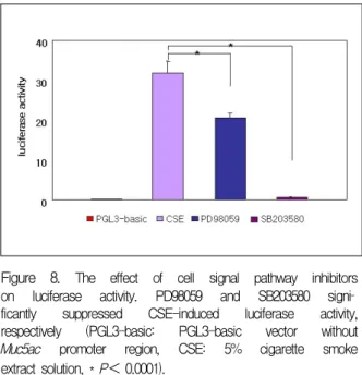
Figure 8. The effect of cell signal pathway inhibitors on luciferase activity. PD98059 and SB203580 signi
ficantly suppressed CSE-induced luciferase activity, respectively (PGL3-basic: PGL3-basic vector without Muc5ac promoter region, CSE: 5% cigarette smoke extract solution, * P< 0.0001).
3. SB203580 처리 시 세포 내 신호전달 경로와 Muc5ac 유전자 발현
담배연기 추출물에 의한 Muc5ac 유전자 발현에 관 여하는 MAPK 중 본 실험에서 ERK1/2와 p38 MAPK 의 인산화가 확인되어 p38 MAPK의 선택적 억제제인 SB203580으로 처리한 후 p38 MAPK 인산화와 Muc5ac promoter 활성도를 관찰하였다. PD98059와 SB203580 은 예상대로 p38 MAPK 인산화를 억제하였으며 (그 림 7) SB203580은PD98059에 비하여 훨씬 강하게 lu
ciferase 활성도를 감소시켰다 (그림 8).
고 찰
기도 내 기류제한과 반복적인 기도염증으로 요약되 는 만성폐쇄성폐질환에서 기도 내 점액분비의 증가는
특징적인 임상양상이다. 만성폐쇄성질환의 원인은 여 러 가지가 알려져 있으나 그 중 흡연이 가장 중요한 원인이며 이는 기도 내 점막의 염증을 일으키고 기도 내 점액분비 증가에 기여한다2,19. 기도점액의 주성분 은 점액소 (mucin)라는 당 단백질 복합체로써 구조의 뼈대를 이루는 여러 단백질이 각각의 특이 유전자에
의해 만들어짐이 최근 밝혀져 있는데 현재까지 모두 15개의 점액 유전자 (Muc gene)가 알려져 있다. 점액 소는 막결합성 점액소와 분비형 점액소로 나누며 이 중 폐 혹은 기도 내에 존재하는 분비형 점액소는 Muc2, Muc5ac, Muc5B, Muc7 그리고 Muc8이고11 이 중 Muc5ac가 흡연에 의해 분비가 유도됨이 알려졌다20. 하지만, 이러한 점액 유전자가 어떠한 세포 내 신호전 달 과정을 통하여 점액분비를 증가시키는지에 대해서 는 명확하게 밝혀진 바가 없으나 최근 호중구의 ela
stase, 흡연, 금속, 세균 감염, 산화제의 자극 등에 의 하여 표피성장인자 수용체가 활성화되고 이어 점액 유전자의 전사가 일어난다는 사실이 알려졌다17,21-23. 본 실험에서도 담배연기로 처리하였을 때 A549 세포 의 Muc5ac mRNA 발현이 증가되었고 표피성장인자 수용체의 활성화를 동반하며 표피성장인자 수용체 인 산화를 억제하는 AG1478 처리 시 Muc5ac 유전자의 발현이 억제되어 유전자 발현에 표피성장인자 수용체 가 관여함을 확인할 수 있었다. 표피성장인자 수용체 가 자극에 의해 활성화 되어 세포 내 신호를 전달하는 데는 ERK1/2, JNK1/2/3, p38 MAPK 혹은 protein ki
nase C를 매개하는 것이 알려져 있는데 Muc2 혹은 Muc5ac 유전자의 발현에 표피성장인자 수용체의 활 성이 먼저 일어나고 이어 ERK 신호전달 체계를 경유 하여 NF-κB나 SP1같은 전사인자가 DNA에 결합하 여 유전자의 발현을 증가시킨다는 보고들이 있다16,18. 세포 내 신호전달에 관여하는 신호전달체계 중 MAPK 군은 proline-targeted serine-threonine kinase에 속 하며 외부 자극을 핵에 전달하는 역할을 하는데 포유 동물에서는 최소 4가지 종류의 MAPK, 즉 ERK1/2, JNK1/2/3, p38 MAPK, ERK5가 존재한다24. 본 연구 자는 담배연기에 의한 Muc5ac 유전자의 발현에 표피 성장인자 수용체의 활성화를 확인하고 그 이하 단계 에서의 신호전달 과정에 여러 MAPK 중 어떤 종류가 관여하는지 확인하였다. 담배연기 자극 시에 ERK1/2 와 p38 MAPK의 인산화는 관찰하였으나 JNK 인산화 는 관찰되지 않았고 MAPK를 활성화 시키는 MAPK kinase (MAPKK) 억제제인 PD98059와 p38 MAPK의 선택적 억제제인 SB203580으로 처리 시 각각 ERK1/2와 p38 MAPK의 인산화가 억제되고 Muc5ac mRNA 발
현이 감소되었다. 이상의 결과로 보아 담배연기에 의 한 Muc5ac 유전자의 발현 증가를 일으키는 세포 내 신호전달에는 MAPK 중 ERK1/2와 p38 MAPK가 관 여하고 JNK는 관계없음을 유추할 수 있다.
담배연기가 표피성장인자 수용체를 활성화시키는 기전은 아직 명확하지 않다. 표피성장인자 수용체의 특이 리간드인 표피성장인자와 transforming growth factor는 직접 수용체에 결합하여 표피성장인자 수용 체 인산화를 유도하지만14 이외에도 외부에서 과산화 수소와 같은 산화제로 자극하거나 활성화 된 호중구 로 자극 시 수용체-리간드 결합과 관계없는 표피성장 인자 수용체 인산화 경로가 보고 되고 있다23. 활성화 호중구 역시 많은 종류의 염증 매개물을 생성하여 DNA 의 산화손상을 일으킨다고 알려져 있어 다양한 자극 에 의해 생성된 산화물질들이 리간드 결합과는 무관 하게 직접적으로 표피성장인자 수용체를 활성화 시키 고 MAPK 경로를 통하여 점액분비를 증가시킨다고 추측된다. 담배연기 내에도 다양한 산화제들이 존재 하므로 이들 산화제에 의해 표피성장인자 수용체 활 성화가 일어난다고 생각할 수 있다. 하지만 흡연은 생 체에서 기도 내 염증을 유발하여 기도 내 세포로부터 다양한 염증매개물들을 분비하게 하는데 특히 호중구 의 동원을 일으키는 IL-8과 같은 시토카인 분비를 자 극함으로써 먼저 호중구가 동원되고 이차적으로 산화 자극에 의해 수용체의 활성화가 이루어질 가능성을 배제할 수는 없다. 그러나 본 실험은 호중구의 동원이 불가능한 폐암 세포주에서 이루어졌으며 호중구의 자 극 혹은 외부에서의 산화제 처리에 의한 점액 유전자 발현을 본 이전의 보고23와 같이 신호전달 과정에 ERK1/2 경로는 관여하나 p38 kinase 경로는 무관하 다고 결론을 내렸다. 연구자의 실험결과 담배연기 자 극으로 나타난 신호전달 경로에는 ERK1/2와 p38 MAPK가 모두 관여하는 결과를 보여 표피성장인자 수용체 인산화는 호중구 동원의 이차적인 결과이거나 단순히 담배연기 내에 포함된 산화 물질에만 의한 것 이라 단정할 수는 없다. 담배연기 내에 들어있는 대표 적 물질인 acrolein으로 기도상피세포를 직접 자극하 였을 때 Muc5ac mRNA의 발현 증가가 보고 되었으 며25,26 이의 기전으로 acrolein이 세포 내 glutathione을
소모시켜 reactive oxygen species를 생성하여 유전자 발현을 증가시킨다는 보고도 있다4,27. 또 acrolein은 생 체에서 염증반응을 일으켜 호중구, 대식세포 및 단핵 구의 활성화를 촉진하고 이들 세포들은 다양한 염증 매개물을 분비하는데 이들 염증 매개물로 기도상피세 포를 자극하였을 때도 역시 Muc5ac mRNA의 발현이 증가되고 단백질 합성이 이루어지는 것으로 보아 acrolein은 직접적으로 Muc5ac 유전자를 활성화시키 고 동시에 염증 반응을 매개하여 간접적으로도 Muc5ac 유전자를 자극하는 것으로 생각된다. 이처럼 다른 신 호 전달 경로를 가진 자극원이 담배연기에 존재하거 나 산화 물질의 종류에 따라 각각 다른 신호전달 경로 를 가질 가능성이 있다. 담배연기 내에 4,000여 가지 물질들이 존재하고 이들의 생물학적 기능이나 영향이 완전히 규명되지 않았기에 이러한 물질들이 직접적으 로 표피성장인자 수용체를 활성화시키고 세포 내 신 호를 전달하는 과정에서 이미 알려진 산화 자극과는 다른 세포 내 신호경로, 즉 p38 MAPK를 경유한다고 생각할 수 있다. 요약하면 담배연기 추출물에 의하여 A549세포의 표피성장인자 수용체가 활성화되고 이어 MAPKK를 통하여 활성화되는 MAPK 중 ERK1/2와 p38 MAPK의 경로를 경유하여 세포 내 신호가 전달 되어 Muc5ac 유전자의 mRNA 발현을 촉진하여 점액 분비 증가가 일어난다고 생각한다.
요 약
연구배경 :
만성폐쇄성폐질환에서 나타나는 기도점액의 과다 분비는 이 질환의 중요한 병리학적 소견이며 호흡곤 란 등 환자의 증상을 악화시키는 요인 중의 하나이다.
기도 점액을 구성하는 여러 성분 중 Muc 유전자에 의 해 만들어지는 당 단백이 흡연에 의해 생성이 증가하 는데 이에 관여하는 세포 내 신호전달 과정에 대하여 확실히 밝혀진 바가 없다. 저자는 Muc 유전자 중 인 체의 기도에 가장 많이 분비되는 Muc5ac 점액 생성 을 담당하는 Muc5ac 유전자의 발현이 흡연에 의하여 증가하는데 관여하는 세포 내 신호전달 과정을 알아 보고자 하였다.
재료 및 방법 :
사람 폐선암 세포주인 A549 세포를 배양하여 Muc5ac 유전자의 promotor를 luciferase reporter plasmid를 사용하여 세포 내에 transfection시키고 5% 담배연기 추출물로 자극하여 배양하였다. 또 세포 내 신호전달 에 관여하는 표피성장인자 수용체 kinase의 억제제인 AG1478, mitogen-activated protein kinase kinase (MAPKK) 억제제인 PD98059, p38 mitogen-activated protein kinase 억제제인 SB203580으로 각각 전 처치 후 역시 5% 담배연기 추출물로 자극 배양하였다. 배 양된 세포에서 단백질을 추출하여 luciferase 분석을 통하여 Muc5ac promoter 활성도를 측정하고 Western blot을 이용하여 표피성장인자 수용체와 mitogen-act
ivated protein kinase (MAPK)인 extracellular signal- related kinase (ERK)1/2, p38 MAPK, c-Jun N-terminal kinase (JNK)의 발현을 확인하였다. 또 세포에서 RNA 를 추출한 후 Muc5ac primer를 이용하여 역전사효소 중합연쇄반응을 수행하여 Muc5ac mRNA 발현을 관 찰하였다.
결 과 :
1. Muc5ac promoter를 삽입한 A549 세포를 5% 담 배연기 추출물로 자극하였을 때 의의 있게 luciferase 활성도가 증가하였고(P<0.001) 자극하는 시간이 3시 간이었을 때 luciferase 활성도가 최고치를 보였다 (P<0.01). 또 담배연기 추출물 자극은 표피성장인자 수 용체를 인산화시켰으며 인산화는 AG1478과 PD98059 에 의하여 억제되었다.
2. AG1478 혹은 PD98059로 전 처치 후 5% 담배연 기 추출물로 자극한 경우 5% 담배연기 추출물 단독으 로 자극한 것에 비하여 유의하게 lucifearse 활성도가 억제되었고 (P<0.01) 세 가지 종류의 MAPK 중 ERK1/2와 p38 MAPK의 인산화는 관찰되었으나 JNK의 인산화 는 관찰되지 않았다. 역전사효소 중합연쇄반응을 이 용하여 관찰한 Muc5ac mRNA 발현은 담배연기 추출 물에 의해 증가되었고 PD98059와 AG1478에 의하여 역시 억제되었다.
3. 담배연기 추출물에 의하여 인산화 된 ERK1/2는 PD98059에 의하여 인산화가 감소하였고 p38 MAPK 의 인산화는 PD98059와 SB203580에 의하여 감소하
였으며 이 두 가지 억제제는 모두 luciferase 활성도를 유의하게 억제시켰다(P<0.0001).
결 론 :
담배연기 추출물은 Muc5ac 유전자의 발현을 증가 시켜 기도 내 점액 분비를 증가시키며 이는 표피성장 인자 수용체를 매개로 ERK1/2와 p38 MAPK를 경유 하여 세포 내 신호전달이 이루어진다고 생각된다. 따 라서 점액 유전자 활성화를 매개하는 신호전달 과정 을 차단하는 약제나 방법이 개발된다면 과도한 점액 분비를 치료할 수 있을 것으로 생각한다.
참 고 문 헌
1. Hogg JC, Macklem PT, Thurlbeck WM. Site and nature of airway obstruction in chronic obstructive lung disease. N Engl J Med 1968;278:1355-60.
2. Higgins M. Epidemiology of COPD: state of the art.
Chest 1984;85:3S-8S.
3. Holbrook JH, Grundy SM, Hennekens CH, Kannel WB, Strong JP. Cigarette smoking and cardiovascular diseases. Circulation 1984;70:1114A-7A.
4. Grafstrom RC, Dypbukt JM, Willey JC, Sundqvist K, Edman C, Atzori L, et al. Pathobiological effects of acrolein in cultured human bronchial epithelial cells. Cancer Res 1988;48:1717-21.
5. Dunnill MS. The pathology of asthma, with special reference to changes in the bronchial mucosa. J Clin Pathol 1960;13:27-33.
6. Widdicombe JG. Airway mucus. Eur Respir J 1989;
2:107-15.
7. Rogers DF. Airway goblet cells: responsive and ada
ptable front-line defenders. Eur Respir J 1994;7:
1690-706.
8. Jeffery PK, Li D. Airway mucosa: secretory cells, mucus and mucin genes. Eur Respir J 1997;10:1655-62.
9. Coles SJ, Levine LR, Reid L. Hypersecretion of mucus glycoproteins in rat airways induced by tobacco smoke.
Am J Pathol 1979;94:459-71.
10. Hoffmann D, Wynder EL. Chemical constituents and bioactivity of tobacco smoke. IARC Sci Publ 1986:
145-65.
11. Leikauf GD, Borchers MT, Prows DR, Simpson LG.
Mucin apoprotein expression in COPD. Chest 2002;121:
166S-82S.
12. Hovenberg HW, Davies JR, Herrmann A, Linden CJ, Carlstedt I. Muc5ac, but not MUC2, is a prominent mucin in respiratory secretions. Glycoconj J 1996;13:
839-47.
13. Zuhdi Alimam M, Piazza FM, Selby DM, Letwin N, Huang L, Rose MC. Muc-5/5ac mucin messenger RNA and protein expression is a marker of goblet cell metaplasia in murine airways. Am J Respir Cell Mol Biol 2000;22:253-60.
14. Takeyama K, Dabbagh K, Lee HM, Agusti C, La
usier JA, Ueki IF, et al. Epidermal growth factor system regulates mucin production in airways. Proc Natl Acad Sci U S A 1999;96:3081-6.
15. Nadel JA. Role of epidermal growth factor receptor activation in regulating mucin synthesis. Respir Res 2001;2:85-9.
16. Li JD, Feng W, Gallup M, Kim JH, Gum J, Kim Y, et al. Activation of NF-kappaB via a Src-dependent Ras-MAPK-pp90rsk pathway is required for Pse
udomonas aeruginosa-induced mucin overproduction in epithelial cells. Proc Natl Acad Sci U S A 1998;
95:5718-23.
17. Kohri K, Ueki IF, Shim JJ, Burgel PR, Oh YM, Tam DC, et al. Pseudomonas aeruginosa induces Muc5ac production via epidermal growth factor receptor. Eur Respir J 2002;20:1263-70.
18. Perrais M, Pigny P, Copin MC, Aubert JP, van Se
uningen I. Induction of MUC2 and Muc5ac mucins by factors of the epidermal growth factor (EGF) family is mediated by EGF receptor/Ras/Raf/extracellular signal-regulated kinase cascade and Sp1. J Biol Chem 2002;277:32258-67.
19. Roth MD, Arora A, Barsky SH, Kleerup EC, Simmons M, Tashkin DP. Airway inflammation in young ma
rijuana and tobacco smokers. Am J Respir Crit Care Med 1998;157:928-37.
20. Takeyama K, Jung B, Shim JJ, Burgel PR, Dao- Pick T, Ueki IF, et al. Activation of epidermal growth factor receptors is responsible for mucin synthesis induced by cigarette smoke. Am J Physiol Lung Cell Mol Physiol 2001;280:L165-72.
21. Voynow JA, Young LR, Wang Y, Horger T, Rose MC, Fischer BM. Neutrophil elastase increases Muc5ac mRNA and protein expression in respiratory epithelial cells. Am J Physiol 1999;276:L835-43.
22. Wu W, Graves LM, Jaspers I, Devlin RB, Reed W, Samet JM. Activation of the EGF receptor signaling pathway in human airway epithelial cells exposed to metals. Am J Physiol 1999;277:L924-31.
23. Takeyama K, Dabbagh K, Jeong Shim J, Dao-Pick T, Ueki IF, Nadel JA. Oxidative stress causes mucin synthesis via transactivation of epidermal growth fa
ctor receptor: role of neutrophils. J Immunol 2000;164:
1546-52.
24. Chang L, Karin M. Mammalian MAP kinase signalling cascades. Nature 2001;410:37-40.
25. Borchers MT, Wert SE, Leikauf GD. Acrolein-induced Muc5ac expression in rat airways. Am J Physiol 1998;
274:L573-81.
26. Borchers MT, Carty MP, Leikauf GD. Regulation of
human airway mucins by acrolein and inflammatory mediators. Am J Physiol 1999;276:L549-55.
27. Lam CW, Casanova M, Heck HD. Depletion of nasal mucosal glutathione by acrolein and enhancement of formaldehyde-induced DNA-protein cross-linking by simultaneous exposure to acrolein. Arch Toxicol 1985;
58:67-71.