• 검색 결과가 없습니다.

Multidisciplinary approaches to recreate a beautiful smile of a patient with bulimia nervosa: a case report

N/A
N/A
Protected

Academic year: 2021

Share "Multidisciplinary approaches to recreate a beautiful smile of a patient with bulimia nervosa: a case report"

Copied!
7
0
0

로드 중.... (전체 텍스트 보기)

전체 글

(1)

 Introduction 

The term ‘eating disorder’ includes bulimia nervosa and anorexia nervosa, and is defined as a persistent disturbance of eating behavior intended to control weight, which significantly impairs physical health or psychosocial functioning.

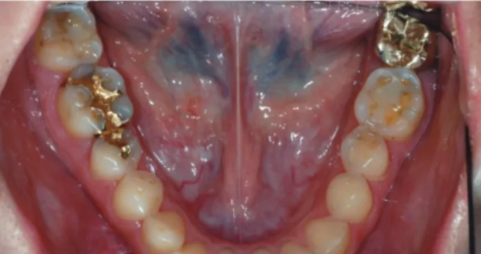
[1] This psychosomatic disease is characterized by conscious starvation, followed by a period of excessive carbohydrate intake, and then, it is often followed by deliberately induced vomiting. Although some patients’ symptoms were improved over time, a substantial proportion continues to have body-image disturbances, disordered eating, and other psychiatric difficulties.[2, 3] The greatest impacts on the oral cavity include severe lingual-occlusal tooth structure

Meng Ding, DDS

1

, Sang-yoon Lee, DDS

2

, Jae-Jun Ryu, DDS, PhD

1

*

1

Department of Esthetic Restorative Dentistry, Graduate School of Clinical Dentistry, Korea University, Seoul, Korea.

2

Department of Prosthodontics, Anam Hospital, Korea University, Seoul, Korea.

The patient was a 26-year old female who was not satisfied with the aesthetic approach of her maxillary incisors.

The treatment goal throughout the process was to manage the risk and recreate the original and youthful appearance of the smile. The biomechanical risk was managed by providing coverage of the exposed dentin and rebuilding the eroded structures with minimal tooth reduction throughout the treatment. Functional risk and prognosis for this patient were both improved by achieving acceptable function. The patient was satisfied with the full-ceramic restorations and the symmetry, harmony gingival architecture. Key words: bulimia nervosa, acid reflux (J Korean Acad Esthet Dent 2014;23(1):7-23)

본 증례는 26세 여성환자로 폭식증과 이로 인한 구토 및 위산역류로 상악 전치부에 광범위한 치질 손상과 더불어 산

부식으로 인해 상아질 노출로 기능뿐만 아니라 심미적인 문제를 주소로 내원했다. 환자는 단시간 내 심미성 회복을 원하

는 상태로 교정 등의 치료는 원치 않았다. 이를 감안해 치료계획은 이미 손상된 치질을 최대한 보존하면서 심미성 및 기

능 회복을 목표로 하였다. 상악 전치부의 치관 비율 및 치은 형태 개선을 위해 치은절제술 및 치관연장술을 시행했으며,

전부도재관을 이용해 수복하였다. 회전 이동되어있는 우측 상악 측절치는 치질 삭제 없이 veneer 형태로 제작했다.

산 부식 및 마모에 강한 지르코니아에 도재로 순측을 veneering해 기능 회복뿐만 아니라 심미적인 면에서도 만족할

만한 결과를 얻을 수 있었다. 추후 미백치료를 고려해 최종보철물의 색상을 결정했으며, 환자는 색상과 형태에 만족했으

며 자신감있는 미소를 회복할 수 있었다. 키워드: 폭식증, 위산역류 (대한심미치과학회지 2014;23(1):27-33)

(2)

approach, which aims to preserve the natural tooth structure, whenever possible, should be the paramount concern.[5]

This report deals with a young female patient who showed severe erosion associated with gastro esophageal reflux caused by anorexia nervosa. She was unsatisfied with her asymmetric gingival contour and anterior teeth appearance.

After evaluation of the patient risk and prognosis, a series treatment was performed to achieve a predictable, long-term, satisfying result, involving crown lengthening procedure(CLP) and all-ceramic restoration. The result was successful in both achieving desired goals while recognizing and accepting that certain areas of the patient’s dentition may need ongoing care.

 Case report 

The patient is a 26-year-old female who was not satisfied with the aesthetic appearance of her maxillary incisors. She has long history of bulimia nervosa, accompanied by vomiting and gastro esophageal reflux. In her clinical examination there was extensive dental erosion on the occlusal, buccal and palatal surfaces of maxillary anterior teeth; buccal and occlusal surfaces of mandibular teeth and cervical lesions were seen(Fig. 1, 2, 3) (smith and knight wear index, score 2-3, Table 1). Right lateral incisor was rotated subsequently result in a cross-bite. Significant maxillary gingival asymmetry and disharmony was also noted. A number of posterior composite fillings were discolored at their edges, with eroded tooth structure surrounding the existing composites (Fig. 2, 3).

Fig. 3. Mandibular occlusal view showing erosion in posterior teeth.

Fig. 1. Severe erosion and attrition were found in maxillary incisors.

Fig. 2. Maxillary occlusal view showing extensive erosion of

virtually all teeth.

(3)

 Treatment Goals 

The following treatment goals were determined:

• Manage the biomechanical risk(bulimia nervosa) and improve the prognosis by both restoring and protecting the structurally compromised teeth.

• Utilize minimally invasive conservative tooth preparation designs wherever possible.

• Establish horizontal symmetry of the maxillary gingival architecture.

• Provide the patient with a smile that she desired.

 Treatment Plan 

• The optimal position of maxillary anterior was established utilizing the facebow and semi-adjustable articulator (Evo7, Kavo)

• Manually veneered zirconia all-ceramic restorations were planned for canines and incisors (central/lateral) in maxilla, with minimal buccal and lingual reduction and subgingival finish lines, right lateral incisor was prepared for a partial coverage crown due to the position restriction.

Fig. 6.

Fig. 4. Diagnostic wax-up Fig. 5. Lateral movement was perform to check the canine

guidance and anteriorteeth interfereance

(4)

Fig. 7. The line of gingivectomy was designed and drew on the cast.

Fig. 9. Gingivectomy performed with an Er,Cr:YSGG, Waterlaser.

Fig. 11. Gingivectomy was completed and hemorrhage control was easily achieved by using waterlaser(photo taken immediately postoperatively).

Fig. 10. Surgical guide was used to check the gingival margin.

Fig. 12. Two-week postsurgical view.

Fig. 8. Fabrication of surgical guide in laboratory.

(5)

sound tooth structure and preparation length to decrease the risk of biologic width violation.

 Treatment

Phases Phase 1: Diagnostic Data

Preoperative photographs, a panorama radiography, a detailed clinical examination, diagnostic and interocclusal records were obtained and forwarded to technician.

Phase 2: Planning and Surgical Crown Lengthening

Plastic surgical guide was fabricated to help to determine the desired gingival margin during operation(Fig.7, 8).

The gingival alteration and associated osseous reduction for each tooth was calculated.[6] A gingivectomy was done to achieve the desired contour, symmetry, and harmony by using a Er,Cr:YSGG laser(Fig. 9, 10, 11). Following surgery, the tissue was allowed to heal for 2 weeks(Fig. 13). It is not enough for a soft tissue healing and mature. However, as the patient asked for a rehabilitation of maxillary teeth as soon as possible, phase 3 was performed at that time to promote Fig. 13. Intro-oral front view after teeth preparation. Fig. 14. Upon healing, the provisional restorations were optimized until the soft tissue contour desired in the final restorations is achieved.

Fig. 15 a, b. Ceramic crowns for maxillary incisors were fabricated on dies.

(6)

Phase 3: Restorative Phase

After 2 weeks of healing time following the crown lengthening, gingival line was corresponded to the surgical guide margin. Master model was fabricated and articulated on a semi-adjustable articulator(Evo 7, KaVo). After diagnostic wax-up(Fig. 4, 5, 6) was performed to visualize and present the ideal maxillary teeth contour and establish a harmonized relationship with canine guidance[7]. Provisional restorations were fabricated(Fig. 14) by duplicating the wax-up model.

These provisional restorations were used for the critical assessment of aesthetics, phonetics and function before moving onto definitive restorations. Once provisional restorations were appropriate, definitive restorations were fabricated using the former as a guide. All ceramic crowns (porcelain veneered to zirconia) were fabricated, tried in, and delivered (Fig. 15, 16, 17). The final occlusion was checked using articulating paper and clinician’s tactile sense. Simultaneous contacts; and a well-managed guidance pathway were achieved in the final result, changing from occlusal dysfunction to acceptable function.

It must be noted that because of patient’s requirement, the restorations were fabricated in a whiter color compare to her natural teeth. However, as the young patient claimed that she was going to bleaching her teeth, these whiter restorations will not compromise the esthetic effect and oral harmony.

 Conclusion 

The treatment goal throughout the process was to manage the risk and recreate the original and youthful appearance of the smile. The biomechanical risk was managed by providing coverage of the exposed dentin and rebuilding the eroded structures with minimal tooth reduction throughout the treatment. Functional risk and prognosis for this patient were both improved by achieving acceptable function. The patient was satisfied with the full-ceramic restorations and the symmetry, harmony gingival architecture.

Fig. 16. Maxillary incisors were restored with ceramic crowns luted with resin composite cement.

Fig. 17. The palatal surface of the right lateral incisor was

partial covered.

(7)

B = buccal or labial L = lingual or palatal O = occlusal I = incisal C = cervical

References

1. Dowson, J.H., Eating disorders and obesity: A comprehensive handbook, 2nd edition. Psychological Medicine, 2003. 33(1): p. 180-181.

2 Hazelton, L.R. and M.P. Faine, Diagnosis and dental management of eating disorder patients. Int J Prosthodont, 1996. 9(1): p. 65-73.

3 Fan-Hsu, J., Evidence linking gastroesophageal reflux disease and dental erosion is not strong. J Am Dent Assoc, 2009. 140(11): p. 1401-2.

4. Barron, R.P., et al., Dental erosion in gastroesophageal reflux disease. J Can Dent Assoc, 2003. 69(2): p. 84-9.

5. Vidal Cde, M., et al., Direct restorative treatment of dental erosion caused by gastroesophageal reflux disease associated with bruxism: a case report. Compend Contin Educ Dent, 2011. 32(7): p. E110-4.

6. Allen, E.P., Use of mucogingival surgical procedures to enhance esthetics. Dent Clin North Am, 1988. 32(2): p.

307-30.

7. Klineberg, I., R. Jagger, and N. Wilson, Occlusion and clinical practice. 2004.

수치

Fig. 2. Maxillary occlusal view showing extensive erosion of  virtually all teeth.
Fig. 4.  Diagnostic wax-up Fig. 5.  Lateral movement was perform to check the canine  guidance and anteriorteeth interfereance
Fig. 7.  The line of gingivectomy was designed and drew on  the cast.
Fig. 15 a, b.  Ceramic crowns for maxillary incisors were fabricated on dies.
+2

참조

관련 문서