64
Introduction
Amyloid light-chain (AL) amyloidosis has an annual inci- dence of 6–10 cases per million in the USA with cardiac in- volvement occurring in 90%.1) The clinical presentation of cardiac amyloidosis (CA) in the form of heart failure due to re- strictive infiltrative cardiomyopathy is well described. Less of- ten patients present with myocardial ischemic syndromes which is thought to be due to amyloid obstruction of the in- tramyocardial coronary arteries.2) Little has been published on the effect of CA on microcirculatory function. Here we dem- onstrate a reduction of myocardial blood flow reserve (MBFR) in a patient with CA using real-time myocardial contrast echo- cardiography (MCE).
Case
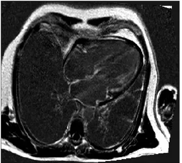
A 52-year-old lady who presented with breathlessness and mild left ventricular impairment with severe biventricular thick- ening on echocardiography. There was no past medical history of note and blood pressure assessment was normal. Haemato- logical investigations revealed systemic light chain (AL) amy- loidosis. Cardiac magnetic resonance imaging (CMR) showed biatrial and biventricular extensive subendocardial delayed en- hancement typical for CA (Fig. 1). Although endomyocardial
pISSN 1975-4612/ eISSN 2005-9655 Copyright © 2016 Korean Society of Echocardiography www.kse-jcu.org http://dx.doi.org/10.4250/jcu.2016.24.1.64
CASE REPORT J Cardiovasc Ultrasound 2016;24(1):64-67
biopsy was not performed, the diagnosis of CA was based on confirmed plasma-cell dyscrasia with presence of amyloid in bone marrow biopsy, and typical amyloid CMR (CMR carries 92% positive predictive value).3) Additionally, repeat echocar- diogram and CMR following chemotherapy showed restora- tion of left ventricular systolic function and reduction in sub- endocardial delayed enhancement. The patient had a normal coronary angiogram one year previously for investigation of chest pain. After developing further atypical chest pains she was referred for dipyridamole stress echocardiography to assess for wall motion abnormality, which was negative. We subse- quently performed vasodilator MCE to assess myocardial blood flow (MBF) at baseline, MBF at peak stress (MBFs) and MBFR.
MBFR is a validated technique in the assessment of microvascu- lar function in the absence of obstructive coronary artery dis- ease first reported by Wei et al.4) in 2001. Real-time MCE was performed according to an established protocol using a com- mercial ultrasound machine (iE33, Philips Medical Systems, Best, the Netherlands) and SonoVue (Bracco Research SA, Ge- neva, Switzerland) contrast agent with a rate set between 0.8–
1.0 mL/min to maximize image quality with minimal attenua- tion.5) Images were recorded in the 3 apical views with low- power settings at a mechanical index of 0.1. Flash-impulse
Abnormal Myocardial Blood Flow Reserve Observed in Cardiac Amyloidosis
Michael Chi Yuan Nam, MD1, Karen Nel, B.Tech1, Roxy Senior, MD2, and Kim Greaves, MD1
1Department of Cardiology, Sunshine Coast Hospital and Health Service, Nambour, QLD, Australia
2Department of Cardiology, Royal Brompton and Harefield NHS Trust, London, United Kingdom
We performed real-time myocardial contrast echocardiography on a patient with cardiac amyloidosis and previous normal coro- nary angiography presenting with atypical chest pain to assess myocardial blood flow reserve (MBFR). Myocardial contrast echo- cardiography was performed and flash microbubble destruction and replenishment analysis was used to calculate myocardial blood flow. Dipyridamole was used to achieve hyperemia. MBFR was derived from the ratio of peak myocardial blood flow at hy- peremia and rest. The results show a marked reduction in MBFR in our patient. Previous reports of luminal obstruction of intra- myocardial rather than epicardial vessels by amyloid deposition may be causing microvascular dysfunction.
KEY WORDS: Myocardial blood flow · Myocardial blood flow reserve · Contrast echocardiography · Cardiac amyloidosis · Echocardiography.
• Received: July 10, 2015 • Revised: January 4, 2016 • Accepted: February 1, 2016
• Address for Correspondence: Michael Chi Yuan Nam, Department of Cardiology, Sunshine Coast Hospital and Health Service, Hospital Road PO BOX 547, Nambour, QLD 4560, Australia Tel: +61-5370-3727, Fax: +61-5470-6084, E-mail: dr.michael.nam@gmail.com
• This is an Open Access article distributed under the terms of the Creative Commons Attribution Non-Commercial License (http://creativecommons.org/licenses/by-nc/3.0) which permits unrestricted non-commercial use, distribution, and reproduction in any medium, provided the original work is properly cited.
Abnormal MBFR in Cardiac Amyloidosis | Michael Chi Yuan Nam, et al.
65 imaging at a high mechanical index (1.0) was performed to
achieve complete myocardial bubble destruction, after which 10 end-systolic frames were recorded digitally in each view.
Imaging was repeated after dipyridamole infusion at 0.56 mg/
kg over a 4-minute period. After an interval of 2 minutes, di- pyridamole vasodilator images were recorded within 3 to 4 minutes. Excellent image quality was obtained with all 16 left ventricular segments adequately visualized. Quantitative anal- ysis was performed by an investigator who was blinded to the CMR data. Standard commercial software (Qlab version 7.0, Q- Laboratory, Philips Medical Systems, Best, the Netherlands) was used to study regions of interest within the myocardium and quantify myocardial replenishment as previously described.4) Basal segments of the 16-segment left ventricular model were not included in the quantitative analysis.
Background-subtracted plots of peak myocardial contrast in- tensity (representing myocardial blood volume A, dB) versus pulsing intervals (representing time) were automatically con- structed by QLab software to fit the monoexponential func- tion conventional equation: y = A (1 - e-βt)2. From these plots, the slope of the replenishment curve was determined (repre- senting myocardial blood velocity β, dB/s). Per segment, the product of A and β yielded rest MBF and post-dipyridamole MBFs (stress MBF, dB2/s), respectively. The replenishment curves are presented in Fig. 2. MBFR was calculated as the ratio of peak MBF to resting MBF.
The replenishment pictures are shown in Fig. 3. Although steady state perfusion at rest and peak stress appears homoge- nous throughout the myocardium the rate of replenishment fol- lowing bubble destruction at rest and peak stress is signifi- cantly reduced. The peak myocardial blood velocity β was therefore less than normal healthy studies. Although the peak
MBF increased (26.9 dB2/s vs. 43.9 dB2/s) the MBFR was only 1.6 (normal > 2).6)
Discussion
CA is a rare condition associated with primary light chain (AL) amyloidosis. The impairment in MBF observed in CA has been described in two small case studies previously and is thought to be due to obstruction of the intramyocardial coro- nary arterial lumen by amyloid deposition.2) In contrast, al- though the larger epicardial arteries show evidence of amyloi- dosis in the arterial walls, the lumen of these vessels are almost always unobstructed. Vascular deposition of amyloid, although not as common as interstitial type, is well described in the lit- erature.2)7) The intramyocardial arteries are selectively affected with deposition first beginning in the media and then progress- ing to the adventitia.1) Eventually, luminal obstruction occurs and in one series was shown to be histologically present in two thirds of patients studied.2) This effect is thought to be the cause of the anginal symptoms described and be responsible for the ischemic histological changes seen in the majority of amyloid
Fig. 1. Cardiac MR four chamber view showing biatrial and biventricular subendocardial delayed enhancement in keeping with a diagnosis of cardiac amyloidosis.
Fig. 2. Replenishment curves following bubble destruction using high amplitude ultrasound pulse at rest (A) and post-dipyridamole (stress) (B).
A normal study curve has also been plotted for comparison, taken from previous published data by our group.5) Peak video intensity A is proportional to and therefore representative of blood volume. Gradient β (dB/s) represents peak blood velocity.
30.0 25.0 20.0 15.0 10.0 5.0 0
0 1 2 3 4 5 6 7 8 9 10 11 Rest
A
β
Pulsing interval (s)
Video intensity (dB)
Normal Amyloid
A
30.0 25.0 20.0 15.0 10.0 5.0 0
0 1 2 3 4 5 6 7 8 9 10 Stress
A
β
Pulsing interval (s)
Video intensity (dB)
Normal Amyloid
B
Journal of Cardiovascular Ultrasound 24 | March 2016
66
hearts. It may also explain the chronically elevated troponin lev- els observed.2)7)8)
Real time MCE with flash bubble destruction is a well-vali- dated diagnostic tool to quantify MBFR.9) Two previous stud- ies have examined MBF in patients with CA. Al Suwaidi et al.10) undertook coronary angiography in five patients with CA and measured coronary flow reserve using adenosine and then acetylcholine. Although all had normal epicardial arteries, both endothelial independent and endothelial dependent functions were significantly impaired. In a second study, a single case of amyloid underwent MCE with vasodilator stress in which MBF was also impaired.11) This report further demonstrates the use of this technique in understanding CA pathophysiology.
In addition we observe that MBFR is lower in our amyloid patient because of peak blood velocity β being lower compared to healthy population data. Overall, microvascular ischaemia resulting from intramyocardial coronary artery obstruction by amyloid deposition can be a probable cause of abnormal MBFR of CA even without epicardial coronary artery disease.
References
1. Narang R, Chopra P, Wasir HS. Cardiac amyloidosis presenting as isch-
emic heart disease. A case report and review of literature. Cardiology 1993;
82:294-300.
2. Neben-Wittich MA, Wittich CM, Mueller PS, Larson DR, Gertz MA, Edwards WD. Obstructive intramural coronary amyloidosis and myocardial ischemia are common in primary amyloidosis. Am J Med 2005;
118:1287.
3. Vogelsberg H, Mahrholdt H, Deluigi CC, Yilmaz A, Kispert EM, Greulich S, Klingel K, Kandolf R, Sechtem U. Cardiovascular mag- netic resonance in clinically suspected cardiac amyloidosis: noninvasive imag- ing compared to endomyocardial biopsy. J Am Coll Cardiol 2008;51:
1022-30.
4. Wei K, Ragosta M, Thorpe J, Coggins M, Moos S, Kaul S. Noninva- sive quantification of coronary blood flow reserve in humans using myocardi- al contrast echocardiography. Circulation 2001;103:2560-5.
5. Rana O, Byrne CD, Kerr D, Coppini DV, Zouwail S, Senior R, Beg- ley J, Walker JJ, Greaves K. Acute hypoglycemia decreases myocardial blood flow reserve in patients with type 1 diabetes mellitus and in healthy hu- mans. Circulation 2011;124:1548-56.
6. Camici PG, Crea F. Coronary microvascular dysfunction. N Engl J Med 2007;356:830-40.
7. Mueller PS, Edwards WD, Gertz MA. Symptomatic ischemic heart dis- ease resulting from obstructive intramural coronary amyloidosis. Am J Med 2000;109:181-8.
8. Apridonidze T, Steingart RM, Comenzo RL, Hoffman J, Goldsmith Y, Bella JN, Landau H, Liu JE. Clinical and echocardiographic correlates of elevated troponin in amyloid light-chain cardiac amyloidosis. Am J Cardiol Fig. 3. Real-time myocardial contrast echo images showing bubble replenishment at rest and stress with a normal study for comparison. Each number in top left of image represents number of frames post bubble destruction. Notice bubble replenishment is more rapid and homogenous in the normal study compared to our patient both at rest and stress.
Patient rest images
Normal rest images
Patient stress images
Normal stress images
Abnormal MBFR in Cardiac Amyloidosis | Michael Chi Yuan Nam, et al.
67 2012;110:1180-4.
9. Senior R, Becher H, Monaghan M, Agati L, Zamorano J, Vanover- schelde JL, Nihoyannopoulos P. Contrast echocardiography: evidence- based recommendations by European Association of Echocardiography. Eur J Echocardiogr 2009;10:194-212.
10. Al Suwaidi J, Velianou JL, Gertz MA, Cannon RO 3rd, Higano ST,
Holmes DR Jr, Lerman A. Systemic amyloidosis presenting with angina pectoris. Ann Intern Med 1999;131:838-41.
11. Abdelmoneim SS, Bernier M, Bellavia D, Syed IS, Mankad SV, Chan- drasekaran K, Pellikka PA, Mulvagh SL. Myocardial contrast echocar- diography in biopsy-proven primary cardiac amyloidosis. Eur J Echocardiogr 2008;9:338-41.