Treatment Response and Long Term Follow-up Results of
Nonspecific Interstitial Pneumonia
The purpose of this study was to investigate the long-term clinical course of non-specific interstitial pneumonia (NSIP) and to determine which factors are associated with a response to steroid therapy and relapse. Thirty-five patients with pathologically proven NSIP were included. Clinical, radiological, and laboratory data were reviewed retrospectively. The male-to-female ratio was 7:28 (median age, 52 yr). Thirty (86%) patients responded to steroid therapy, and the median follow-up was 55.2 months (range, 15.9-102.0 months). Five patients (14%) showed sustained disease progression and three died despite treatment. In the five with sustained disease progression, NSIP was associated with various systemic conditions, and the seropositivity of fluorescent antinuclear antibody was significantly associated with a poor response to steroids (P = 0.028). The rate of relapse was 25%, but all relapsed patients improved after re-treatment. The initial dose of steroids was significantly low in the relapse group (P = 0.020). In conclusion, progression is associated with various systemic conditions in patients who show progression. A low dose of initial steroids is significantly associated with relapse.
Key Words: Idiopathic Interstitial Pneumonias; Lung Diseases, Interstitial; Pulmonary Fibrosis; Disease Progression; Mortality; Prednisolone; Prognosis; Recurrence; Steroids; Drug Therapy
Ji Yeon Lee1, Sang-Man Jin1, Byoung Jun Lee1, Doo Hyun Chung2, Bo-Gun Jang2, Heae Surng Park2, Sang-Min Lee1, Jae-Joon Yim1, Seok-Chul Yang1, Chul-Gyu Yoo1, Sung Koo Han1, Young-Soo Shim1, and Young Whan Kim1
1Division of Pulmonary and Critical Care Medicine,
Department of Internal Medicine and Lung Institute, and 2Department of Pathology, Seoul National
University College of Medicine, Seoul, Korea Received: 7 August 2011
Accepted: 13 March 2012 Address for Correspondence: Young Whan Kim, MD
Division of Pulmonary and Critical Care Medicine, Department of Internal Medicine and Lung Institute of Medical Research Center, Seoul National University College of Medicine, 101 Daehak-ro, Jongno-gu, Seoul 110-744, Korea Tel: +82.2-2072-2856, Fax: +82.2-762-9662 E-mail: ywkim@snu.ac.kr
This study was supported by a grant of Korea Healthcare Technology R&D Project, Ministry of Health & Welfare, Repulblic of Korea (A100190).
http://dx.doi.org/10.3346/jkms.2012.27.6.661 • J Korean Med Sci 2012; 27: 661-667
INTRODUCTION
Non-specific interstitial pneumonia (NSIP) has been proposed as a histological subtype of idiopathic interstitial pneumonia and is characterised by varying degrees of alveolar wall inflam-mation and fibrosis in a pattern that suggests temporal homo-geneity (1). NSIP is associated with various medical conditions such as human immunodeficiency virus infection, hypersensi-tivity pneumonitis, and several collagen vascular diseases (CVDs) including polymyositis-dermatomyositis, rheumatoid arthritis, Sjögren’s syndrome, and systemic sclerosis. The diagnosis of id-iopathic NSIP is made when no specific cause can be detected (1-7).
The overall prognosis and response to steroid therapy is gen-erally favourable in patients with NSIP (1, 8-12). However, not all patients respond well to treatment. In some patients, relapse can occur when steroids are tapered or stopped. In others, ste-roids do not modify disease activity, leading to disease progres-sion and death. Travis et al. (9) suggested that separating the cellular from the fibrotic patterns of NSIP is important because survival might differ between these two groups. Patients with
cellular pattern NSIP have 5- and 10-yr survival rates similar to those with desquamative interstitial pneumonia, whereas those with fibrotic pattern NSIP have a worse 5-yr survival rate, which was similar to those with usual interstitial pneumonia (9). Park et al. (13) also reported that patients with fibrotic NSIP are fre-quently hospitalised with a recurrence rate of 36%. However, there are relatively few data on the clinical course and outcomes other than mortality. It is also unclear which factors affect the responsiveness to steroids and relapse.
The purpose of this study was to investigate the clinical course of NSIP after treatment and to determine which factors are as-sociated with the responsiveness to steroids and relapse.
MATERIALS AND METHODS Study subjects
We reviewed the medical records of patients admitted to a ter-tiary referral hospital (Seoul National University Hospital) from March 1995 to July 2007. We found 41 patients who were diag-nosed clinically with NSIP at discharge who had undergone a surgical lung biopsy for a histological examination. The biopsy
slides were reviewed by three pathologists according to the Amer-ican Thoracic Society/European Respiratory Society consensus classification of interstitial pneumonia (14); and 37 patients with pathologically confirmed NSIP were selected. We excluded two patients lost to follow-up, so 35 patients were included.
Variables and definitions
A retrospective study was performed to identify factors associ-ated with the responsiveness to steroids and relapse. Clinical, radiological, and laboratory data were reviewed, including age, gender, smoking history, symptoms on admission, comorbidi-ties, initial oxygen tension in arterial blood (PaO2), results of
pulmonary function tests, and steroid doses.
The response to steroid was assessed from the clinical records of the patients and included improvement in symptoms, radio-graphic findings, and pulmonary function tests (PFTs) after ini-tial corticosteroid therapy. The timing for assessing the response was 1 yr after the initiation of treatment. Improvement was de-fined as > 10% increase in forced vital capacity (FVC) and dif-fusing capacity of the lung for carbon monoxide (DLCO).
Progres-sion was defined as > 10% decline in FVC and DLCO from
base-line and/or death. When the PFT data could not be obtained because of worsening of respiratory symptoms accompanied by continued worsening of findings on chest radiography (e.g. acute exacerbation), the case was also defined as disease pro-gression. Patients who did not demonstrate improvement or progression were considered stable. Finally, we classified the patients who showed improvement or remained stable after the initial steroid therapy as steroid responders, and those who showed progression as steroid non-responders.
Relapse was defined as an aggravation of symptoms and lung function (and/or radiological abnormalities) after complete dis-continuation of treatment and treatment with prednisolone and/ or cytotoxic agent was restarted.
Analysis
Continuous variables were analysed in the univariate analysis using non-parametric tests (Mann-Whitney U- or Kruskal-Wal-lis tests), and categorical variables were compared using Pear-son’s chi-squared or Fisher’s exact test, as appropriate. Survival was evaluated using the Kaplan-Meier method. A survival anal-ysis was performed on the total sample, including patients with cellular or fibrotic NSIP, because the number of patients was small, only three deaths occurred, and the histological subtypes could not be distinguished in many patients. SPSS version 17.0 (Chicago, IL, USA) was used for all analyses. A P value < 0.05 was considered significant.
Ethics statement
The study was approved by the institutional review board of Seoul National University Hospital (Approval No.
H-1008-095-328). The board waived informed consent from the subject pa-tients. This study was conducted in accordance with the Decla-ration of Helsinki.
RESULTS
Twenty-eight women and seven men (median age, 52 yr) were included in the analysis. The demographic and clinical charac-teristics of the enrolled patients are described in Table 1. Most patients presented with subacute onset of dyspnea or cough. Ground-glass opacity was the most frequent abnormality (88.6%), Table 1. Baseline characteristics
Parameters Findings Total (number) 35 (100%) Age (yr) 52 (35-68) Female sex 28 (80%) Smoking status (n = 26) Never smoker Ex-smoker Current smoker 17 (65.4%) 7 (26.9%) 2 (7.7%) Symptoms Dyspnea Cough Sputum Weight loss Fever 29 (82.9%) 27 (77.1%) 20 (57.1%) 6 (17.1%) 5 (14.3%) Duration of symptoms, months (n = 33) 2 (0.3-120) Radiological feature Ground-glass opacity Consolidation Traction bronchiectasis Reticulation Honeycombing 31 (88.6%) 14 (40.0%) 12 (34.3%) 8 (22.9%) 6 (17.1%) Histological pattern Cellular Fibrotic Indeterminate 17 (48.6%) 10 (28.6%) 8 (22.9%) Comorbidities
Collagen vascular disease* Diabetes mellitus Tuberculosis Malignancy Hypertension 9 (25.7%) 7 (20.0%) 7 (20.0%) 5 (14.3%) 4 (11.4%) Initial BAL finding % (n = 18)
Total cells × 105 (counts/µL) Macrophages (%) Neutrophils (%) Lymphocytes (%) Eosinophils (%) T4/T8 ratio (n = 16) 3.99 (0.67-27.4) 27 (0-90) 6.5 (0-35) 34.5 (6-76) 0.5 (0-44) 0.38 (0.10-2.52) Initial pulmonary function tests
FVC, % pred (n = 34) DLCO, % pred (n = 32) 63.5 (32-103) 46.5 (26-95) Initial PaO2, mmHg (n = 27) 73.1 (45.3-112.0) Follow-up duration, months (range) (n = 32)† 55.2 (15.9-102.0) Values in parentheses represent the range or percentage. *Sjogren syndrome (n = 4, 11.4%), mixed connective tissue disease (n = 2, 5.7%), polymyositis (n = 1, 2.9%), diffuse scleroderma (n = 1, 2.9%), rheumatoid arthritis (n = 1, 2.9%), dermatomyo-sitis (n = 1, 2.9%); †Follow-up duration of survivors. BAL, bronchoalveolar lavage; FVC, forced vital capacity; pred, predicted; DLCO, diffusing capacity of the lung for carbon monoxide; PaO2, oxygen tension in arterial blood.
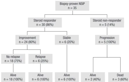
Fig. 1. Clinical courses of the patients with nonspecific in-terstitial pneumonia (NSIP).
No relapse n = 18 (75%) n = 6 (25%)Relapse Alive n = 18 (100%) n = 6 (100%)Alive Steroid responder n = 30 (86%) Improvement n = 24 (80%) Biopsy-proven NSIP n = 35 Stable n = 6 (20%) Alive n = 6 (100%) n = 2 (40%)Alive n = 3 (60%)Dead Progression n = 5 (100%) Steroid non-responder n = 5 (14%)
Table 2. Comparison between steroid responders and non-responders
Variables Steroid responders (n = 30) Steroid non-responders (n = 5) P value
Age (yr) 53.5 (35-68) 51 (49-58) 0.962
Female sex 24 (80.0%) 4 (80.0%) > 0.999
Duration of symptoms, months (n = 33) 2.0 (0.3-120.0) 12.0 (2.0-36.0) 0.221
Never-smoker (n = 26) 16 (66.7%) 1 (50.0%) > 0.999 Histologic pattern (n = 27) Cellular Fibrotic 15 (65.2%) 8 (34.8%) 2 (50.0%) 2 (50.0%) 0.613
Collagen vascular disease 6 (20.0%) 3 (60.0%) 0.095
Presence of systemic condition* 15 (50.0%) 5 (100%) 0.057
Radiological feature Ground-glass opacity Consolidation Traction bronchiectasis Reticulation Honeycombing 27 (90.0%) 11 (36.7%) 9 (30.0%) 7 (23.3%) 5 (16.7%) 4 (80.0%) 3 (60.0%) 3 (60.0%) 1 (20.0%) 1 (20.0%) 0.477 0.369 0.313 > 0.999 > 0.999 Initial BAL finding % (n = 18)
Total cells × 105 (counts/µL) Macrophages (%) Neutrophils (%) Lymphocytes (%) Eosinophils (%) T4/T8 ratio (n = 16) 4.15 (0.67-25) 18 (0-90) 7 (0-33.0) 40 (6-76) 1 (0-44) 0.46 (0.10-2.52) 3.30 (2.61-27.4) 46 (30-67) 6 (2-35) 27 (23-40) 0 (0-2) 0.31 (0.21-0.55) 0.594 0.373 0.905 0.635 0.485 0.638 Initial laboratory findings, median (range)
WBC (counts/µL) Cholesterol, mg/dL (n = 34) Albumin, g/dL (n = 34) CRP, mg/dL (n = 31) ESR, mm/hr (n = 20) RF positive, n (%) (n = 24) FANA positive, n (%) (n = 24) 8,275 (3,630-13,480) 175 (116-312) 3.9 (2.5-4.6) 0.47 (0.01-9.91) 27 (1-78) 8 (36.4) 5 (23.8) 8,200 (5,480-37,700) 186 (147-191) 3.4 (2.9-4.0) 0.51 (0.01-7.98) 55.5 (47-72) 1 (50.0) 3 (100.0) 0.556 0.957 0.179 0.86 0.053 > 0.999 0.028 Initial pulmonary function tests
FVC, % pred (n = 34)
DLCO, % pred (n = 32) 64 (41-103) 46.5 (26-95) 62 (32-90)53.5 (37-77) 0.5110.732 Initial PaO2, mmHg (n = 27) 75 (45.3-99.1) 66.4 (57.0-112.0) 0.339 Values in parentheses represent the range or percentage. *The systemic conditions included collagen vascular disease, malignancy, diabetes mellitus, pulmonary tuberculosis, hepatitis B, and sustained exposure to drugs or chemicals (Table 3, E1, E2). BAL, bronchoalveolar lavage; WBC, white blood cells; CRP, C-reactive protein; ESR, erythrocyte sedimentation rate; RF, rheumatoid factor; FANA, fluorescent antinuclear antibody; FVC, forced vital capacity; pred, predicted; DLCO, diffusing capacity of the lung for carbon monoxide; PaO2, oxygen tension in arterial blood.
followed by consolidation (40.0%) on the initial chest comput-ed tomography (CT) scans. A histological analysis showcomput-ed the cellular pattern in 17 patients (48.6%) and the fibrotic pattern in
10 patients (28.6%); the histological NSIP subtype was indeter-minate in eight patients (22.9%). The median follow-up dura-tion for survivors was 55.2 months (range, 15.9-102.0 months).
The follow-up results of the enrolled patients are summarised in Fig. 1. All 35 patients received corticosteroid therapy alone as the initial treatment, and cytotoxic agents were added in seven patients (four patients cyclophosphamide; three patients aza-thioprine) who showed rapid disease progression or steroid dependency. The median dose of initial prednisolone was 0.54 mg/kg/day. A higher dose of initial steroids (> 1.5 mg/kg/day) was used in only two patients, 9.5 mg/kg/day in one patient who showed rapid disease progression, and 1.8 mg/kg/day in another who showed arterial hypoxemia on room air (PaO2, 54.3 mmHg).
The range of initial prednisolone doses used in patients except these two was 0.4-1.1 mg/kg/day. The prednisolone dose was slowly tapered after 4-6 weeks based on the clinical and PFT evaluation of early response. Thirty (86%) patients responded to steroid therapy, and five (14%) were considered non-respond-ers. Three of the 5 non-responders died of disease progression with combined pneumonia, although all had received addition-al cyclophosphamide pulse therapy. The patients died 1.6, 1.7, and 5.2 months after beginning treatment. In contrast, steroid responders were all alive at the end of the follow-up period. Six (20%) steroid responders were stable but were considered ste-roid dependent because the disease worsened when steste-roids were reduced. Therefore, steroid treatment was maintained in these six patients for the entire follow-up period, and azathio-prine was added in two patients. The maintenance dose of pred-nisolone was 5 mg in five patients and 15 mg in one patient. The 1- and 5-yr survival rates in all patients were 97.1% and 93.9%, respectively.
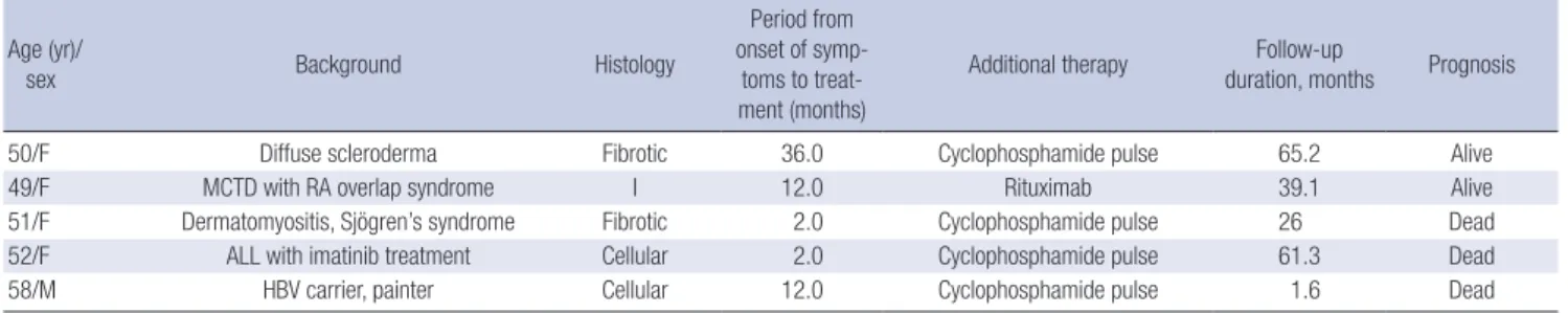
Table 2 shows a comparison of various parameters between steroid responders and non-responders. Histological subtype was not related to steroid response. Smoking history, period from the onset of symptoms to treatment, initial chest CT ings, initial PFT results, and initial bronchoalveolar lavage find-ings did not differ between the groups. However, seropositivity for the fluorescent antinuclear antibody (FANA) was significant-ly associated with a poor response to steroids. Higher erythro-cyte sedimentation rate (ESR) tended to be related to disease progression. Table 3 shows the clinical characteristics of steroid non-responders. Most of the patients with poor treatment out-comes had underlying systemic conditions associated with NSIP,
such as CVD or exposure to drugs.
Twenty-four (80%) of the 30 steroid responders showed clini-cal improvement after initial treatment, and 18 (75%) remained relapse free during the median follow-up of 41.5 months (range, 5.1-76.4 months) after discontinuing the steroids. Relapses oc-curred in six (25%) of 24 patients within a median of 8.2 months (range, 3.0-16.4 months) after withdrawal of steroids. Patients who relapsed were all retreated with a median steroid dose of 30 mg (range, 25-30 mg), and azathioprine was added in one patient. The median duration of retreatment was 4.3 months (range, 3.5-10.7 months) in four patients, and steroid treatment was maintained in another two patients. Patients with and with-out relapse were compared to search for predictive factors (Tables 4 and 5). The initial dose of prednisolone was significantly low-er in the relapse group. Additionally, the duration of initial treat-ment tended to be shorter in the relapse group (4.7 vs 7.7 months),
Table 3. Characteristics of the progression group Age (yr)/
sex Background Histology
Period from onset of symp-toms to treat-ment (months)
Additional therapy Follow-up
duration, months Prognosis
50/F Diffuse scleroderma Fibrotic 36.0 Cyclophosphamide pulse 65.2 Alive
49/F MCTD with RA overlap syndrome I 12.0 Rituximab 39.1 Alive
51/F Dermatomyositis, Sjögren’s syndrome Fibrotic 2.0 Cyclophosphamide pulse 26 Dead 52/F ALL with imatinib treatment Cellular 2.0 Cyclophosphamide pulse 61.3 Dead
58/M HBV carrier, painter Cellular 12.0 Cyclophosphamide pulse 1.6 Dead
MCTD, mixed connective tissue disease; RA, rheumatoid arthritis; I, indeterminate; ALL, acute lymphoblastic leukaemia; HBV, hepatitis B virus.
Table 4. Comparison of clinical and radiological features between the no relapse and relapse groups
Variables No relapse (n = 18) Relapse (n = 6) P value
Age (yr) 55.5 (35-68) 45 (39-59) 0.109 Female sex 14 (77.8%) 5 (83.3%) > 0.999 Duration of symptoms, months (n = 22) 2.0 (0.3-120.0) 4.0 (2.0-12.0) 0.372 Never smoked (n = 22) 10 (62.5%) 5 (83.3%) 0.616 Histological pattern (n = 18) Cellular Fibrotic 6 (50.0%) 6 (50.0%) 5 (83.3%) 1 (16.7%) 0.316 Collagen vascular disease 4 (22.2%) 1 (16.7%) > 0.999 Presence of systemic condition* 8 (44.4%) 3 (50.0%) > 0.999 Radiological feature Ground-glass opacity Consolidation Traction bronchiectasis Reticulation Honeycombing 17 (94.4%) 9 (50.0%) 4 (22.2%) 4 (22.2%) 4 (22.2%) 5 (83.3%) 1 (16.7%) 2 (33.3%) 2 (33.3%) 0 (0%) 0.446 0.341 0.618 0.618 0.539 Initial treatment
Initial dose of prednisolone (mg) Initial dose of prednisolone (mg/kg) Total duration (months)
38 (25-100) 0.6 (0.5-1.8) 7.7 (3.3-24.9) 30 (25-35) 0.5 (0.5-0.5) 4.7 (2.5-12.2) 0.028 0.020 0.182 Follow-up duration, months (range) 53.4 (15.9-94.7) 37.9 (18.9-78.2) 0.257 Values in parentheses represent the range or percentage. *The systemic conditions included collagen vascular disease, malignancy, diabetes mellitus, pulmonary tuber-culosis, hepatitis B, and sustained exposure to drugs or chemicals (Table E2).
but no statistical significance was observed. Smoking history, histological pattern, presence of CVD, initial chest CT, and PFT and laboratory findings did not differ significantly between the groups. The detailed lists of underlying systemic conditions in steroid responders are summarized in Tables E1 and E2. Both FVC and DLCO improved gradually in both the cellular
and fibrotic NSIP groups. DLCO tended to be higher in patients
with cellular NSIP than in those with fibrotic NSIP throughout the follow-up period, although the difference was not signifi-cant. A comparison of the PFT results between patients with and without CVD showed that FVC and DLCO tended to be
low-er in patients with CVD than in those without CVD but without statistical significance. Patients with CVD tended to have fewer changes in PFT results, however this difference was not signifi-cant (data not shown).
DISCUSSION
The NSIP population showed clinical similarities to previous study populations with a predominance of never-smoking, mid-dle-aged females (15, 16). In our study, the overall prognosis of patients with NSIP was generally favourable with an initial re-sponse rate to steroid therapy of 86% and a 5-yr survival rate of 93.9%. The overall 5-yr survival rate varied in previous studies (1, 3, 8, 17-19); and this variation could be explained by the het-erogeneity of the populations studied in terms of histological subtypes and the underlying conditions associated with NSIP.
The survival rates in our study were higher than those reported previously in patients with fibrotic NSIP but were lower than those in patients with cellular NSIP (3, 9, 13, 18).
The presence of an underlying systemic condition might be relevant to a poor prognosis for patients with NSIP. Notably, all five patients in the progression group had chronic diseases or associated conditions such as CVD and sustained exposure to drugs or chemicals. Among the three patients who died, one had dermatomyositis and Sjögren’s syndrome and another had a history of sustained exposure to paint fumes for > 40 yr. An-other patient had acute lymphoblastic leukaemia and had been treated with imatinib for about 15 months before NSIP was di-agnosed. She died of progressive NSIP, although complete re-mission of leukaemia had been achieved. FANA seropositivity was also significantly associated with progression, and the ini-tial ESR level tended to be higher in the progression group. An-ti-nuclear antibodies are found in patients not only with auto-immune diseases but also those with various non-rheumato-logical conditions associated with tissue damage such as infec-tions, cancer, and hormonal or blood diseases. Recent studies have shown that even idiopathic NSIP might be associated with an autoimmune background that later reveals itself as an organ-specific or a systemic autoimmune disease (20, 21). Thus, we presumed that the presence of ANA might be an early manifes-tation of this systemic background and that simultaneous sys-temic manifestations might be related to a poor prognosis. This possibility parallels our results showing the patients with NSIP and various systemic conditions had worse prognoses. Unfor-tunately, we found no statistically significant relationship be-tween the systemic conditions and poor prognosis for NSIP, probably because of the small sample size and the heterogene-ity of associated conditions. The pathophysiological responses to injury grade might differ between patients with various con-ditions associated with NSIP. Felício et al. (22) detected signifi-cantly greater collagen and elastic fibre proliferation in the lungs of patients with CVD and NSIP compared with those with idio-pathic NSIP. The increased elastosis may have been caused by major repair and remodelling processes following septal inflam-mation and consequent fiber fragmentation. These processes might also be responsible for the loss of the normal alveolar wall architecture, which contributes to alveoli collapse, thereby im-pairing the mechanisms of inflammatory resolution (22). Re-cently, a case series of progressive NSIP revealed a change in histology to a more fibrous pattern through sequential biopsies, and half of the patients had CVD (23). Progression of fibropro-liferation is associated with poor outcomes in response to ste-roid treatment (24).
Among the steroid responders in our study, up to 40% of pa-tients suffered relapse or steroid dependency. Park et al. (13) reported that relapse occurred in 36% of patients with fibrotic NSIP who improved or were stable after initial treatment, and Table 5. Laboratory findings and pulmonary function tests in the no relapse and
re-lapse groups
Variables No relapse (n = 18) Relapse (n = 6) P value Initial BAL finding % (n = 11)
Total cells × 105, counts/µL Macrophages Neutrophils Lymphocytes Eosinophils T4/T8 ratio 4.50 (0.67-25) 24 (0-90) 7 (0-21) 46 (6-76) 0 (0-33) 0.27 (0.10-2.52) 3.99 (1.68-4.32) 14 (6-44) 6 (3-16) 39 (25-73) 6.5 (1-44) 0.50 (0.23-0.75) 0.705 0.570 0.850 0.636 0.065 0.705 Initial laboratory findings,
median (range) WBC (counts/µL) Cholesterol (mg/dL) Albumin (g/dL) CRP, mg/dL (n = 23) ESR, mm/hr (n = 15) RF positive, No. (%) (n = 19) FANA positive, No. (%) (n = 18)
8,945 (3,630-13,480) 189 (122-312) 3.8 (2.5-4.6) 0.47 (0.01-9.91) 24 (1-55) 5 (33.3) 3 (20.0) 7,200 (4,440-10,300) 161 (116-197) 4.1 (3.7-4.2) 0.26 (0.14-1.72) 74 (22-78) 1 (25.0) 2 (66.7) 0.317 0.125 0.131 0.575 0.083 > 0.999 0.172 Initial PaO2, mmHg (n = 19) 72.8 (45.3-99.1) 86.8 (80.2-94.0) 0.057 Initial pulmonary function tests
FVC, % pred (n = 23)
DLCO, % pred (n = 22) 65 (41-103)55 (26-95) 42.5 (33-92)64 (53-79) > 0.9990.740 Values in parentheses represent the range or percentage. BAL, bronchoalveolar la-vage; WBC, white blood cells; CRP, C-reactive protein; ESR, erythrocyte sedimentation rate; RF, rheumatoid factor; FANA, fluorescent antinuclear antibody; FVC, forced vital capacity; pred, predicted; DLCO, diffusing capacity of the lung for carbon monoxide.
the disease-related mortality was 30% in this subgroup. In con-trast, all of the relapsed patients in our study showed improve-ment after re-treatimprove-ment with steroids, and none of the patients in this subgroup died. We also found that a lower dose of initial steroid was associated with a relapse of NSIP, a finding that con-trasts with that of Park et al. (13). However, the initial doses of steroid in the study by Park et al. (13) were higher than the dos-es used in both the relapse and no relapse groups in our study. The study by Park et al. (13) included only patients with fibrotic NSIP, whereas we included both cellular and fibrotic subtypes in the analysis of relapse. The optimal dose and duration of glu-cocorticoid therapy is still unclear because most studies of pa-tients with NSIP used a variety of regimens in a small number of patients (8, 13, 19, 22, 24, 25).
This study had some limitations. First, it was a retrospective study with a small sample size, so a multivariate analysis could not be performed. Second, the pathological subtypes could not be confirmed in all patients, although we attempted to identify the pathological patterns in all patients. Third, a possibility of selection bias existed between the biopsy group and the non-biopsy group in patients with CVD and NSIP. Surgical non-biopsy was performed in most patients with atypical radiological fea-tures other than usual interstitial pneumonia. However, we be-lieve that it is difficult to make a correct clinical diagnosis of the various types of CVD based solely on CT findings (26). Finally, our study population included a heterogeneous group with var-ious underlying conditions associated with NSIP.
In conclusion, we investigated the treatment course of NSIP and identified several prognostic factors for treatment response and relapse. In patients who show sustained disease progres-sion despite treatment, the progresprogres-sion is associated with vari-ous systemic conditions such as CVD, malignancy, or exposure to drugs or chemicals. A lower dose of initial steroids is signifi-cantly associated with relapse.
REFERENCES
1. Katzenstein AL, Fiorelli RF. Nonspecific interstitial pneumonia/fibrosis. Histologic features and clinical significance. Am J Surg Pathol 1994; 18: 136-47.
2. Suffredini AF, Ognibene FP, Lack EE, Simmons JT, Brenner M, Gill VJ, Lane HC, Fauci AS, Parrillo JE, Masur H, et al. Nonspecific interstitial pneumonitis: a common cause of pulmonary disease in the acquired immunodeficiency syndrome. Ann Intern Med 1987; 107: 7-13. 3. Cottin V, Donsbeck AV, Revel D, Loire R, Cordier JF. Nonspecific
intersti-tial pneumonia. Individualization of a clinicopathologic entity in a series of 12 patients. Am J Respir Crit Care Med 1998; 158: 1286-93.
4. Fujita J, Yamadori I, Suemitsu I, Yoshinouchi T, Ohtsuki Y, Yamaji Y, Kamei T, Kobayashi M, Nakamura Y, Takahara J. Clinical features of non-specific interstitial pneumonia. Respir Med 1999; 93: 113-8. 5. Douglas WW, Tazelaar HD, Hartman TE, Hartman RP, Decker PA,
Schroeder DR, Ryu JH. Polymyositis-dermatomyositis-associated
inter-stitial lung disease. Am J Respir Crit Care Med 2001; 164: 1182-5. 6. Kim DS, Yoo B, Lee JS, Kim EK, Lim CM, Lee SD, Koh Y, Kim WS, Kim
WD, Colby TV, et al. The major histopathologic pattern of pulmonary fibrosis in scleroderma is nonspecific interstitial pneumonia. Sarcoidosis Vasc Diffuse Lung Dis 2002; 19: 121-7.
7. Yoshinouchi T, Ohtsuki Y, Fujita J, Yamadori I, Bandoh S, Ishida T, Ueda R. Nonspecific interstitial pneumonia pattern as pulmonary involve-ment of rheumatoid arthritis. Rheumatol Int 2005; 26: 121-5.
8. Nagai S, Kitaichi M, Itoh H, Nishimura K, Izumi T, Colby TV. Idiopathic nonspecific interstitial pneumonia/fibrosis: comparison with idiopathic pulmonary fibrosis and BOOP. Eur Respir J 1998; 12: 1010-9.
9. Travis WD, Matsui K, Moss J, Ferrans VJ. Idiopathic nonspecific intersti-tial pneumonia: prognostic significance of cellular and fibrosing patterns: survival comparison with usual interstitial pneumonia and desquama-tive interstitial pneumonia. Am J Surg Pathol 2000; 24: 19-33.
10. Latsi PI, du Bois RM, Nicholson AG, Colby TV, Bisirtzoglou D, Nikola-kopoulou A, Veeraraghavan S, Hansell DM, Wells AU. Fibrotic idiopathic interstitial pneumonia: the prognostic value of longitudinal functional trends. Am J Respir Crit Care Med 2003; 168: 531-7.
11. Flaherty KR, Toews GB, Travis WD, Colby TV, Kazerooni EA, Gross BH, Jain A, Strawderman RL 3rd, Paine R, Flint A, et al. Clinical significance of histological classification of idiopathic interstitial pneumonia. Eur Respir J 2002; 19: 275-83.
12. Riha RL, Duhig EE, Clarke BE, Steele RH, Slaughter RE, Zimmerman PV. Survival of patients with biopsy-proven usual interstitial pneumonia and nonspecific interstitial pneumonia. Eur Respir J 2002; 19: 1114-8. 13. Park IN, Jegal Y, Kim DS, Do KH, Yoo B, Shim TS, Lim CM, Lee SD, Koh Y,
Kim WS, et al. Clinical course and lung function change of idiopathic nonspecific interstitial pneumonia. Eur Respir J 2009; 33: 68-76. 14. Shimizu S, Yoshinouchi T, Ohtsuki Y, Fujita J, Sugiura Y, Banno S,
Yama-dori I, Eimoto T, Ueda R. The appearance of S-100 protein-positive den-dritic cells and the distribution of lymphocyte subsets in idiopathic non-specific interstitial pneumonia. Respir Med 2002; 96: 770-6.
15. Park JH, Kim DS, Park IN, Jang SJ, Kitaichi M, Nicholson AG, Colby TV. Prognosis of fibrotic interstitial pneumonia: idiopathic versus collagen vascular disease-related subtypes. Am J Respir Crit Care Med 2007; 175: 705-11.
16. Travis WD, Hunninghake G, King TE Jr, Lynch DA, Colby TV, Galvin JR, Brown KK, Chung MP, Cordier JF, du Bois RM, et al. Idiopathic nonspe-cific interstitial pneumonia: report of an American Thoracic Society proj-ect. Am J Respir Crit Care Med 2008; 177: 1338-47.
17. Bjoraker JA, Ryu JH, Edwin MK, Myers JL, Tazelaar HD, Schroeder DR, Offord KP. Prognostic significance of histopathologic subsets in idiopath-ic pulmonary fibrosis. Am J Respir Crit Care Med 1998; 157: 199-203. 18. Daniil ZD, Gilchrist FC, Nicholson AG, Hansell DM, Harris J, Colby TV,
du Bois RM. A histologic pattern of nonspecific interstitial pneumonia is associated with a better prognosis than usual interstitial pneumonia in patients with cryptogenic fibrosing alveolitis. Am J Respir Crit Care Med 1999; 160: 899-905.
19. Nicholson AG, Colby TV, du Bois RM, Hansell DM, Wells AU. The prog-nostic significance of the histologic pattern of interstitial pneumonia in patients presenting with the clinical entity of cryptogenic fibrosing alveo-litis. Am J Respir Crit Care Med 2000; 162: 2213-7.
20. Romagnoli M, Nannini C, Piciucchi S, Girelli F, Gurioli C, Casoni G, Ravaglia C, Tomassetti S, Gurioli Ch, Gavelli G, et al. Idiopathic
nonspe-cific interstitial pneumonia: an interstitial lung disease associated with autoimmune disorders? Eur Respir J 2011; 38: 384-91.
21. Kinder BW, Collard HR, Koth L, Daikh DI, Wolters PJ, Elicker B, Jones KD, King TE Jr. Idiopathic nonspecific interstitial pneumonia: lung man-ifestation of undifferentiated connective tissue disease? Am J Respir Crit Care Med 2007; 176: 691-7.
22. Felicio CH, Parra ER, Capelozzi VL. Idiopathic and collagen vascular disease nonspecific interstitial pneumonia: clinical significance of remod-eling process. Lung 2007; 185: 39-46.
23. Schneider F, Hwang DM, Gibson K, Yousem SA. Nonspecific interstitial pneumonia: a study of 6 patients with progressive disease. Am J Surg Pathol 2012; 36: 89-93.
24. Meduri GU, Chinn AJ, Leeper KV, Wunderink RG, Tolley E,
Winer-Mu-ram HT, Khare V, Eltorky M. Corticosteroid rescue treatment of progres-sive fibroproliferation in late ARDS. Patterns of response and predictors of outcome. Chest 1994; 105: 1516-27.
25. Nakamura Y, Chida K, Suda T, Hayakawa H, Iwata M, Imokawa S, Tsuchi-ya T, Ida M, Gemma H, Yasuda K, et al. Nonspecific interstitial pneumo-nia in collagen vascular diseases: comparison of the clinical characteris-tics and prognostic significance with usual interstitial pneumonia. Sar-coidosis Vasc Diffuse Lung Dis 2003; 20: 235-41.
26. Daimon T, Johkoh T, Honda O, Sumikawa H, Ichikado K, Kondoh Y, Taniguchi H, Fujimoto K, Yanagawa M, Inoue A, et al. Nonspecific inter-stitial pneumonia associated with collagen vascular disease: analysis of CT features to distinguish the various types. Intern Med 2009; 48: 753-61.