Single-Bundle Anterior Cruciate Ligament Reconstruction with Semitendinosus Tendon Using the PINN-ACL
CrossPin System: Minimum 4-Year Follow-up
Hee-Soo Kyung, MD, Seung-Gil Baek, MD, Byoung-Joo Lee, MD, and Chang-Hwa Lee, MD
Department of Orthopaedic Surgery, Kyungpook National University Hospital, Kyungpook National University School of Medicine, Daegu, Korea
Purpose: This study evaluated mid-term results of anterior cruciate ligament (ACL) reconstruction using the PINN-ACL CrossPin system that allowed for short graft fixation.
Materials and Methods: Forty-three patients underwent single-bundle ACL reconstruction with a 4-strand semitendinosus tendon graft using the PINN-ACL CrossPin system. Femoral fixation was done using the PINN-ACL CrossPin system, and the tibial side was fixed with post-tie and a bioabsorbable interference screw. The mean follow-up period was 50 months. Evaluation was done using the Lachman test, pivot-shift test, International Knee Documentation Committee (IKDC) score and grade. Anterior displacement was assessed.
Results: There was improvement in the Lachman test and pivot-shift test at final follow-up, form grade II (n=40) or III (n=3) to grade I (n=3) or 0 (n=40) and from grade I (n=20) or II (n=10) to grade I (n=8) or 0 (n=22), respectively. The mean IKDC score was 88.7, and grade A and B were 93.0% at final follow-up. Side-to-side difference was improved from 6.7 mm to 2.1 mm at final follow-up. Complications occurred in 3 patients, a re- ruptured due to trauma at 2 years after surgery and a deep infection and a superficial infection.
Conclusions: The mid-term follow-up results of ACL reconstruction with the PINN-ACL CrossPin system were satisfactory. The PINN-ACL CrossPin can be considered as a useful instrument for short graft fixation.
Keywords: Anterior cruciate ligament, Reconstruction, Semitendinosus tendon, PINN-ACL CrossPin pISSN 2234-0726 · eISSN 2234-2451
Knee Surgery & Related Research
BPTB grafts and 24-week period for complete bone-to-tendon healing3). Adequate fixation has been considered essential for good outcomes of ACL reconstruction and a variety of graft fixa- tion methods have been introduced. Fixation methods can be classified into compression, expansion, and suspension methods.
The compression method allows for early firm fixation and heal- ing with tight bone-tendon interface and enables close fixation to the ACL footprint, but it has low failure load and stability4,5). The expansion fixation mechanism can be advantageous in obtaining secure fixation because two cross pins transversely inserted through a graft provides a centrifugal pressure on the femoral tunnel, but treatment results depend on the press-fit of the graft, bone density around the femoral tunnel, and correct placement of cross pins through the graft tendon6-8). The suspen- sion methods are sub-classified into cortical, cancellous and, cortio-cancellous suspension methods9). The cortical suspension method provides good fixation strength, but it has a bungee cord effect10) and a windshield wiper effect11) due to the long fixation point from the articular surface. The cortico-cancellous suspen-
Introduction
In anterior cruciate ligament (ACL) reconstruction, the use of bone patellar tendon bone graft (BPTB) has been decreasing due to increased anterior knee pain, weakness of extensor power and difficulty of kneeling position. Instead, the use of hamstring tendon has been increasing1,2); however, the disadvantages of hamstring grafts include weaker fixation strength compared to
Received July 14, 2014; Revised (1st) August 18, 2014;
(2nd) September 17, 2014; Accepted September 23, 2014 Correspondence to: Hee-Soo Kyung, MD
Department of Orthopaedic Surgery, Kyungpook National University Hospital, 130 Dongdeok-ro, Jung-gu, Daegu 700-721, Korea Tel: +82-53-420-5636, Fax: +82-53-422-6605
E-mail: hskyung@knu.ac.kr
43
This is an Open Access article distributed under the terms of the Creative Commons Attribution Non-Commercial License (http://creativecommons.org/licenses/by-nc/3.0/) which permits unrestricted non-commercial use, distribution, and reproduction in any medium, provided the original work is properly cited.
Copyright © 2015 KOREAN KNEE SOCIETY www.jksrr.org
sion method has strong stability and stiffness due to the use of a metaphyseal crosspin. To et al.12) reported that stiffness of the graft fixation complex was more affected by fixation method than the graft type in a cadaver study. Intercondylar cortico-cancellous fixation close to the articular surface is expected to offer better results than the EndoButton fixation.
Speirs et al.13) reported that the cortico-cancellous suspensory fixation method required a short graft length due to fixation of the cross pin within the metaphysis, the lowest creep and cyclic elongation amplitude, and the highest strength and stiffness of all the tested devices. In particular, the PINN-ACL CrossPin (ConMed Linvatec, Largo, FL, USA) system was found to be the most rigid and strongest of all the tested reconstruction systems.
Therefore, the cortico-cancellous fixation method seems to have the advantages of short graft, stability, and stiffness, and the PINN-ACL CrossPin implant features the proprietary self-rein- forced poly-L-lactide acid polymer enabling it to be the strongest bioabsorbable implant. The cross pin absorption begins in vivo approximately within 15 to 24 weeks after insertion, the continu- ous loop is composed of high strength polyethylene fiber, and the ultimate pullout tensile strength is 1700N14) (Fig. 1).
In this study, we analyzed the mid-term results of ACL re- construction using the PINN-ACL CrossPin system, a cortico- cancellous suspension method device. We hypothesized that ACL reconstruction using the PINN-ACL CrossPin system would significantly improve manual stability, anterior-posterior laxity measured by instrument, and functional score.
Materials and Methods
From June 2007 to July 2008, 43 of 46 patients with ruptured ACLs were evaluated. The patients underwent single-bundle ACL reconstruction using a 4-strand semitendinosus tendon (semi-T) with the PINN-ACL CrossPin system. Three patients were excluded due to conditions that might affect the results:
articular cartilage damage and osteoarthritis in 1 patient, subtotal meniscectomy in 1 patient, and total meniscectomy in 1 patient.
All included patients were male with a mean age of 28.7 years (range, 18 to 54 years). The ACL reconstruction was performed on the right knee in 24 patients and on the left knee in 19 pa- tients. The mean follow-up period was 50 months (range, 48 to 61 months). Associated lesions were meniscal tear in 15 patients, which required meniscus repair in 5 and partial meniscectomy in 10, and medial collateral ligament injury in 2 patients, which was treated by conservative methods.
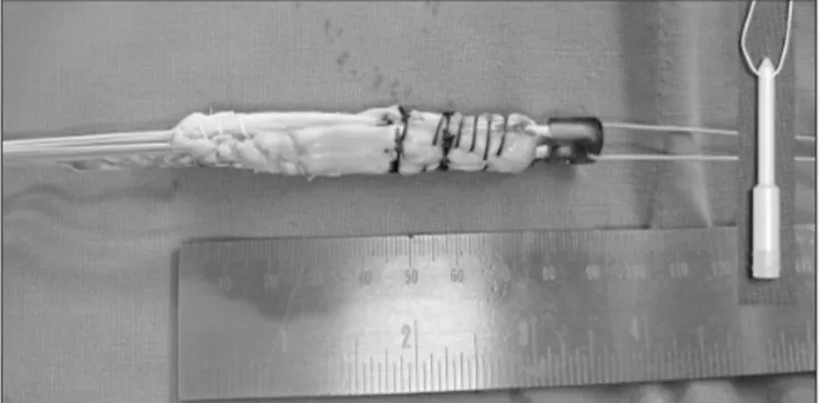
A 4-strand semi-T was used as a graft. A longer than 28 cm semi- T was harvested with an additional 2 cm of periosteum extension and was folded twice to be a 4-strand graft15). The mean length of the graft was 7.2 cm and the diameter was 8.2 cm (Fig. 2). A modified trans-tibial method was used for femoral tunneling16).
Depending on the diameter of the femoral tunnel, a position- ing rod (8 mm or 9 mm) of appropriate size was selected and assembled onto the U-Guide. A disposable transverse cannula was slid onto the U-Guide body. With the U-Guide assembled, the positioning rod was inserted. When the U-Guide assembly was fully inserted, the laser etch marks on the positioning rod indicated the length of the femoral tunnel. After the U-Guide was fully inserted into the tunnel, the U-Guide body was rotated
Fig. 1. The PINN-ACL CrossPin is composed of a CrossPin implant and a graft harness with a loop. The photograph also shows the CrossPin U- guide, a drill bit, a positioning rod, and a transverse cannula.
Fig. 2. The photograph shows the PINN-ACL CrossPin-graft complex.
A single 4-strand semitendinosus tendon was used as a graft. The mini- mum length of the semitendinosus tendon for the PINN-ACL CrossPin- graft complex was 28 cm, which resulted in a 4-strand graft with a length of 7 cm.
until the transverse cannula mounted on the U-Guide body was directed toward the lateral condyle. A transverse tunnel was drilled from the lateral to the medial condyle. Each cross pin had a cortical length designed to occupy the cortical side of the transverse tunnel. A cross pin of proper length was determined as the one whose cortical length was less than, or equal to, the measured cortical tunnel length. After selecting the proper size cross pin, the hamstring graft construct (i.e., Graft Harness and graft bundle assembly) was drawn into the knee using the graft passing guide pin. The lead suture was passed on the Graft Har- ness through the eyelet of the graft passing guide pin and, while maintaining lateral to medial alignment of the axis of the eyelet in the Graft Harness, the graft construct was passed into the tibial tunnel. It was firmly pulled on the graft construct until fullly seated in the femoral socket. A sheathed scope could be placed into the Transverse Cannula to visualize the alignment of the axis of the Graft Harness eyelet with the axis of the transverse tunnel.
The CrossPin Driver was inserted into the proximal end of the implant and tapped with a mallet to move the implant through the Transverse Cannula and into the transverse tunnels. The CorssPin implant, pressed into the lateral transverse tunnel, was advanced with the Driver and a mallet until it stops while pulling out the transverse cannula.
The position of the femoral tunnel was at the center of the foot- print, directed 10:30 (or 1:30) o’clock position. Fixation of the femoral tunnel was done using the PINN-ACL CrossPin system.
The position of the tibial was at the center of the footprint (Fig. 3).
We tried to preserve the remnants of ACL as much as possible.
Tibial side fixation was done using a bioabsorbable screw and then post-tied with a washer and screw.
Postoperative rehabilitation started with quadriceps strength ex- ercises immediately after surgery. Range of motion exercises were allowed at 2 weeks after surgery with extension locking braces applied. Weight-bearing was allowed as tolerated. At 2 weeks after surgery, up to 90° of active range of motion was permitted for 4 weeks and full range of motion exercise was performed thereafter. From 6 weeks after surgery, patients followed a usual rehabilitation program17). In patients who had undergone con- comitant meniscal repair, the rehabilitation program was delayed by 2 weeks.
Evaluation was done as follows. Anterior instability was evalu- ated on the day of admission using the Lachman test and the KneeLax3 arthrometer (Monitored Rehab Systems, Haarlem, Netherlands)18). Rotational instability was evaluated using the pivot-shift test with the patient under anesthesia immediately before surgery. Functional knee score was evaluated using the International Knee Documentation Committee (IKDC) subjec- tive score and objective grade. Data from both sides and pre- and postoperative data were compared.
Student’s t-test was used to analyze parametric continuous data and chi-square test was used for non-parametric data. Statistical significance was accepted for p-values of <0.05, and SPSS ver.
21.0 (IBM Corp., Armonk, NY, USA) was used for all analysis.
Results
The range of motion was improved to normal without limita- tion at final follow-up. The Lachman test results were improved form grade II (n=40) or III (n=3) to grade I (n=3) or grade 0 (n=40) at final follow-up (p=0.001). The pivot-shift test results were improved from grade I (n=20) or II (n=10) to grade I (n=8) or grade 0 (n=22) at final follow-up (p=0.001). The IKDC subjec- tive score was improved to 88.7 and the objective grades A and B were noted in 93% of the patients at final follow-up (p=0.039, 0.001). Anterior laxity measured by the KneeLax3 arthrometer was improved from 6.7±4.5 mm preoperatively to 2.1±1.0 mm at final follow-up (p=0.021) (Table 1). Three cases of complications occurred: a graft re-rupture was treated with revision reconstruc- tion at 2 years after surgery; a superficial infection on the tibial side was improved after debridement; and a deep knee infection on the tibial side found at 2 weeks after surgery was identified as methicillin resistance Staphylococcus aureus and treated with thorough arthroscopic debridement, massive irrigation, and 4 Fig. 3. Schematic drawing of anterior cruciate ligament reconstruction
using the PINN-ACL CrossPin system.
weeks of antibiotic injection.
Discussion
In this study, anterior instability evaluated using the Lachman test and KneeLax3 arthrometer, and rotational instability evalu- ated by the pivot-shift test were significantly improved after sur- gery. The functional knee score evaluated using the IKDC score
& grade was also remarkably improved after surgery.
Seo et al.19) reported on 56 cases of ACL reconstruction using the PINN-ACL CrossPin system. In the study, the side-to-side difference measured by the KT-1000 arthrometer was 2.4 mm at a mean of 14.5-month follow-up and the mean IKDC score was 87.3. Kong et al.20) reported on 56 cases of ACL reconstruction using RigidFix, another CrossPin system. In their study, the mean side-to-side difference was 2.1 mm and 98.2% of the cases had IKDC grades A or B. Streich et al.21) reported about 25 cases of single bundle ACL reconstruction with a single semi-T: the mean side-to-side difference was 0.94 mm, the pivot-shift test was grade 0 in 19 cases, more than grade 1 in 6 cases, and the mean
IKDC score was 88.6. The results of our study were comparable to those demonstrated in the abovementioned studies.
Seo et al.19) reported that the incidence of CrossPin-femoral tunnel mismatch was high. To prevent this problem, they tried to firmly fix the drill guide sheath to the femur or create a short femoral tunnel to perform drilling at almost perpendicular di- rection to the cortical bone, but this technique requires further improvement of tools for minimization of complications. In our study, we encountered a mismatch between the harness hole within the femoral tunnel and the CrossPin tunnel caused by rotation of the harness in bone tunnel. We solved this problem by firmly fixing the guide assembly and switching from a small sized stick to a larger one of the same size of the harness hole to make 90% of the harness hole coincide with the CrossPin tunnel. Then, the CrossPin system was inserted.
Yamazaki et al.22) and Zantop et al.23) reported on the optimal length of the soft-tissue graft within a bone tunnel. They con- cluded that a graft length of over 15 mm does not influence the kinematic or structural properties of the knee joint. Although Lipscomb et al.24) indicated that slight or no deficits were ob- served in the knee flexor strength, most reports suggested that deficits of 10% to 20% in the knee flexor strength are common after ACL reconstruction using both semi-T and gracilis tendon autograft25,26). The single 4-strand semi-T tendon ACL graft was shorter but could be made thicker, had biomechanical benefits, and decreased donor site morbidity by not harvesting the gracilis tendon27). For a four-strand hamstring graft, at least a graft length of 7 cm is recommended15) (Fig. 2). Thus, the minimum required semi-T tendon length is 28 cm for a four-strand semi-T graft.
It is possible to obtain an additional 2 cm of semi-T tendon by including the periosteum15). In this study the mean graft length was 7.2 cm, the mean diameter was 8.2 mm, which was thicker than the semi-T/gracilis 4-strand graft. So, we could expect less decrease of the knee flexor strength without harvesting of the gracilis tendon, enhanced tendon healing to the bone tunnel due to inclusion of the periosteum in the graft and a thicker graft28).
The PINN-ACL CrossPin instrument could be one of the useful cortio-cancellous suspensory devices for femoral fixation, allow- ing for easy fixation with a shorter graft (single semi-T 4-strand) and reducing donor site morbidity. The other CrossPin, RigidFix system, requires a 3-cm long graft in the femoral bone tunnel for adequate fixation. However, the PINN-ACL CrossPin needs a 1.5–2 cm graft for adequate fixation. So it is useful for shorter graft fixation.
In this study, one case of graft re-rupture occurred, but it was not related to the fixation method. Regarding the one case of Table 1. Clinical Results of Anterior Cruciate Ligament Reconstruction
Using Hamstring Tendon with the PINN-ACL Crosspin System Variable Preoperative Final follow-up p-value Lachman test grade
0 0 40 0.001
1 0 3
2 40 0
3 3 0
Pivot-shift test (+)
0 13 35 0.001
1 20 8
2 10 0
3 0 0
IKDC subjective score 70±9.2 88.7±6.1 0.039 IKDC objective grade
A 0 27 0.001
B 0 13
C 33 3
D 13 0
SSD by KneeLax3
arthrometer (mm) 6.7±4.5 2.1±1.0 0.021
Values are presented as number or mean±standard deviation.
IKDC: International Knee Documentation Committee, SSD: side to side difference.
deep infection and another case of superficial infection, these two infections were improved after debridement. We suspect that the cause of infection might have been contamination of the guide assembly because the PINN-ACL CrossPin instrument was com- plex and composed of several small parts and guide assembly. So, we believe there is a need for thorough cleansing and steriliza- tion including foreign body particle removal from the guide as- sembly before surgery. Maletis et al.29) evaluated the incidence of postoperative ACL reconstruction infections in the total 10,626 cases and concluded that graft choice would make a difference.
The overall incidence of surgical site infection (SSI) was 0.48%
(n=51), with 17 (0.16%) superficial infections and 34 (0.32%) deep infections. Hamstring tendon autografts (0.61%) had the highest incidence of deep SSIs of the total graft types (BPTB autograft 0.07% vs. allograft 0.27%). After adjusting for age, sex, and body mass index, the likelihood of a patient with a hamstring autograft having a deep SSI was 8.24 times higher than someone receiving a BPTB autograft. The risk of infections in allografts was not statistically significantly higher than BPTB autografts.
Van Tongel et al.30) reported the incidence of septic arthritis after ACL reconstruction using semi-T/gracilis autograft was 0.51%.
The graft can be retained during treatment of septic arthritis after ACL reconstruction.
The limitations of this study are no inclusion of a control group, retrospective study design, and no performance of radiologic evaluation.
Conclusions
This study demonstrated that good results can be obtained after single-bundle ACL reconstruction using 4-strand semi-T tendon with the PINN-ACL CrossPin system at a minimum follow-up of 48 months. We believe the PINN-ACL CrossPin system is a use- ful instrument for shorter graft fixation.
Conflict of Interest
No potential conflict of interest relevant to this article was re- ported.
References
1. Kim HS, Seon JK, Jo AR. Current trends in anterior cruciate ligament reconstruction. Knee Surg Relat Res. 2013;25:165- 73.
2. Lind M, Menhert F, Pedersen AB. The first results from the
Danish ACL reconstruction registry: epidemiologic and 2 year follow-up results from 5,818 knee ligament reconstruc- tions. Knee Surg Sports Traumatol Arthrosc. 2009;17:117- 24.
3. Brand J Jr, Weiler A, Caborn DN, Brown CH Jr, Johnson DL. Graft fixation in cruciate ligament reconstruction. Am J Sports Med. 2000;28:761-74.
4. Charlton WP, Randolph DA Jr, Lemos S, Shields CL Jr. Clini- cal outcome of anterior cruciate ligament reconstruction with quadrupled hamstring tendon graft and bioabsorbable interference screw fixation. Am J Sports Med. 2003;31:518- 21.
5. Nebelung W, Becker R, Merkel M, Ropke M. Bone tunnel enlargement after anterior cruciate ligament reconstruction with semitendinosus tendon using Endobutton fixation on the femoral side. Arthroscopy. 1998;14:810-5.
6. Ahmad CS, Gardner TR, Groh M, Arnouk J, Levine WN.
Mechanical properties of soft tissue femoral fixation devices for anterior cruciate ligament reconstruction. Am J Sports Med. 2004;32:635-40.
7. Kousa P, Jarvinen TL, Vihavainen M, Kannus P, Jarvinen M.
The fixation strength of six hamstring tendon graft fixation devices in anterior cruciate ligament reconstruction. Part I:
femoral site. Am J Sports Med. 2003;31:174-81.
8. Zantop T, Weimann A, Rummler M, Hassenpflug J, Petersen W. Initial fixation strength of two bioabsorbable pins for the fixation of hamstring grafts compared to interference screw fixation: single cycle and cyclic loading. Am J Sports Med.
2004;32:641-9.
9. Milano G, Mulas PD, Ziranu F, Piras S, Manunta A, Fab- briciani C. Comparison between different femoral fixation devices for ACL reconstruction with doubled hamstring ten- don graft: a biomechanical analysis. Arthroscopy. 2006;22:
660-8.
10. Hocher B, Zart R, Braun N, Schwarz A, van der Woude F, Rohmeiss P, Koppenhagen K. The Endothelin system in polycystic kidneys of Han: SPRD rats. J Cardiovasc Pharma- col. 1998;31 Suppl 1:S342-4.
11. Peyrache MD, Djian P, Christel P, Witvoet J. Tibial tunnel enlargement after anterior cruciate ligament reconstruction by autogenous bone-patellar tendon-bone graft. Knee Surg Sports Traumatol Arthrosc. 1996;4:2-8.
12. To JT, Howell SM, Hull ML. Contributions of femoral fixa- tion methods to the stiffness of anterior cruciate ligament replacements at implantation. Arthroscopy. 1999;15:379-87.
13. Speirs A, Simon D, Lapner P. Evaluation of a new femoral
fixation device in a simulated anterior cruciate ligament re- construction. Arthroscopy. 2010;26:351-7.
14. Chang HC, Nyland J, Nawab A, Burden R, Caborn DN.
Biomechanical comparison of the bioabsorbable RetroScrew system, BioScrew XtraLok with stress equalization tensioner, and 35-mm Delta Screws for tibialis anterior graft-tibial tunnel fixation in porcine tibiae. Am J Sports Med. 2005;33:
1057-64.
15. Kyung HS, Kim TG, Oh CW, Yoon SH. Anterior cruciate ligament reconstruction with a four-strand single semitendi- nosus tendon autograft. J Korean Arthrosc Soc. 2009;13:138- 42.
16. Lee JK, Lee S, Seong SC, Lee MC. Anatomic single-bundle ACL reconstruction is possible with use of the modified transtibial technique: a comparison with the anteromedial transportal technique. J Bone Joint Surg Am. 2014;96:664- 72.
17. Shelbourne KD, Nitz P. Accelerated rehabilitation after ante- rior cruciate ligament reconstruction. J Orthop Sports Phys Ther. 1992;15:256-64.
18. Paine R, Lowe W. Comparison of Kneelax and KT-1000 knee ligament arthrometers. J Knee Surg. 2012;25:151-4.
19. Seo SS, Kim CW, Nam TS, Choi SY. ACL reconstruction with autologous hamstring tendon: comparison of short term clinical results between Rigid-fix and PINN-ACL cross pin. Knee Surg Relat Res. 2011;23:208-12.
20. Kong CG, In Y, Kim GH, Ahn CY. Cross Pins versus Endo- button Femoral Fixation in Hamstring Anterior Cruciate Ligament Reconstruction: minimum 4-year follow-up. Knee Surg Relat Res. 2012;24:34-9.
21. Streich NA, Friedrich K, Gotterbarm T, Schmitt H. Recon- struction of the ACL with a semitendinosus tendon graft: a prospective randomized single blinded comparison of dou- ble-bundle versus single-bundle technique in male athletes.
Knee Surg Sports Traumatol Arthrosc. 2008;16:232-8.
22. Yamazaki S, Yasuda K, Tomita F, Minami A, Tohyama H.
The effect of intraosseous graft length on tendon-bone heal- ing in anterior cruciate ligament reconstruction using flexor
tendon. Knee Surg Sports Traumatol Arthrosc. 2006;14:
1086-93.
23. Zantop T, Ferretti M, Bell KM, Brucker PU, Gilbertson L, Fu FH. Effect of tunnel-graft length on the biomechanics of an- terior cruciate ligament-reconstructed knees: intra-articular study in a goat model. Am J Sports Med. 2008;36:2158-66.
24. Lipscomb AB, Johnston RK, Snyder RB, Warburton MJ, Gilbert PP. Evaluation of hamstring strength following use of semitendinosus and gracilis tendons to reconstruct the anterior cruciate ligament. Am J Sports Med. 1982;10:340-2.
25. Nakamura N, Horibe S, Sasaki S, Kitaguchi T, Tagami M, Mitsuoka T, Toritsuka Y, Hamada M, Shino K. Evaluation of active knee flexion and hamstring strength after anterior cruciate ligament reconstruction using hamstring tendons.
Arthroscopy. 2002;18:598-602.
26. Tashiro T, Kurosawa H, Kawakami A, Hikita A, Fukui N. In- fluence of medial hamstring tendon harvest on knee flexor strength after anterior cruciate ligament reconstruction. A detailed evaluation with comparison of single- and double- tendon harvest. Am J Sports Med. 2003;31:522-9.
27. Yang DL, Cheon SH, Oh CW, Kyung HS. A comparison of the fixation strengths provided by different intraosseous ten- don lengths during anterior cruciate ligament reconstruc- tion: a biomechanical study in a porcine tibial model. Clin Orthop Surg. 2014;6:173-9.
28. Kyung HS, Oh CW, Lee HJ. Clinical evaluation of anterior cruciate ligament reconstruction with remnant-preserving technique: method using single four-strand semitendinosus tendon. J Korean Orthop Assoc. 2011;46:60-7.
29. Maletis GB, Inacio MC, Reynolds S, Desmond JL, Maletis MM, Funahashi TT. Incidence of postoperative anterior cru- ciate ligament reconstruction infections: graft choice makes a difference. Am J Sports Med. 2013;41:1780-5.
30. Van Tongel A, Stuyck J, Bellemans J, Vandenneucker H.
Septic arthritis after arthroscopic anterior cruciate ligament reconstruction: a retrospective analysis of incidence, man- agement and outcome. Am J Sports Med. 2007;35:1059-63.