Korean J Gastroenterol Vol. 76 No. 1, 49-51 https://doi.org/10.4166/kjg.2020.76.1.49 pISSN 1598-9992 eISSN 2233-6869
IMAGE OF THE MONTH
Korean J Gastroenterol, Vol. 76 No. 1, July 2020 www.kjg.or.kr
장기 추적 관찰 중 제균치료로 퇴행된 과형성 위 용종
강수진, 신철민
분당서울대학교병원 내과
Regression of a Large Hyperplastic Polyp after Helicobacter pylori Eradication Treatment
Soo Jin Kang and Cheol Min Shin
Department of Internal Medicine, Seoul National University Bundang Hospital, Seoungnam, Korea
CC This is an open access article distributed under the terms of the Creative Commons Attribution Non-Commercial License (http://creativecommons.org/licenses/
by-nc/4.0) which permits unrestricted non-commercial use, distribution, and reproduction in any medium, provided the original work is properly cited.
Copyright © 2020. Korean Society of Gastroenterology.
교신저자: 신철민, 13620, 성남시 분당구 구미로 173번길 82, 분당서울대학교병원 내과
Correspondence to: Cheol Min Shin, Department of Internal Medicine, Seoul National University Bundang Hospital, 82 Gumi-ro 173beon-gil, Bundang-gu, Seongnam 13620, Korea. Tel: +82-31-787-7057, Fax: +82-31-787-4052, E-mail: [email protected], ORCID: https://orcid.org/0000-0003-2265-9845
Financial support: None. Conflict of interest: None.
증례: 59세 남자가 건강검진으로 시행한 상부위장관 내시 경에서 위선종이 확인되어 추가적인 치료를 위해 의뢰되었다.
환자는 내원 5-6년 전부터 식후 복부 불편감이 있었으나 그 외 특이 증상은 없었으며 과거력과 가족력에서 특이 소견은 없었다. 내원 당시 신체검진 상 특이 소견은 없었으며, 실험실 적 검사에서도 백혈구 4,200/mm3, 혈색소 13.6 g/dL, 혈소판 145,000/mm3, 총 단백 7.3 g/dL, 알부민 4.6 g/dL, 콜레스테롤 204 mg/dL, 총 빌리루빈 1.2 mg/dL, AST/ALT 18/26 IU/L로 이상 소견은 없었다.
본원에서 시행한 상부위장관 내시경 소견에서 전정부 후벽 부에 약 2 cm 크기의 변연부의 융기를 동반하는 함몰형 병변 (type 0-IIc+IIa)이 관찰되었고 해당 병변에 대해서 고장성 생 리식염수를 주입하고 내시경 점막 절제술(endoscopic mu- cosal resection)을 시행하였다(Fig. 1). 최종 병리 결과 상 2.2×1.3×0.05 cm 크기의 저등급 관상선종이 확인되었으며 절제면 침범 소견은 없었다. 이후 1-2년 간격으로 이시성 위 병변(metachronous gastric neoplasm)에 대한 선별 검사로 위 내시경을 시행하였다.
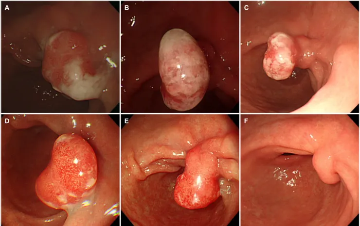
내시경 점막 절제술 2년 후 시행한 상부위장관 내시경에서 반흔 부위에 발적을 동반하는 용종 모양의 병변이 관찰되기 시작하였다(Fig. 2A). 이후 1-2년 간격으로 시행한 상부위장 관 내시경 상에서 용종은 약 2 cm로 크기 변화 없이 유지되
었다(Fig. 2B-D) 그러나 시술 9년 후 시행한 상부위장관 내시 경 소견에서 용종의 목 부위에 결절성 변화가 관찰되었고, 조 직 검사 상 헬리코박터 파일로리 연관 만성 위염(
Helicobacter pylori-
associated chronic active gastritis) 소견이 확인되 었다(Fig. 2E). 이에 표준 3제요법으로 7일간 제균 치료를 시 행하였으며 요소호기 검사(urea breath test)로 성공적인 제 균을 확인하였으며, 제균 1년 후에 시행한 상부위장관 내시경 소견에서 전정부 후벽의 반흔 부위에서 관찰되던 발적을 동반 하는 용종이 거의 소실된 것을 확인할 수 있었다(Fig. 2F).진단: 장기 추적 관찰 중 제균 치료로 소실된 과형성 위 용종 과형성 용종(hyperplastic polyp)은 위 용종 중 흔한 양성 상피성 용종으로1 용종성 소화상피 과증식과 염증성 용종을 의미하며,2 이형성증(dysplasia)이나 위암으로 진행할 위험성 은 0.6-19%로 보고되고 있다.3크기가 작고 증상이 없는 경우 에는 추적 관찰하지만 크기가 1-2 cm 이상인 경우나 출혈, 위 배출 장애 등의 증상이 동반된 경우에는 내시경적 절제술 등의 조치가 필요하다.4
헬리코박터 파일로리(
Helicobacter pylori
)균 감염은 과형 성 용종과 연관이 높다고 알려져 있으며,5일본 및 영국 가이 드라인에서는 과형성 용종 환자에서 헬리코박터균 감염이 있 다면 헬리코박터 제균 치료를 권고하고 있다.3,6 그러나 우리50
강수진, 신철민. 제균치료로 퇴행된 과형성 위 용종The Korean Journal of Gastroenterology
A B
Fig. 1. Endoscopic treatment of gastric adenoma. (A) A 2.5 cm sized slightly depressed lesion with marginal elevation (type 0-IIc+IIa) was observed at the posterior wall of the antrum. (B) An endoscopic mucosal dissection was performed for the lesion. The final pathologic diagnosis was a 2.2×1.3 cm-sized tubular adenoma, low-grade dysplasia. The resection margin was negative for the tumor.
A B C
D E F
Fig. 2. Long-term follow-up of the hyperplastic polyp. Endoscopic surveillance was performed annually or biennially after the endoscopic treatment. Two years after the endoscopic treatment, a 2 cm sized hyperplastic polyp was noted on the endoscopic resection scar (A). There was no change in size 4, 6, and 8 years (B-D, respectively) after the endoscopic treatment. Nine years after the endoscopic treatment, nodularity was observed at the neck of the polyp. Thus, a biopsy was done to rule out malignancy (E). The pathologic diagnosis was Helicobacter pylori (H. pylori)-associated chronic active gastritis. (F) One year after the H. pylori eradication. The hyperemic polyp disappeared, and only its neck portion remained.
Kang SJ and Shin CM. Regression of Hyperplastic Polyp after Helicobacter pylori Eradication
51
Vol. 76 No. 1, July 2020
나라는 아직 과형성 위 용종과 관련된 헬리코박터 제균 치료 가이드라인은 없는 상태이다.
하지만 국내에서도 이에 대한 연구가 보고되었으며, 한 연 구에서는 183명의 헬리코박터 감염을 동반한 과형성 위 용종 환자를 평균 2.2년간 추적 관찰하였고 헬리코박터 제균 치료 를 실시한 군이 제균 치료를 실시하지 않은 군에 비해 용종의 소실이 확연하게 나타났으며(83.7% vs. 34.1%, p=0.001), 성 공적인 제균 치료가 과형성 위 용종의 소실 가능성을 더 높인 다는 것을 보여주었다.7이 외에도 헬리코박터 양성인 과형성 용종 환자에서 헬리코박터 제균 치료가 용종 퇴화의 중요한 예측인자이며,8 과형성 용종에 대한 내시경적 절제 후 헬리코 박터 감염이 과형성 위 용종의 재발과 연관성이 높았다는 연 구 보고가 있으며,9 최근에는 32명의 환자를 대상으로 한 무 작위 임상시험에서도 헬리코박터 제균 치료가 과형성 용종의 퇴화를 유도한다는 결과를 보였다.10
이번 증례는 내시경 점막절제술 후 시술 반흔에 생긴 과형 성 위 용종을 10년간 장기 추적 관찰한 결과를 보여주고 있 다. 드물지 않게 시술 이후에 반흔 부위에 크기가 큰 과형성 위 용종이 발생하여 치료 및 경과 관찰 여부를 결정하는데 난감한 상황이 있을 수 있다. 본 증례에는 1 cm 이상으로 큰 과형성 용종을 장기간 추적하였을 때 양호한 예후를 보이는 것을 확인하였다. 한편 2018년 1월 이후 우리나라 헬리코박 터 제균 치료 적응증이 확대되면서, 과형성 용종에 대해서도 인정비급여로 헬리코박터 제균 치료를 시행해 볼 수 있게 되 었는데, 본 증례에서는 헬리코박터균 감염과 연관된 과형성 위 용종이 제균 치료로 퇴화되는 것을 확인할 수 있었으며, 이에 대한 국내 연구 결과에 대한 문헌고찰과 함께 보고하는 바이다.
REFERENCES
1. Stolte M, Sticht T, Eidt S, Ebert D, Finkenzeller G. Frequency, loca- tion, and age and sex distribution of various types of gastric polyp. Endoscopy 1994;26:659-665.
2. Shaib YH, Rugge M, Graham DY, Genta RM. Management of gas- tric polyps: an endoscopy-based approach. Clin Gastroenterol Hepatol 2013;11:1374-1384.
3. Goddard AF, Badreldin R, Pritchard DM, Walker MM, Warren B;
British Society of Gastroenterology. The management of gastric polyps. Gut 2010;59:1270-1276.
4. Ji F, Wang ZW, Ning JW, Wang QY, Chen JY, Li Y. Effect of drug treat- ment on hyperplastic gastric polyps infected with Helicobacter pylori: a randomized, controlled trial. World J Gastroenterol 2006;12:1770-1773.
5. Nam SY, Park BJ, Ryu KH, Nam JH. Effect of Helicobacter pylori infection and its eradication on the fate of gastric polyps. Eur J Gastroenterol Hepatol 2016;28:449-454.
6. Asaka M, Kato M, Takahashi S, et al. Guidelines for the manage- ment of Helicobacter pylori infection in Japan: 2009 revised edition. Helicobacter 2010;15:1-20.
7. Nam SY, Park BJ, Ryu KH, Nam JH. Effect of Helicobacter pylori eradication on the regression of gastric polyps in National Cancer Screening Program. Korean J Intern Med 2018;33:506-511.
8. Lim SA, Yun JW, Yoon D, et al. Regression of hyperplastic gastric polyp after Helicobacter pylori eradication. Korean J Gastrointest Endosc 2011;42:74-82.
9. Kang KH, Hwang SH, Kim D, et al. The effect of Helicobacter pylori infection on recurrence of gastric hyperplastic polyp after endo- scopic removal. Korean J Gastroenterol 2018;71:213-218.
10. Nam SY, Lee SW, Jeon SW, Kwon YH, Lee HS. Helicobacter pylori eradication regressed gastric hyperplastic polyp: a randomized controlled trial. Dig Dis Sci 2020 Jan 23. [Epub ahead of print]