pISSN: 0378-6471 eISSN: 2092-9374 DOI : 10.3341/jkos.2011.52.5.633
= 증례보고 =
유리체강 내 실리콘기름의 대뇌로의 이동으로 인한 반대편안의 이측 시야결손
이성복1,2⋅신경섭1⋅조영준1,2
충남대학교 의과대학 안과학교실1, 충남대학교 의학연구소2
목적: 유리체강 내 실리콘기름이 대뇌의 외측뇌실 및 시신경교차부로 이동하여 반대편안의 이측 시야결손이 발생한 증례를 경험하였 기에 이를 보고하고자 한다.
증례요약: 56세 남자가 10일 전부터 발생한 좌안의 이측반맹을 주소로 내원하였다. 3개월 전부터 우안 시력저하, 우안통 및 두통이 있었다. 타 병원에서 8년 전 우안 망막박리로 유리체절제술을 시행 받았으며, 수술 직후 재발한 망막박리에 대하여 두 차례 재수술 후 실리콘기름 삽입술을 시행 받은 상태였다. 안저검사상 우안의 녹내장성 시신경유두 변화와, 안압이 54 mmHg로 높은 상태였다.
시야검사상 좌안의 이측반맹이 관찰되었고, 대뇌 병변이 의심되어 뇌자기공명영상 촬영을 시행하였다. 뇌하수체 상부와 시신경교차부 의 오른쪽 위에 우안 유리체강 내의 실리콘기름과 동일한 신호강도를 보이는 물질이 관찰되었고, 추적관찰 중 실리콘기름의 외측뇌실 로의 이동 및 자세변화에 따른 위치 변화가 관찰되었다.
결론: 유리체절제술 후 실리콘기름 삽입술을 시행 받은 환자에서, 유리체강 내 실리콘기름이 시신경교차부로 이동하여 반대편안의 시야결손이 발생하였고, 뇌실로의 이동으로 시야결손이 호전된 증례를 경험하였기에 이를 보고하는 바이다.
<대한안과학회지 2011;52(5):633-638>
■ 접 수 일: 2010년 8월 13일 ■ 심사통과일: 2010년 10월 24일
■ 게재허가일: 2011년 3월 8일
■ 책 임 저 자: 조 영 준
대전시 중구 대사동 640번지 충남대학교병원 안과
Tel: 042-220-7607, Fax: 042-255-3745 E-mail: youngjoon@cnu.ac.kr
실리콘기름은 생체 내에서 분해, 흡수가 이루어지지 않 아 유리체절제술 후 장기간의 망막압박이 필요한 경우 유 리체 대용물로 사용되는 물질이다. 1960년대 초에 Cibis et al1이 처음 사용하였으며, 이후 Scott2와 Zivojnović3에 의해 실리콘기름을 사용한 유리체절제술의 좋은 수술 결과 발표 가 있었다. 현재 실리콘기름을 사용한 안내충전술은 증식당 뇨망막병증, 증식유리체망막병증, 거대열공박리, 안외상에 의한 망막박리 등 다양한 경우에 안전하게 사용되고 있 다.4-10하지만 장기간 실리콘기름의 사용으로 인한 합병증 으로 이차 녹내장, 백내장, 각막내피부전, 실리콘기름의 유 화, 망막전막, 견인망막박리 등이 또한 보고되고 있다.11-23 망막의 유착이 안정적으로 이루어진 이후에는 실리콘기름 을 제거하는 것이 좋지만, 제거 후 망막박리가 재발할 수 있어 어떤 경우는 실리콘기름을 제거하지 못하는 경우도 있다. 따라서 적절한 시기에 선택적으로 시행되어야 한 다.24-28
저자들은 열공 망막박리의 치료로 유리체절제술과 함께
실리콘기름을 삽입한 환자에서, 실리콘기름이 중추신경계 내로 이동하여 시신경교차부위의 압박을 일으켜 반대편안 의 시야결손을 일으킨 것을 뇌자기공명영상 및 뇌전산화단 층촬영을 통해 증명하였다. 이러한 증례는 외국에 몇 몇 보 고된 적이 있으나,29,30국내에서는 아직 보고된 바가 없기에 이를 보고하고자 한다.
증례보고
56세 남자가 10일 전부터 발생한 좌안의 이측반맹의 시 야결손을 주소로 내원하였다. 3개월 전부터 우안 시력저하, 우안통 및 두통이 있었다. 내원 시 나안시력은 우안 광각없 음, 좌안 1.0이었다. 과거력상 타병원에서 8년 전 우안 망막 박리 진단받고, 유리체절제술 시행받았으며, 이후 재발한 망막박리에 대하여 두 차례 더 유리체절제술 및 실리콘기 름 삽입술을 시행받았다. 이후 환자가 자의적으로 병원에 내원하지 않아 정기적 추적관찰은 되지 않았다고 한다. 환 자의 기억으로 수술 후 시력은 안전수지였고, 내원 3개월 전부터 광각없음의 시력으로 떨어졌다고 한다. 고혈압과 당 뇨로 약물치료 중이었으나 좌안의 안저검사상 특이 소견은 없었고, 특별한 안외상이나 이외의 다른 안과적 수술 기왕 력은 없었다. 전안부 검사상 우안에 홍채절제술이 시행되어 있었고, 무수정체안 상태 외에는 특별한 이상소견은 없었
Figure 1. Color fundus photograph shows elevated retina and
the cup-disc ratio is 1.0 in the right eye. There is the reflex on the retinal surface by the silicone oil. There were no abnormal findings in the left eye.A B C
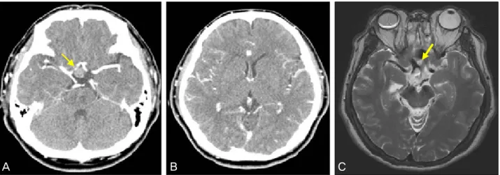
Figure 3. (A) Computed tomography (CT) of the head shows 1.4 cm sized no enhanced mass in the suprasellar area (arrow). (B)
There was no mass in the ventricle. (C) A axial T2-weighted magnetic resonance imaging (MRI) scan of the head shows hetero- genous signal intensity in the silicone filled right eye as well as in the right side of the optic chiasm (arrow).A
B
Figure 2. (A) Left temporal hemianopia of the left eye. Visual
acuity was 1.0. (B) 1 month later, after migration of silicone oil into lateral ventricle, the visual field defect improved.다. 내원시 안압은 우안 54 mmHg, 좌안 13 mmHg였고, 안 저소견상 우안은 실리콘기름이 들어있었으며, 하부에 일부 망막이 박리된 소견을 보였고, 시신경유두비가 1.0에 가깝게 증가되어 있었다. 좌안은 시신경유두비 0.4이고, 전안부 및 안저검사상 특별한 이상소견은 보이지 않았다(Fig. 1).
시야검사에서 좌안의 이측반맹 소견을 보였고(Fig. 2), 만성 두통이 동반되어 있어 중추신경계 병변에 의한 질환 을 배제하기 위해 뇌전산화단층촬영을 시행하였다. 안장위 부위에 1.4 cm의 조영증강되지 않는 종괴가 발견되었으며, 뇌자기공명영상에서 T1, T2 강조영상에서 모두 우안 유리 체강내 실리콘기름과 같은 신호강도를 가지는 이질성의 1.4 cm의 종괴가 보였다. 이는 안장위 부분, 시신경교차부 의 오른쪽 위에 위치하고 있어, 시신경 압박에 의한 시야장 애를 유발한 것으로 생각하였다(Fig. 3). 내원 직후 Mannitol 정맥주사 및 Acetazolamide (Diamox®) 복용, Dorzolamide- Timolol (Cosopt®) 점안을 통하여 우안 안압은 12 mmHg 로 조절되었다.
4일 뒤 뇌전산화단층촬영 및 뇌자기공명영상 추적검사를 시행하였으며, 앞에서 보였던 병변외에 좌측의 외측뇌실의 앞부분에 약 1 cm 크기의 새로운 병변이 발견되었고, 이는 안구내의 실리콘기름과 같은 밀도 및 신호강도를 보였다.
저자들은 Eller et al31이 보고한 증례에서처럼 뇌전산화단 층촬영 검사를 앙와위 자세와 함께, 엎드린 자세에서 촬영 을 시행하였으며, 실리콘기름이 외측 뇌실 내에서 부력에 의해 위쪽인 뇌실의 뒤부분으로 이동한 것을 확인할 수 있 었다(Fig. 4).
A
B C
Figure 4. (A) Computed tomography (CT) scan of the head, about 1 cm sized hyperdense mass is newly seen in the left lateral
ventricle. The material filling the right eyeball, suprasellar area and the new lesion in the frontal horn of the left lateral ventricle showed identical CT density (arrow). (B) T2-weighted magnetic resonance imaging (MRI) scan shows a new lesion (arrow). (C) Computed tomography (CT) scan of the head, axial section, with the patient in a prone position. Silicone oil shifts to the posterior portion of the lateral ventricle (arrow).고 찰
실리콘기름은 고분자 물질인 Oligodimethylsiloxane 중 합체로 사슬 길이에 따라 분자량 차이가 나며, 1,000- 12,500 CST까지 다양한 점성을 가지고 있고,32 비중이 물 보다 작아 부력과, 높은 표면장력으로 안내 충전물로 사용 시 효과가 뛰어나다. 망막수축에 대해 높은 저항력을 가지 고, 섬유혈관증식으로 인한 망막의 재박리를 기계적으로 제 한하는 효과가 있어 증식당뇨망막병증, 재출혈의 위험성이 큰 경우, 증식유리체망막병증, 거대열공망막박리, 안구위축 의 방지를 위해 영구적인 안내충전이 필요한 경우, 환자의 상태가 한 자세를 유지하기 힘든 경우,4,5 망막박리를 동반 한 안 외상 등 심한 망막박리에 효과적인 수술적 치료법으 로 널리 사용되고 있다.7-10하지만 단점으로 추후 안내에서 제거하기 위한 수술이 또 한번 필요하며, 이외에도 백내장, 안압 상승 및 녹내장,각막병증,굴절력의 원시화, 실리콘기
름의 유화현상(emulsification), 재발성 망막박리, 망막전 막, 저안압, 주변부 안와조직으로의 이동 등이 실리콘기름 안내 충전과 관련된 합병증으로 보고되고 있다.11-23 이런 다양한 합병증 중에 실리콘기름의 대뇌로의 이동에 대해서 는 해외에서 드물게 몇몇 발표가 보고되고 있다.29,30
실리콘기름이 대뇌로 이동하는 기전에 대해서는 잘 알려 진 바가 없다. 본 환자는 특별한 외상력은 없었으며, 요추천 자를 포함한 뇌척수액 안으로 이물질이 들어갈 만한 다른 처치는 시행하지 않았다. 해외의 증례발표및 문헌을 보면, 시신경유두소와 등의 시신경 기형에서 유리체강 내와 지주 막하 공간과 연결통로가 있을 것이라는 가설이 있고,33 시 신경 내로 실리콘기름이 직접 침투하여 뇌실 내로 들어갔 을 것이라는 가설이 있다.34 그 기전으로는 지속적인 안압 상승에 의해 체판뒤부분의 시신경이 허혈성 괴사에 빠지게 되고 해면성 변성이 온다. 이러한 시신경의 낭성공간에 유 리체가 채워지게 되고, 작은 기름방울이 연뇌막을 통과하여
지주막하공간으로 들어가 뇌척수액으로 유입되어 뇌실 내 로 들어갈 수 있다는 것이다. 또한 이는 조직학적 검사로도 증명이 되었는데, 실리콘기름이 충전된 녹내장성 시신경 손 상이 있는 안구의 체판뒤부분에서 실리콘이 채워진 구멍이 발견되었다.35
본 증례의 환자는 실리콘기름 삽입술을 시행하고 자의로 정기적 추적관찰 없이 그냥 지냈으며, 상당기간 동안 안압 이 높은 상태로 지냈을 것으로 추측된다. 증거로 녹내장성 시신경유두변화와, 시신경의 손상을 안저검사 및 빛간섭단 층촬영에서 확인할 수 있었다. 환자는 현재 Acetazolamide (Diamox®)복용, Dorzolamide-Timolol (Cosopt®)점안을 통하여 안압을 정상 범위 내로 조절하고 있다.
높은 안압으로 인한 시신경의 위축은 유리체강 내 실리 콘기름의 중추신경계 내로의 이동을 유발할 수 있다. 환자 의 경우 실리콘기름이 시신경을 따라 이동하던 중 안장위 부분, 시신경교차부 오른쪽 위에 모이게 되어 시신경 압박 증상을 일으켜 좌안 이측반맹의 시야결손을 유발하였다. 처 음 촬영한 뇌전산화단층촬영 및 뇌자기공명영상에서는 뇌 실에는 특이소견이 없었으나, 추적관찰 시 시행한 영상검사 에서는 외측 뇌실에 실리콘기름과 동일한 밀도 및 신호강 도를 보이는 새로운 병변이 있었고, 이는 체위변화에 따라 이동하는 경향을 보였다. 이후 1개월 뒤 추적관찰을 시행하 였을 때에는 시야검사가 많이 호전된 소견을 보였는데(Fig.
2), 환자가 경제적 사정으로 추적 뇌영상 검사를 거부하여 정확한 이유는 알 수 없지만, 압박증상을 일으키던 시신경 교차부위의 실리콘기름이 외측뇌실 및 다른 곳으로 이동한 결과로 추측해 볼 수 있겠다. 하지만 이미 들어간 실리콘기 름이 어느 위치로 들어가는지, 중추신경계로 추가로 이동하 는지 여부에 대하여 관찰하기 위해 정기적인 뇌영상검사의 추적관찰이 필요하다.
Eckle et al29이 보고한 증례에서는 신경외과적 수술인 전두엽 하방 개두술을 통하여 실리콘기름을 직접 흡입 및 세척술을 시행하였고 좋은 결과를 발표하였다. 하지만 침습 적인 술기이기 때문에 이에 수반되는 합병증 역시 고려해 야 한다. 본 증례에서는 현재 망막박리가 동반되어 있어 실 리콘기름을 제거하기 어려운 상태인 점과, 실리콘기름의 이 동으로 수반되는 특별한 중추신경계의 합병증이 관찰되지 않은 점, 실리콘기름의 이동으로 시야결손이 호전된 점을 고려할 때 침습적 술기보다는 안압조절을 통한 추가적인 실리콘기름의 이동을 막는 것이 적절한 치료방침이라 생각 된다.
아직 실리콘기름의 이동의 정확한 과정 및 기전에 대해 서 정확히 밝혀진 바가 없지만, 높은 안압이 녹내장성 시신 경유두변화를 일으키고, 시신경에 손상을 일으키며 이것이
유리체강 내의 실리콘기름을 두개 내의 시신경을 따라 대 뇌의 외측뇌실로, 즉 중추신경계 안으로 이동시킬 수 있는 요인이라는 것을 알았다. 따라서 성공적으로 망막이 유착된 경우 실리콘기름 제거술의 시행여부나, 제거의 적절한 시기 에 대해서 고려해보아야 한다. 만약 실리콘을 오래 유지해 야 하거나 제거할 수 없는 경우에는 환자의 자각증상이 없 어도, 시력이 나쁘거나 광각이 없는 실명한 눈이라도 철저 한 안압 조절이 중요하다는 것을 본 증례를 통해 알 수 있 었다.
참고문헌
1) Cibis PA, Becker B, Okun E, Canaan S. The use of liquid silicone in retinal detachment surgery. Arch Ophthalmol 1962;68:590-9.
2) Scott JD. The treatment of massive vitreous retraction by the sepa- ration of pre-retinal membranes using liquid silicone. Mod Probl Ophthalmol 1975;15:185-90.
3) Zivojnović R, Mertens DA, Baarsma GS. Fluid silicon in detach- ment for surgery (author's transl). Klin Monbl Augenheilkd 1981;179:17-22.
4) Bodanowitz S, Kir N, Hesse L. Silicone oil for recurrent vitreous hemorrhage in previously vitrectomized diabetic eyes. Ophthalmologica 1997;211:219-22.
5) Heimann K, Dahl B, Dimopoulos S, Lemmen KD. Pars plana vi- trectomy and silicone oil injection in proliferative diabetic retinopathy. Graefes Arch Clin Exp Ophthalmol 1989;227:152-6.
6) The Silicone Study Group. Vitrectomy with silicone oil or sulfur hexafluoride gas in eyes with severe proliferative vitreoretinop- athy: results of a randomized clinical trial. Silicone Study Report 1.
Arch Ophthalmol 1992;110:770-9.
7) The Silicone Study Group. Vitrectomy with silicone oil or per- fluoropropane gas in eyes with severe proliferative vitreoretinop- athy: results of a randomized clinical trial. Silicone Study Report 2.
Arch Ophthalmol 1992;110:780-92.
8) Yeo JH, Glaser BM, Michels RG. Silicone oil in the treatment of complicated retinal detachments. Ophthalmology 1987;94:1109- 13.
9) Seong MC, Chung HW, Lee SY, et al. The clinical results of sili- cone oil tamponade in pars plana vitrectomy for various vitreor- etinal diseases. J Korean Ophthalmol Soc 2007;48:1057-66.
10) Gallemore RP, McCune II BW. Silicone oil in vitreoretinal surgery.
In: Ryan SJ, ed. Retina, 4th ed. St. Louis: Elsevier Mosby, 2006; v.
3. chap 130.
11) Gonvers M. Temporary silicone oil tamponade in the management of retinal detachment with proliferative vitreoretinopathy. Am J Ophthalmol 1985;100:239-45.
12) Nguyen QH, Lloyd MA, Heuer DK, et al. Incidence and manage- ment of glaucoma after intravitreal silicone oil injection for com- plicated retinal detachments. Ophthalmology 1992;99:1520-6.
13) Beekhuis WH, van Rij G, Zivojnović R. Silicone oil keratopathy:
indications for keratoplasty. Br J Ophthalmol 1985;69:247-53.
14) Abrams GW, Azen SP, Barr CC, et al. The incidence of corneal ab- normalities in the Silicone Study. Silicone Study Report 7. Arch Ophthalmol 1995;113:764-9.
15) Valone J Jr, McCarthy M. Emulsified anterior chamber silicone oil and glaucoma. Ophthalmology 1994;101:1908-12.
16) Yoon TJ, Oum BS. Factors for epiretinal membrane formation after retinal detachment surgery with silicone oil tamponade. J Korean Ophthalmol Soc 2004;45:1681-8.
17) Gonvers M, Andenmatten R. Temporary silicone oil tamponade and intraocular pressure: An 11-year retrospective study. Eur J Ophthalmol 1996;6:74-80.
18) Federman JL, Schubert HD. Complications associated with the use of silicone oil in 150 eyes after retina-vitreous surgery.
Ophthalmology 1988;95:870-6.
19) Riedel KG, Gabel VP, Neubauer L, et al. Intravitreal silicone oil in- jection: complications and treatment of 415 consecutive patients.
Graefes Arch Clin Exp Ophthalmol 1990;228:19-23.
20) Oh TS, Kim SY. Complications associated with intravitreal sili- cone oil injection. J Korean Ophthalmol Soc 1993;34:1012-22.
21) Stefánsson E, Anderson MM Jr, Landers MB 3rd, et al. Refractive changes from use of silicone oil in vitreous surgery. Retina 1988;8:20-3.
22) Barr CC, Lai MY, Lean JS, et al. Postoperative intraocular pressure abnormalities in the Silicone Study. Silicone Study Report 4.
Ophthalmology 1993;100:1629-35.
23) Quintyn JC, Genevois O, Ranty ML, Retout A. Silicone oil migra- tion in the eyelid after vitrectomy for retinal detachment. Am J Ophthalmol 2003;136:540-2.
24) Scholda C, Egger S, Lakits A, Haddad R. Silicone oil removal: re- sults, risks and complications. Acta Ophthalmol Scand 1997;75:
695-9.
25) Jonas JB, Budde WM, Knorr HL. Timing of retinal redetachment
after removal of intraocular silicone oil tamponade. Am J Ophthalmol 1999;128:628-31.
26) Kampik A, Höing C, Heidenkummer HP. Problems and timing in the removal of silicone oil. Retina 1992;12:S11-6.
27) Halberstadt M, Domig D, Kodjikian L, et al. PVR recurrence and the timing of silicon oil removal. Klin Monbl Augenheilkd 2006;223:361-6.
28) Yoon JS, Lee SY, Lee SC, Kwon OW. Clinical outcomes after sili- cone oil removal. J Korean Ophthalmol Soc 2003;44:642-8.
29) Eckle D, Kampik A, Hintschich C, et al. Visual field defect in asso- ciation with chiasmal migration of intraocular silicone oil. Br J Ophthalmol 2005;89:918-20.
30) Yu JT, Apte RS. A case of intravitreal silicone oil migration to the central nervous system. Retina 2005;25:791-3.
31) Eller AW, Friberg TR, Mah F. Migration of silicone oil into the brain: a complication of intraocular silicone oil for retinal tamponade. Am J Ophthalmol 2000;129:685-8.
32) Parel JM, Milne P, Gautier S, et al. Silicone oils: Physicochemical properties. In: Ryan SJ, ed. Retina, 4th ed. St. Louis: Elsevier Mosby, 2006: v. 3. chap. 129.
33) Friberg TR, McLellan TG. Vitreous pulsations, relative hypotony, and retrobulbar cyst associated with a congenital optic pit. Am J Ophthalmol 1992;114:767-9.
34) Shields CL, Eagle RC Jr. Pseudo-Schnabel's cavernous degener- ation of the optic nerve secondary to intraocular silicone oil. Arch Ophthalmol 1989;107:714-7.
35) Manschot WA. Intravitreal silicone injection. Adv Ophthalmol 1978;36:197-207.
=ABSTRACT=
Temporal Hemianopsia of Healthy Eye in a Patient with Contralateral Silicone Oil Filled Eye
Sung Bok Lee, MD1,2, Kyung Sup Shin, MD1, Young Joon Jo, MD1,2
Department of Ophthalmology, Chungnam National University College of Medicine1, Daejeon, Korea Institute for Medical Sciences, Chungnam National University Research2, Daejeon, Korea
Purpose: To report a case of temporal hemianopsia of a healthy eye occurring in the contralateral silicone oil-filled eye due to migration of silicone oil into the optic chiasm and lateral ventricle.
Case summary: A 56-year-old man visited our clinic with temporal hemianopsia for 10 days in the left eye. Three months before, the patient had presented with decreased vision and ocular pain in the right eye as well as a headache. The patient underwent vitrectomy at another hospital for the management of retinal detachment occurring in the right eye 8 years earlier. In addition, for recurred retinal detachment, reoperations were performed twice with silicone oil injection.
Funduscopy revealed findings such as glaucomatous optic disc and an intraocular pressure of 54 mmHg in the right eye.
On visual field examination, the temporal hemianopsia was detected in the left eye. Under the suspicion of cerebral le- sions, a magnetic resonance imaging (MRI) examination was performed. On the right side of the optic chiasm and the su- prasellar region, materials were present whose signal intensity was identical to silicone oil in the right vitreal cavity. During a follow-up, the migration of silicone oil into the lateral ventricle and the alteration of its location with the positional change were observed.
Conclusions: In a patient who received silicone oil injection following vitrectomy, the silicone oil migrated to the optic chiasm and induced the occurrence of visual field defect in the contralateral eye. The visual field defect improved because of the migration into the lateral ventricle.
J Korean Ophthalmol Soc 2011;52(5):633-638
Key Words: Glaucoma, Migration, Silicone oil, Visual field defect
Address reprint requests to Young Joon Jo, MD
Department of Ophthalmology, Chungnam National University Hospital
#640 Daesa-dong, Jung-gu, Daejeon 301-721, Korea
Tel: 82-42-220-7607, Fax: 82-42-255-3745, E-mail: youngjoon@cnu.ac.kr