http://dx.doi.org/10.12790/jkssh.2014.19.2.57
THE HAND
서론
척 골 감 입 증 후 군 (ulnar impaction syndrome)은 Friedman과 Palmer1은 수근부 척측에 과도한 힘이 가해질 때, 척수근관절의 통증, 부종, 관절운동 제한 등의 증상이 발
생하는 퇴행 상태라고 정의하였다. 척골 단축술은 1950년 Milch2가 원위요골 부정 유합의 치료 방법으로 최초로 제안하 였고, 1985년 Darrow 등3이 금속판과 나사못을 이용한 척골 단축술을 처음으로 보고하였다. 1993년 Chun과 Palmer4가 척골감입증후군에서 척골 단축술의 치료 결과를 보고한 이래
Factors Affecting the Occurrence of Distal Radioulnar Joint Arthritis after Ulnar Shortening Osteotomy
Chol-Jin Kim
1, Ho-Jin Gil
2, Yang-Guk Chung
2,
Seung-Han Shin
2,
Dong-Hyun Kim
2, Jun-Soo Park
2, Hyun-Chul Choi
21Department of Orthopedic Surgery, The Armed Forces Dae Jeon Hospital, Daejeon, Korea
2Department of Orthopedic Surgery, Seoul St.
Mary’s Hospital, The Catholic University of Korea College of Medicine, Seoul, Korea
Received:June 13, 2014 Revised:June 15 , 2014 Accepted:June 16, 2014
Correspondence to:Yang-Guk Chung Department of Orthopedic Surgery, Seoul St.
Mary’ Hospital, The Catholic University of Korea, 222 Banpodaero, Seocho-gu, Seoul 137-701, Korea
TEL:+82-2-2258-2837 FAX:+82-2-535-9834
E-mail:ygchung@catholic.ac.kr
*This research was supported by the 2013 Seoul St.
Mary’s Hospital Clinical Medicine Research Program through the Catholic University of Korea.
Purpose: Ulnar shortening osteotmy is a common operation for the treatment of ulnar impaction syndrome. The purpose of this study was to evaluate factors that may affect the occurrence of distal radioulnar joint (DRUJ) arthritis after ulnar shortening osteotomy.
Methods:From September 2005 to August 2012, we performed 81 ulnar short- ening osteotomies for ulnar impaction syndrome, and evaluated occurrence or deterioration of DRUJ arthritis in 58 patients with a minimum follow-up of 1 year. We analyzed potential factors that may affect the occurrence of DRUJ arthritis, such as, age, sex, hand dominance, pre- and postoperative ulnar vari- ance, preexisting DRUJ arthritis, types of radial sigmoid notch, amount of ulnar shortening, and follow up period.
Results:DRUJ arthritis occurred or deteriorated in 32 out of the 58 patients.
Regression analysis indicated a significant correlation between the type of radi- al sigmoid notch (type 1) and DRUJ arthritis. Other factors were not found to be correlated with occurrence or deterioration of DRUJ arthritis.
Conclusion:This study suggests that patients with type 1 radial sigmoid notch (ulnar inclination of more than 10 degrees) are more likely to develop DRUJ arthritis after ulnar shortening osteotomy.
Keywords:Ulnar impaction syndrome, Ulnar shortening, Distal radioulnar joint arthritis, Pain
This is an Open Access article distributed under the terms of the Creative Commons Attribution Non-Commercial License (http://creativecommons.org/ licenses/by- nc/3.0/) which permits unrestricted noncommercial use, distribution, and reproduction in any medium, provided the original work is properly cited.
로 척골 단축술은 척수근관절의 압력을 줄일 수 있는 술식으 로 척골감입증후군의 표준적인 치료 방법으로 받아들여진다.
그러나 양성 척골변이가 큰 경우나 척골 단축술의 결과로 음 성 척골변이가 지나치게 증가한 경우 원위 요척 관절의 부조 화가 발생할 수 있으며 원위 요척 관절 및 수근관절의 퇴행성 변화가 발생될 수 있다5. 저자들은 척골감입증후군에 대한 척 골 단축술 후 원위 요척 관절 관절염의 발생에 영향을 미치는 요인들에 대하여 알아보고자 하였다.
대상 및 방법
2005년 9월 이후부터 2012년 8월까지 척골감입증후군에 대하여 척골 단축술을 시행 받은 81예 중 1년 이상 추시된 58 예를 대상으로 하였다. 척골감입증후군의 진단은 이학적 검사 와 방사선 검사를 바탕으로 하였다. 이학적 검사는 수근관절 을 척측 굴곡 후 회내전 또는 회외전을 시행할 때 통증을 유발 하는 경우를 양성으로 판단하였다. 방사선 검사는 견관절 90�
외전, 주관절 90�굴곡 상태에서 수근관절 전후면, 측면을 촬 영하였으며 Kreder method6를 이용하여 척골의 변위를 측정 하였다. 척골 단축술은 척골 경상돌기로부터 약 5 cm 근위부 의 척골에 사선으로 절골술을 시행 후 척골 변위가 중립에서 음성 1 mm가 되도록 수술 전에 계측한 길이만큼 단축술을 시 행하였으며 척골의 내고정은 3.5 mm 피질골 나사 및 재건 금 속판을 사용하였다. 내고정 장치의 제거는 수술 후 1년 이상 경과한 시점에 단축술 시행부위의 골유합과 재형성 변화가 충
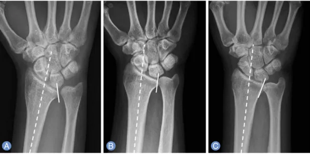
분히 진행된 경우에 시행하였다. 환자는 남자가 14명, 여자가 44명이었으며 평균 나이는 47.7세(범위, 21-80세)였다. 평균 추시 기간은 24.1개월(범위, 12-97개월)이었으며 추시 기간 중 2-3개월 간격으로 단순방사선 검사를 시행하였다. 진단 당 시의 나이(50세 미만/이상), 성별, 우세손 여부, 수술 전 척골 변이의 정도(4 mm 미만/이상), 수술 전 원위 요척 관절의 관 절염 유무, 요골 S 절흔의 형태7(I형, 요골의 장축을 기준으로 S 절흔의 기울기가 10�이상의 척측 경사; II형, 10�미만의 요 측 또는 척측 경사; III형, 10�이상의 요측 경사) (Fig. 1), 척골 단축의 정도(4.6 mm 미만/이상), 수술 후 척골 변이의 정도(2 mm 미만/이상), 추시 기간(24개월 미만/이상)이 원위 요척 관 절의 관절염의 진행이나 발생에 영향을 미치는지를 분석하였 다. 원위 요척 관절의 관절염의 진행이나 발생에 대하여는 단 순방사선 검사 소견을 근거로 평가하였으며, 평가기준은 하지 관절염 평가기준인 Kellgren과 Lawrence8의 관절염 분류법 을 상지에 적용토록 수정하여 사용하였다(Table 1). 통계적 유 의성 평가를 위해 단순 회귀 분석, 다중 회귀 분석 및 로지스 틱 회귀분석을 시행하였다. 단순 및 다중, 로지스틱 회귀분석 의 독립변수는 진단 당시의 나이, 성별, 우세손 여부, 수술 전 척골변이의 정도, 수술 전 원위 요척 관절의 관절염 유무, 요 골 S 절흔의 형태, 척골 단축의 정도, 수술 후 척골변이의 정 도, 추시 기간이었고, 결과 변수는 관절염의 진행 정도였다.
임상적인 평가를 위해 수술 전과 수술 후 최종 추시 시 visual analogue scale (VAS) score와 pain grade (0, no pain; 1, pain on excertion; 2, pain on rest)를 측정하였다.
J Korean Soc Surg Hand Vol. 19, No. 2, June 2014
J
OURNAL OF THEK
OREANS
OCIETY FORS
URGERY OF THEH
ANDFig. 1.
Types of the sigmoid notchs of distal radioulnar joints.
(A)Type I is a hemispherical type that sigmoid notch is tilt-
ed to the ulnar side, more than 10 degrees.
(B)Type II is a cylinderical type. Sigmoid notch is tilted less than 10 degrees
to the radial or ulnar side.
(C)Type III is a conical type that sigmoid notch tilted to radial side more than 10 degrees.
수술 전 후 통증 정도의 차이에 대한 통계적 유의성 평가를 위 해 VAS score에 대하여는 대응 표본 T검정을 시행하였으며, pain grade에 대하여는 Wilcoxon signed rank test 분석을 시행하였다(SPSS ver. 19, SPSS Inc., Chicago, IL, USA).
관절염이 진행되지 않은 군과 관절염이 진행되거나 수술 후 새롭게 발생한 군의 수술 전 및 최종 추시 시 VAS score와 pain grade의 차이에 대한 통계적 유의성 평가를 위해 독립
표본 T검정 분석을 시행하였다.
결과
진단 당시의 나이, 성별, 우세손 여부, 수술 전 척골변이의 정도, 수술 전 원위 요척 관절의 관절염 유무, 요골 S 절흔의 형태, 척골 단축의 정도, 수술 후 척골 변이의 정도, 추시 기간 등 변수의 분포는 다음의 표와 같다(Table 2).
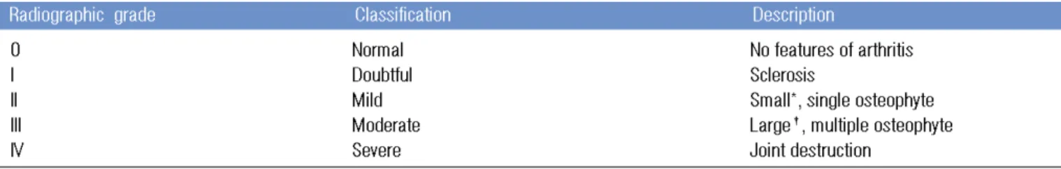
총 58예 중 32예(55%)에서 수술 전 원위 요척관절의 관절염 이 진행되거나 수술 후 새롭게 발생하였다. 원위 요척 관절염 에 영향을 미칠 수 있는 변수에 대한 단순 회귀 분석에서는 요 골 S 절흔의 형태(p=0.005)가 원위 요척관절 관절염의 진행이 나 발생과 유의한 상관성을 가지는 것으로 나타났으며, 진단 당시의 나이, 성별, 우세손 여부, 수술 전 척골변이 정도, 수술 전 원위 요척 관절의 관절염 유무, 척골 단축 정도, 수술 후 척 골변이 정도, 추시 기간 등의 다른 인자들은 유의한 상관관계 를 보이지 않았다(Table 3). 다중 회귀 분석에서는 요골 S 절 흔의 형태가 유의한 상관성을 보였으며(p=0.003), 로지스틱 회귀 분석에서도 요골 S 절흔의 형태만이 유의한 상관성을 보 였다(p=0.007). 척측 경사를 보이는 제I형 요골 S 절흔을 가지 는 환자 17명 중 15명(88%)과 10�미만의 요측 또는 척측 경사 를 보이는 제II형에서는 20명 중 11명(55%), 요측 경사를 보이 는 제III형에서는 21명 중 9명(42%)에서 관절염의 진행이나 Table 1.
Radiographic criteria for assessment of distal radioulnar joint arthritis
0 Normal No features of arthritis
I Doubtful Sclerosis
II Mild Small*, single osteophyte
III Moderate Large
�, multiple osteophyte
IV Severe Joint destruction
*Small, less than 2 mm in size;
�Large, greater than 2 mm in size.
Radiographic grade Classification Description
Table 2.
Independent variable of distal radioulnar joint arthritis Age (yr)
<50 29
≥
50 29
Sex Male 14
Female 44
Dominant hand
Dominant 31
Non-dominant 27
Preoperative ulnar variance
<4 mm 10
≥
4 mm 48
Preoperative arthritis
0 36
I 7
II 13
III 2
IV 0
Types of sigmoid notch
I 17
II 20
III 21
Ulnar shortening length
<4.6 mm 14
≥
4.6 mm 44
Postoperative ulnar variance
<2 mm 54
≥
2 mm 4
Follow-up term
<24 mo 32
≥
24 mo 26
Independent variable Number of cases
Table 3.Results of simple linear regression analysis for factors affecting on distal radioulnar joint arthritis
Age 0.186 0.176
Sex 0.339 0.128
Dominant hand 0.877 0.021
Preoperative ulnar variance 0.168 0.183
Preoperative arthritis 0.168 0.183
Types of sigmoid notch 0.005 0.368
Ulnar shortening length 0.339 0.128
Postoperative ulnar variance 0.139 0.197
Follow-up term 0.488 0.071
Independent variable p -value Correlation coefficient
J Korean Soc Surg Hand Vol. 19, No. 2, June 2014
J
OURNAL OF THEK
OREANS
OCIETY FORS
URGERY OF THEH
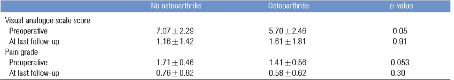
AND발생을 보였다. 수술 전 VAS score는 6.29±2.38에서 최종 외래 추시 시 1.63±1.65으로, pain grade 또한 1.53±0.54에 서 0.65±0.62로 호전되어 통계학적으로 유의한 차이를 보였 다(Table 4). 관절염이 진행되지 않은 군과 관절염이 진행되거 나 수술 후 새롭게 발생한 군 간의 수술 전, 최종 추시 시 VAS score와 pain grade는 통계학적으로 유의한 차이를 보이지 않았다(Table 5).
1. 증례
11)) 증증례례 11
21세 여자 환자로 내원 1년 전부터 시작된 우측 수근관절 통 증을 주소로 내원하였다. 이학적 검사에서 척수근관절의 압통 및 회내전, 회외전 시 통증을 호소하였다. 방사선 검사상 7 mm 척골 양성변이 소견 및 제II형의 요골 S 절흔 소견을 보였 다(Fig. 2A). 척골감입증후군 진단하에 척골 단축술을 시행하 Table 4.
The outcomes of VAS score and pain grade
VAS score 6.29
±2.38 1.63
±1.65 <0.05
Pain grade* 1.53
±0.54 0.65
±0.62 <0.05
Values are presented as mean
±standard deviation.
VAS, visual analogue scale.
*Pain grade: 0, no pain; 1, pain on excertion; 2, pain on rest.
Preoperative At last follow-up p -value
Table 5.
Comparision of VAS score and pain grade with/without DRUJ arthritis Visual analogue scale score
Preoperative 7.07
±2.29 5.70
±2.46 0.05
At last follow-up 1.16
±1.42 1.61
±1.81 0.91
Pain grade
Preoperative 1.71
±0.46 1.41
±0.56 0.053
At last follow-up 0.76
±0.62 0.58
±0.62 0.300
Values are presented as mean
±standard deviation.
VAS, visual analogue scale; DRUJ, distal radioulnar joint.
No osteoarthritis Osteoarthritis p -value
Fig. 2.
The patient with a complaining of right wrist pain for the past 1 year. (
A)The radiograph showed 7 mm ulnar pos-
itive variance and sigmoid notch tilt to ulnar side (type II sigmoid notch).
(B)Ulnar impaction syndrome was diagnosed
and a ulnar shortening surgery was performed. The postoperative radiograph showed neutral ulnar variance.
(C)Postoperative 14 months, there was no evidence of arthritic change.
(D)Postoperative 27 months, there was little pro-
gression of arthritic change at distal radioulnar joint with sclerosis (white arrow). Visual analogue scale score was
improved from 3 preoperatively to 0 at the last follow up and pain grade from 2 to 0 at the last follow-up.
였으며 수술 후 척골변이는 중립이었다(Fig. 2B). 수술 후 12 개월에 시행한 방사선 검사상 관절염 소견 보이지 않았으나 (Fig. 2C) 수술 후 27개월에 시행한 방사선 검사상 원위 요척 관절 간격 감소 및 경화 소견을 보였다(Fig. 2D). VAS score 는 수술 전 3에서 수술 후 0으로, pain grade는 수술 전 1에서 수술 후 0으로 호전되었다.
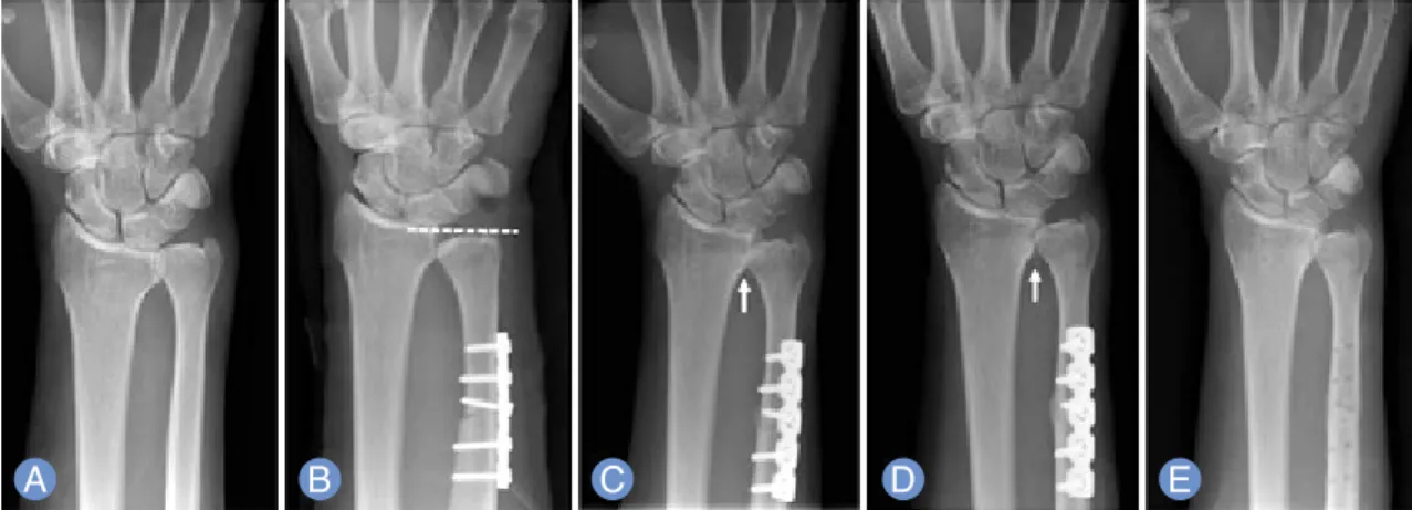
2 2)) 증증례례 22
62세 남자 환자로 내원 5개월 전부터 시작된 좌측 수근관절 통증을 주소로 내원하였다. 이학적 검사에서 척수근관절의 압 통 및 회내전, 회외전 시 통증을 호소하였다. 방사선 검사상 3 mm 척골 양성변이 소견 및 제I형의 요골 S 절흔 소견을 보였 다(Fig. 3A). 척골감입증후군 진단하에 척골 단숙출 시행하였 으며 수술 후 2 mm 음성 척골변이 소견을 보였다(Fig. 3B).
수술 후 5개월에 시행한 방사선 검사상 작은 크기의 골극 형성 소견 보였으며(Fig. 3C) 수술 후 12개월에 시행한 방사선 검사 상 관절 간격 감소 및 골극 형성 진행 소견을 보여(Fig. 3D) 골 극 절제술을 시행하였다(Fig. 3E). VAS score는 수술 전 8에 서 골극 절제술 후 18개월의 최종추시상 2로, pain grade는 수술 전 2에서 최종추시상 1로 호전되었다.
고찰
척골감입증후군은 수근관절의 척측 굴곡 상태에서 회외전 또는 회내전을 할 때 통증이 발생하는 것이 특징이며 척수근
관절의 압력 증가로 인하여 발생하는 것으로 알려져 있다. 척 골감입증후군의 진단은 이학적 검사, 방사선 검사, 자기공명 영상 검사 등의 방법을 통해서 이루어진다. 수근 관절의 요골 의 길이보다 척골의 길이가 상대적으로 긴 양성 척골 변위 (positive ulnar variance)에서 호발하며1,5, 특히 동양인에게 서 양성 척골 변위가 많다9.
원위 요척관절은 척골 두와 원위 요골의 S 절흔이 이루는 관 절이며 방사선 영상에서 요골 S 절흔의 경사각(sigmoid notch inclination)과 척골 석 경사각(ulnar seat inclination) 을 확인할 수 있다10.
저자들의 연구에서는 척골감입증후군에 대해서 척골 단축 술을 시행한 환자 58예 중 32예에서 수술 전 원위 요척관절의 관절염이 진행되거나 수술 후 새롭게 발생하였다. Table 1에 서 제시한 기준대로 관절염의 발생과 진행을 판단하였으며 단 순 방사선 검사상 작은 변화에도 엄격한 기준을 적용하여 타 연구11에 비교하여 관절염의 발생 빈도가 높게 평가된 원인으 로 판단된다. 제I형 요골 S 절흔을 보이는 환자의 88%, 제II형 요골 S 절흔을 보이는 환자의 55%, 제III형 요골 S 절흔을 보 이는 환자의 42%에서 관절염의 진행이나 새롭게 발생한 소견 을 보였다. 단순 회귀 분석에서 요골 S 절흔의 형태에 따라 수 술 후 원위 요척관절 관절염의 발생률에 유의한 차이가 있는 것으로 나타났으며, 다중 회귀 분석에서도 요골 S 절흔의 형 태가 척골 단축술 후 원위 요척관절 관절염의 발생과 진행에 영향을 미치는 것으로 나타났다. 로지스틱 회귀 분석에서도 요골 S 절흔의 형태가 상관관계를 보여(p=0.007) 수술 후 관
Fig. 3.
The patient has been complaining with a left wrist pain for five months.
(A)The radiograph showed 5 mm ulnar
positive variance and sigmoid notch tilt to ulnar side (type I).
(B)A ulnar shortening surgery was performed. The postop-
erative radiograph showed 2 mm ulnar negative variance.
(C)Postoperative 5 months, there was little osteophyte forma-
tion at distal radioulnar joint (white arrow).
(D)Postoperative 9 months, progression of arthritic changes and more osteo-
phyte formation were found (white arrow), which resulted in painful pronation/supination motion.
(E)The osteophytes
were excised. Visual analogue scale score was improved from 8 preoperatively to 2 at the last follow up and pain grade
from 2 to 1 at postosteophyte-excision 18 months.
절염의 발생 및 진행에 위험 인자가 될 수 있는 것으로 판단되 었다.
Minami와 Kato12는 척골 단축술 후 원위 요척 관절염이 28% 발생한다고 보고하였고, Koppel 등13도 38%에서 척골 단 축술 후 원위 요척 관절의 방사선학적인 변화를 보고하였다.
Nishiwaki 등14은 척골단축 정도가 클수록 이후 원위 요척 관 절염이 발생할 확률이 높다고 보고하였고, Baek 등11은 척골단 축 정도와 수술 전 척골변이의 정도 가 원위 요척 관절염의 발 생률과 유의한 상관관계를 보인다고 보고하였다.
원위 요척관절의 안정성은 관절막, 수장 및 수배측 요척인 대, 삼각섬유연골, 척수근 신전건의 부건막, 척수근 인대 등의 연부조직에서 기인한다14-17. 척골 단축술과 원위 요척관절의 안정성은 밀접한 관련성이 있다. Baek 등18은 척골감입증후군 에서 단축술을 시행한 후 척골 두의 후방 전위가 개선되었으 며 척수근 인대가 긴장되어 원위 요척관절의 안정성이 증가한 다고 보고하였다. 하지만 Nishiwaki 등14은 원위 요척인대의 완전 파열이 발생한 상태에서 척골 단축술을 시행할 경우 원 위 요척관절의 안정성을 얻을 수 없다고 보고하였다.
Sagerman 등10은 척골 단축술을 시행하게 될 경우 요골 S 절 흔의 경사각과 척골 석 경사각의 변화가 유발되어 원위 요척 관절의 관절염을 유발할 수 있다고 하였다. 원위 요척관절은 적절한 긴장이 유지되어야 하지만 척골 단축술을 시행하였을 때 원위 요척관절의 압력이 증가할 경우 긴장성이 높아지며19, 반대로 요척 인대의 이완이 발생할 경우 긴장성이 낮아지게 된다. 본 연구에서 제I형 요골 S 절흔 형태를 보이는 환자에서 제II, III형의 환자에 비해 관절염의 발생 빈도가 상대적으로 높았다. 이는 척골 단축술을 시행하게 되면 척골두가 근위부 로 이동하게 되면서 척수근 인대 및 원위 요척인대가 긴장되 고, 원위 요척관절의 요골 S 절흔의 경사각과 척골 석 경사각 의 변화가 유발되어 원위 요척관절의 관절염이 발생하는 빈도 가 높아진 것으로 판단된다.
하지만 본 연구는 단순방사선 검사를 이용하여 관절염의 발 생 유무를 평가하였으나 원위 요척관절 주변 인대의 긴장도나 관절내 압력의 변화에 대한 평가를 하지 못했다는 것이 한계 점으로 남는다.
요골단축술 후 원위 요척관절의 관절염이 진행하거나 새로 발생한 군과 관절염이 발생하지 않은 군간의 VAS score나 pain grade의 차이가 없는 것으로 나타났는데 이는 추시 기간 이 평균 24.1개월로 짧아 진행되는 관절염에도 임상적으로 증 상이 아직 나타나지 않았을 수 있으므로 추가적인 장기간의 추시가 요망된다고 하겠다.
결론
10�이상의 척측 경사를 보이는 제I형 요골 S 절흔 형태를 가지는 환자에서의 척골 단축술은 원위 요척인대의 긴장도와 원위 요척관절의 압력을 증가시켜 퇴행성 관절염을 일으킬 수 있으므로 이에 대한 고려가 필요할 것으로 생각된다. 병발된 원위 요척관절 관절염의 임상적 결과에 대하여는 장기간의 경 과 관찰이 필요할 것이다.
REFERENCES
1. Friedman SL, Palmer AK. The ulnar impaction syn- drome. Hand Clin. 1991;7:295-310.
2. Milch H. Colles’ fracture. Bull Hosp Joint Dis. 1950;
11:61-74.
3. Darrow JC Jr, Linscheid RL, Dobyns JH, Mann JM 3rd, Wood MB, Beckenbaugh RD. Distal ulnar recession for disorders of the distal radioulnar joint. J Hand Surg Am.
1985;10:482-91.
4. Chun S, Palmer AK. The ulnar impaction syndrome:
follow-up of ulnar shortening osteotomy. J Hand Surg Am. 1993;18:46-53.
5. Palmer AK, Werner FW. Biomechanics of the distal radioulnar joint. Clin Orthop Relat Res. 1984;(187):26- 35.
6. Kreder HJ, Hanel DP, McKee M, Jupiter J, McGillivary G, Swiontkowski MF. X-ray film measurements for healed distal radius fractures. J Hand Surg Am. 1996;
21:31-9.
7. Tolat AR, Stanley JK, Trail IA. A cadaveric study of the anatomy and stability of the distal radioulnar joint in the coronal and transverse planes. J Hand Surg Br.
1996;21:587-94.
8. Kellgren JH, Lawrence JS. Radiological assessment of osteo-arthrosis. Ann Rheum Dis. 1957;16:494-502.
9. Nakamura R, Tanaka Y, Imaeda T, Miura T. The influ- ence of age and sex on ulnar variance. J Hand Surg Br.
1991;16:84-8.
10. Sagerman SD, Zogby RG, Palmer AK, Werner FW, Fortino MD. Relative articular inclination of the distal radioulnar joint: a radiographic study. J Hand Surg Am.
1995;20:597-601.
11. Baek GH, Lee HJ, Gong HS, et al. Long-term outcomes of ulnar shortening osteotomy for idiopathic ulnar impaction syndrome: at least 5-years follow-up. Clin J Korean Soc Surg Hand Vol. 19, No. 2, June 2014
J
OURNAL OF THEK
OREANS
OCIETY FORS
URGERY OF THEH
ANDOrthop Surg. 2011;3:295-301.
12. Minami A, Kato H. Ulnar shortening for triangular fibrocartilage complex tears associated with ulnar posi- tive variance. J Hand Surg Am. 1998;23:904-8.
14. Nishiwaki M, Nakamura T, Nakao Y, Nagura T, Toyama Y. Ulnar shortening effect on distal radioulnar joint sta- bility: a biomechanical study. J Hand Surg Am. 2005;30:
719-26.
13. Koppel M, Hargreaves I, Herbert T. Ulnar shortening osteotomy for ulnar carpal instability and ulnar carpal impaction. J Hand Surg Eur Vol. 1997;22:451-6.
15. Kleinman WB, Graham TJ. The distal radioulnar joint capsule: clinical anatomy and role in posttraumatic limitation of forearm rotation. J Hand Surg Am. 1998;
23:588-99.
16. Schuind F, An KN, Berglund L, et al. The distal radioul- nar ligaments: a biomechanical study. J Hand Surg Am.
1991;16:1106-14.
17. Ward LD, Ambrose CG, Masson MV, Levaro F. The role of the distal radioulnar ligaments, interosseous mem- brane, and joint capsule in distal radioulnar joint stabil- ity. J Hand Surg Am. 2000;25:341-51.
18. Baek GH, Chung MS, Lee YH, Gong HS, Lee S, Kim HH.
Ulnar shortening osteotomy in idiopathic ulnar impaction syndrome. J Bone Joint Surg Am. 2005;87:
2649-54.
19. Miura T, Firoozbakhsh K, Cheema T, Moneim MS, Edmunds M, Meltzer S. Dynamic effects of joint-level- ing procedure on pressure at the distal radioulnar joint.
J Hand Surg Am. 2005;30:711-8.
J Korean Soc Surg Hand Vol. 19, No. 2, June 2014
J
OURNAL OF THEK
OREANS
OCIETY FORS
URGERY OF THEH
AND척골 단축술 후 원위 요척 관절 관절염의 발생에 영향을 미치는 요인
김철진
1∙길호진
2∙정양국
2∙신승한
2∙김동현
2∙박준수
2∙최현철
21국군대전병원 정형외과, 2가톨릭대학교 의과대학 서울성모병원 정형외과
목적: 척골감입증후군의 치료로 시행하는 척골 단축술 후 원위 요척 관절 관절염의 발생에 영향을 미치는 요인에 대하 여 알아보고자 하였다.
방법:2005년 9월부터 2012년 8월까지 척골감입증후군에 대하여 척골 단축술을 시행 받은 81예 중 1년 이상 추시된 58 예를 대상으로 하였다. 나이, 성별, 우세손 여부, 수술 전 척골변이 정도, 수술 전 원위 요척 관절의 관절염 유무, 요골 S 절흔 형태, 척골 단축 정도, 수술 후 척골변이 정도, 추시 기간이 관절염의 진행이나 발생에 미치는 영향을 분석하였다.
결과:58예 중 32예에서 수술 전의 관절염이 진행되거나 수술 후 새롭게 발생하였다. 회귀 분석에서 요골 S 절흔의 형 태가 관절염의 진행이나 발생과 유의한 상관성을 보였다. 다른 인자들은 관절염의 진행이나 발생에 유의한 상관성을 보 이지 않았다.
결론:10�이상 척측 경사를 보이는 요골 S 절흔을 가지는 환자는 척골 단축술 후 원위 요척 관절의 관절염이 발생할 가 능성이 있으므로 주의를 요한다.
색인단어:척골감입증후군, 척골 단축술, 원위 요척관절 관절염, 통증
접수일2014년 6월 13일수정일2014년 6월 15일 게재확정일2014년 6월 16일
교신저자정양국
서울시 서초구 반포대로 222 가톨릭대학교 의과대학 정형외과학교실 TEL02-2258-2837 FAX02-535-9834 E-mailygchung@catholic.ac.kr