COMPARATIVE ELECTROMYOGRAPHIC
ANALYSIS OF MASTICATORY MUSCLES BETWEEN BILATERAL AND UNILATERAL MASTICATORS
Sun- Hye Na, D.D.S., M.S.D., Dong-Wan Kang, D.D.S., M.S.D., Ph.D.
Department of Prosthodontics, College of Dentistry, Chosun University
There are several variations in normal mastication. In them, unilateral mastication is chewing, predominantly on a preferred side of the dentition and hardly on the non-preferred side. Continual unilateral mastication may alter the coordination of masticatory muscles. Although they studied about these EMG of masticatory muscles, there were no information about characteristics of masticatory muscle activity in unilateral mastication. Therefore, In this study, we investi- gated the activity of the masseter and anterior temporal muscles during rest, clenching in max- imum intercuspation and gum chewing in habitually unilateral mastication group com- pared with normal group and tried to know effects of continual unilateral mastication on activ- ity of masticatory muscles.
The results of this study were as follows
1. In electromyographic activity during rest, in bilateral mastication group pattern of muscle activity of right and left side was symmetrical. But, in unilateral mastication group, records of anterior part of temporal muscle was higher than that of bilateral mastication group (p<.01) and patterns of muscle activity of right and left side in both muscle were asymmetri- cal.(p<.05)
2. In electromyographic activity during clenching in maximum intercuspation, records of super- ficial part of masseter muscle were higher than anterior part of temporal muscle in both group.
Muscle activity of temporal muscle in unilateral mastication group was a little higher than bilateral mastication group and asymmetry of activity pattern in temporal and masseter muscle was shown but these differences were not statistically significant. (p< .05) 3. In electromyographic activity during gum chewing, temporal muscle was activated earli-
er than masseter muscle and maximum bite force is derived from masseter muscle in both group. In unilateral mastication group, electromyographic activity of masseter and tem- poral muscle of preferred chewing side, regardless of right or left side chewing, was high- er than that of bilateral mastication group and especially, difference in masseter muscle was statistically significant. (p< .01)
Based on the above results, our study suggested that recording of masticatory muscle activity will be helpful in the effective diagnosis and treatment of some types of the parafunctional habits.
Key Words
Bilateral mastication, Electromyographic, Masseter, Temporal, Unilateral mastication J Korean Acad Prosthodont : Volume 40, Number 6, 2002
O
ne of the most important functions of the stomatognathic system is mastication1. Mastication is a rhythmic movement which depends on coor- dination of the masticatory apparatus, the affer- ent sensory receptors and the central nervous system. Sensory feedback from receptors of the trigeminal nerve affects chewing behaviour by modifying the output of the central nervous sys- tem. The inter-relations among occlusion, mas- ticatory muscles and temporomandibular joints are intricate and must be precisely coordinated for functional mastication.2The mechanism of mas- tication is highly flexible, and system deficiencies are readily compensated for.1Mastication consists of vigorous alternating contractions, in which the elevator muscles produce both forces and movement while the depressor muscles contract only to move the mandible.3There are several variations from normal mas- tication. Among them, unilateral mastication is chewing, predominantly on a preferred side of the dentition and hardly on the non-preferred side.4 Continual unilateral mastication may alter the coor- dination of masticatory muscles. It produces strain in a single muscle unlike bilateral mastication and contributes to more severe damage to the stomatognathic system.4Therefore, the balance between left and right masticatory muscle activ- ities is important to the normal function of the jaw musculature. Pathophysiology of the stomatog- nathic dysfunction are complex, but can be sep- arated into masticatory muscle disorders.
Furthermore, epidemiological studies of the dis- order suggested that large numbers of the pop- ulation have clinically detectable dysfunctions.
However, they exhibit no significant symptoms and did not require any treatment. Therefore, to study these disorders, various pathophysiologic conditions of stomatognathic dysfunction must be considered.
The anterior and posterior temporal muscles and superficial and deep masseter muscles start their activity at the point around first tooth contact. The contralateral masseter muscle is activated before the deep masseter and anterior temporal muscle is activated at the onset of tooth contact.5 Coordination and timing of the muscles during mastication in fully dentate subjects has been evaluated previously. In general, the ipsilateral anterior temporal muscle is activated before the contralateral temporal muscle is activated, where- as masseter muscle is activated on the contralat- eral side and shows greatest strength on the ipsi- lateral side.5
Thus recording electromyographic activity dur- ing mastication is a very useful tool to evaluate the functional integrity of the masticatory system.
Surface electromyography(EMG) of the masticatory muscles has been widely used in the diagnosis of the stomatognathic disorder.5Although its role as an evaluation instrument in the diagnosis of stomatognathic disorder has been questioned by a couple of groups, it has substantially increased the knowledge of function and dysfunction of the masticatory system(Dahlstrom,1989), with some value in the investigation of clinical problems (Schroeder et al,1991). For example, EMG is use- ful for physiotherapists, with its application to the study of the muscular function, in the charac- terization of muscular unbalance and in the eval- uation of treatment.6
Many researchers have described in detail the kinematics events associated with chewing.7Jaw tracking and electromyography(EMG) have been essential tools for documentation of these events.8 More specially, the use of the chewing pathway and shape and timing of the EMG signal during chewing have been used to classify these behav- iors into normal and abnormal.7Unfortunately, by its very nature, chewing movements include substantial randomness and, therefore, have low
reproducibility and limited diagnostic useful- ness when attempts are made to apply a group- derived definition for bilateral mastication chew- ing. The study of muscle action in humans has been limited to an estimation of the contractile force by recording electromyographic activity.
Although they studied about these EMG of masticatory muscles, there were no information about characteristics of masticatory muscle activ- ity in unilateral mastication. Therefore, in this study, we investigated the activity of the masseter and anterior temporal muscles during rest, clenching in maximum intercuspation and gum chewing in habitually unilateral mastication group com- pared with normal group and tried to know effects of continual unilateral mastication on activity of masticatory muscles.
MATERIAL AND METHODS SUBJECTS
To check the masticatory habit, existence of TMD, past dental history and other dental con- dition, 350 students in Chosun University answered questionnaires designed by the author. And then, they were interviewed for verification.
Final 20 subjects based on interview results were selected. All participants were male aged between 20 and 27years old. They were composed of two groups, bilateral mastication group(10) and uni- lateral mastication group(10) by questionnaire answer and oral examination result.The subjects of bilateral mastication group were selected when they fulfill all the following conditions.
∙Angle class Ⅰ molar relations with com- plete permanent dentitions
∙No pain on masticatory muscle palpation
∙No sign and symptom in TMJ
∙No limitation of mandibular movement All of unilateral mastication group had right-side
preference during mastication which was derived from habit.
ELECTRODES POSITION AND PLACEMENT
Electromyographic activity was recorded using BIO-PAK system(Bio-Research Associates Inc.
USA). Calibration was 200㎶ and a speed of 2000ms/division depending on the recordings.9 The instrument was directly connected to a com- puter which presented the data graphically and recorded them on a magnetic media for later reference and analysis. Disposable bipolar surface disc electrodes made of Ag/Agcl(Myotronics Research Inc, USA) were used to record EMG.10To locate the areas for electrode placement the sub- jects were asked to clench the jaws and the most prominent part of the major muscle mass of the anterior temporal was located superior and pos- terior to the lateral part of the orbital rim and the superficial masseter muscle was palpated. The long axis of each muscle was located by palpation during clenching. Each pair of electrodes were placed at a distance of 22mm(from electrode center to electrode center) which was parallel to the main direction of the bilateral anterior temporal and masseter muscles by anatomic landmarks.3,11The ground electrode was placed on the front of neck. The skin area, where the elec- trodes are to be placed, was cleaned with alcohol or an appropriate cleanser, to reduce the imped- ance and to enhance signal conductivity.
Recordings were done 5~6minutes later to allow the conductive paste to adequately moisten the skin surface. The electrodes were placed on an area without hair to minimize noise on the elec- tromyographic recording. The electrodes were attached with adhesive strips and were pressed firmly in place. The subjects were sitting straight on an ordinary upright chair without head sup- port. All the recordings were done three times.
There was short rest between each recording to avoid muscular fatigue. Resting periods were recorded before and during the experiment.
RECORDING PROCEDURE
The subjects sat relaxed and upright in a straight- backed chair without head support and were asked not to look at the monitor. The patients were asked to make no head and body movement which doing the asked tasks which includes only the mandibular movement.
To measure the electromyographic activity during rest, subjects were asked to relax and keep maxillary and mandibular teeth remain uncontacted. Recording was done three times successively, with 3-minutes interval between each recording. To measure the electromyo- graphic activity during clenching, subjects were asked to bite maximally bilaterally. Subjects were asked to clench as hard as possible and maintain it for 2seconds. Recording was done three times successively, with 3- minutes interval between each recordings to avoid muscle fatigue. To measure the electromyographic activity during chewing, Subjects were asked to chew half a stick of chew- ing gum(2×3cm) until it gets homogeneous con- sistency. The subjects were asked to chew the gum on one side for 30seconds. Recordings were done three times successively on both the right side and on the left side, with 3- minutes interval between each recording.
EMG DATA ANALYSIS
Three data were analysed in the following fashion visual investigation was done to find any characteristic pattern during rest, clenching in maximum intercuspation and gum chewing EMG of each group. The EMG data were analysed by finding the total cumulative EMG level from the onset to the offset of the rest, clenching in max-
imum intercuspation and gum chewing period for each recorded muscle. During rest and clenching, these electromyographic data were then used to calculate the asymmetry index of right and left side and the activity index of temporal to masseter muscle of each group, During gum chewing, these electromyographic data were then used to calculate the activity index of temporal to masseter muscle and an asymmetry index to determine the work efforts of chewing side ver- sus non-chewing side activity for each muscle. To evaluate the muscle activity balance of the left and right side, the asymmetry index(AI), advocated by Naeije, McCarroll & Weijs(1989), was modified for this study. When the subject did not have a pre- ferred chewing side(PCS), the asymmetry index was calculated replacing PSC electromyograph- ic activity with right electromyographic activity and non-preferred chewing side (NPCS) elec- tromyographic activity with left electromyo- graphic activity. The absolute asymmetry index was used during investigation of deteriorating mus- cle symmetry regardless of whether the PCS or NPCS was originally dominant. These values were statistically tested by using the independent sample t-test. To avoid TypeⅡ errors, all proba- bility levels below 5% and 1% were accepted as statistically significant.
RESULTS
The electromyographic activities of left, right mas- seter and anterior temporal muscle during rest, clenching in maximum intercuspation were illus- trated in table Ⅰ-1, Ⅰ-2. Twenty subjects were asked to relax and clench maximally three times and the mean of 3 recordings was illustrated.
In order to quantitatively analyze the degree of asymmetry in temporal and masseter muscle, asymmetry index(%)11-13in masticatory muscles was introduced which is mathematically equivalent to the activity index.
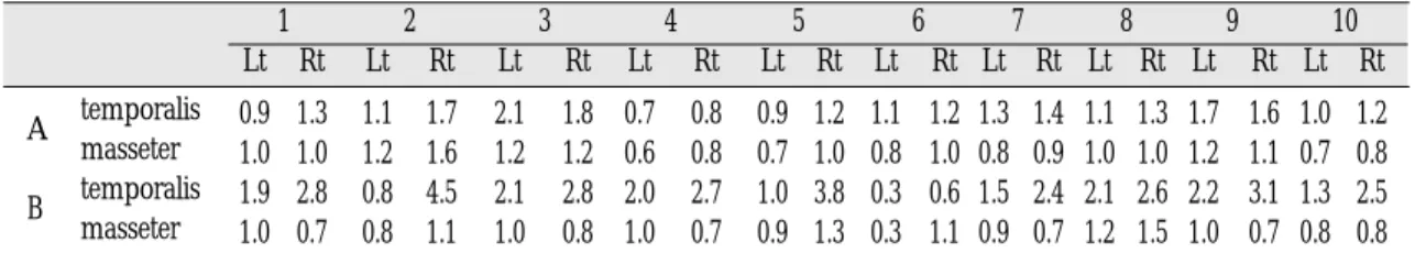
Table Ⅰ-1. The electromyographic activity during rest(Unit:㎶)
0.9 1.3 1.1 1.7 2.1 1.8 0.7 0.8 0.9 1.2 1.1 1.2 1.3 1.4 1.1 1.3 1.7 1.6 1.0 1.2 1.0 1.0 1.2 1.6 1.2 1.2 0.6 0.8 0.7 1.0 0.8 1.0 0.8 0.9 1.0 1.0 1.2 1.1 0.7 0.8 1.9 2.8 0.8 4.5 2.1 2.8 2.0 2.7 1.0 3.8 0.3 0.6 1.5 2.4 2.1 2.6 2.2 3.1 1.3 2.5 1.0 0.7 0.8 1.1 1.0 0.8 1.0 0.7 0.9 1.3 0.3 1.1 0.9 0.7 1.2 1.5 1.0 0.7 0.8 0.8 (A: bilateral mastication group, B: unilateral mastication group)
A temporalis masseter B temporalis
masseter
1 2 3 4 5 6 7 8 9 10
Lt Rt Lt Rt Lt Rt Lt Rt Lt Rt Lt Rt Lt Rt Lt Rt Lt Rt Lt Rt
Table Ⅰ-2. The electromyographic activity during clenching in maximal intercuspation(Unit:㎶)
55.2 57.0 54.6 51.8 58.5 52.4 29.0 31.4 40.5 41.7 38.5 38.3 55.5 57.1 42.0 37.1 40.2 51.3 42.7 47,0 70.2 67.2 64.3 57.2 81.4 83.9 59.8 61.3 54.4 55.1 52.3 53.1 60.7 64.4 55.1 51.4 69.8 54.1 67.9 75.3 31.0 42.2 32.1 40.0 16.2 21.5 34.0 56.3 37.4 63.7 39.5 57.2 11.4 21.3 30.0 37.7 28.9 40.3 50.0 57.4 26.8 47.7 26.0 47.2 34.3 39.6 50.0 93.8 87.4 99.4 73.3 93.8 27.9 48.8 26.4 44.7 26.3 46.0 54.9 60.9 (A: bilateral mastication group, B: unilateral mastication group)
A temporalis masseter B temporalis
masseter
1 2 3 4 5 6 7 8 9 10
Lt Rt Lt Rt Lt Rt Lt Rt Lt Rt Lt Rt Lt Rt Lt Rt Lt Rt Lt Rt
Table Ⅱ-1. The asymmetry index(%) of masseter and temporal muscle electromyographic activity between left and right side during rest(Unit:%)
AI A -8.18 -11.43 +7.69 -6.67 -14.29 -4.35 -3.70 -8.33 +3.03 -9.09
in temporalis
B +19.15 +9.81 +4.29 +14.89 +8.33 +33.33 +23.08 +10.64 +16.98 +31.58 muscle
AI A 0.00 +4.29 0.00 +10.29 +7.65 +11.11 -5.88 0.00 -4.35 +6.67
in masseter
B -17.65 +15.79 -11.11 -17.65 +18.18 +53.85 -12.50 +11.11 +35.48 +46.67 muscle
(A: bilateral mastication group, B: unilateral mastication group)
1 2 3 4 5 6 7 8 9 10
Table Ⅱ-2. The asymmetry index(%) of masseter and temporal muscle electromyographic activity between left and right side during clenching in maximum intercuspation(Unit:%)
AI A -1.60 +2.63 +5.50 -3.97 -1.46 +0.26 -1.42 +6.19 -12.13 -4.79
in temporalis
B +15.30 +10.96 +14.06 +24.70 +26.01 +18.30 +30.28 +11.37 +16.47 +6.89 muscle
AI A -2.18 -5.84 +1.51 +1.24 +0.64 +0.76 +2.96 -3.47 +12.67 +5.17
in masseter
B +28.05 +28.96 +15.63 +30.46 +11.18 +12.27 +56.95 +25.74 +27.25 +5.18 muscle
(A: bilateral mastication group, B: unilateral mastication group)
1 2 3 4 5 6 7 8 9 10
EMG(right)-EMG(left) Asymmetry index(%)=————————————
EMG(right)+EMG(left)
×100
Mean values of asymmetry index(%) of temporal and masseter muscle in each group during rest were shown in table Ⅱ-1 and during clenching in table Ⅱ-2.
Table Ⅱ-1 and Ⅱ-2 represented the average of the index in each condition. When an index score is close to 0%, this means that muscles on the right and left sides were activated symmetrically.
Index score close to +100%, means that is activated the right muscle solely and index score close to - 100%, means that is activated in the left muscle sole- ly. Table Ⅱ-1 and Ⅱ-2 showed substantially large asymmetry indices of masseter muscle and anterior temporal muscles in unilateral masti- cation group than bilateral mastication group in both conditions.
EMG(masseter)-EMG(temporal) Activity index(%) =—————————————————
EMG(masseter)+EMG(temporal)
×100
In order to quantitatively compare the contri- bution of the masseter and anterior temporal muscles to the bite force the average masseter mus- cle and temporal muscle activity were calculated in each subject. The following activity index(%)7,12 was used to indicate the relative contribution of the masseter muscle and temporal muscle to the contraction effort.
Mean values of asymmetry index(%) of temporal and masseter muscle in each group during rest were shown in Table Ⅲ-1 and during clench- ing in Table Ⅲ-2.
When an index score is close to 0%, this means that temporal and masseter muscles were activated symmetrically on the right and left sides respec- tively. Index score close to +100%, means that is activated in the masseter muscle solely and index
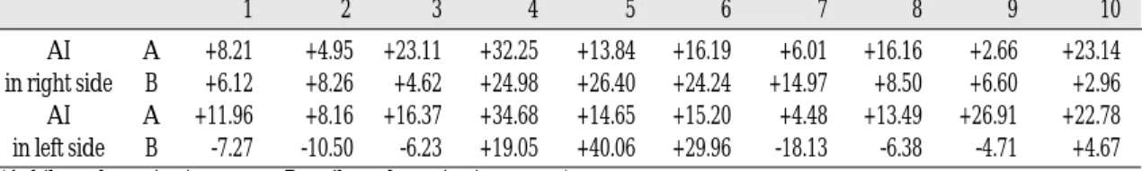
Table Ⅲ-1. The Activity index(%) of temporal to masseter electromyographic activity during rest (Unit:%)
AI A -13.04 -3.03 -20.00 0.00 -9.09 -9.09 -27.27 -13.04 -18.52 -20.00
in right side B -60.00 -60.71 -55.56 -58.82 -49.02 -25.00 -54.84 -26.83 -19.23 -6.38
AI A +5.26 +4.35 -27.27 -7.69 -12.50 -15.79 -18.18 -4.76 -17.24 -17.65
in left side B -31.03 0.00 -35.48 -33.33 -5.26 0.00 -25.00 -27.27 -37.50 -23.81 (A: bilateral mastication group, B: unilateral mastication group)
1 2 3 4 5 6 7 8 9 10
Table Ⅲ-2. The Activity index(%) of temporal to masseter electromyographic activity during clenching in maximum intercuspation ( Unit: % )
AI A +8.21 +4.95 +23.11 +32.25 +13.84 +16.19 +6.01 +16.16 +2.66 +23.14 in right side B +6.12 +8.26 +4.62 +24.98 +26.40 +24.24 +14.97 +8.50 +6.60 +2.96 AI A +11.96 +8.16 +16.37 +34.68 +14.65 +15.20 +4.48 +13.49 +26.91 +22.78 in left side B -7.27 -10.50 -6.23 +19.05 +40.06 +29.96 -18.13 -6.38 -4.71 +4.67 (A: bilateral mastication group, B: unilateral mastication group)
1 2 3 4 5 6 7 8 9 10
score close to -100%, means that is activated in the temporal muscle solely.
Table Ⅲ-1 showed negative values of activity index predominantly in bilateral mastication group, therefore remarkable negative values of activity index in unilateral mastication group and this means tonus of temporal muscle is far more than masseter muscle during rest. Table Ⅲ- 2 revealed relatively somewhat positive values of activity index in bilateral mastication group, therefore activity indices in unilateral mastication
group are not uniformed. This means that major- ity of contraction effort during clenching in max- imal intercuspation is derived masseter muscle than temporal muscle. But this characteristics is not clear in unilateral mastication group.
The electromyographic activities of left, right mas- seter and anterior temporal muscle during chew- ing at left and right sides were illustrated in Table Ⅳ. All of subjects with unilateral mastica- tion habit to participate in this study were unilateral masticators on right. Therefore, that is to say,
Table Ⅳ. The electromyographic activity during chewing in left and right side(Unit:㎶)
16.5 20.1 29.1 36.5 26.3 32.6 30.4 34.1 22.4 29.8 20.1 34.7 20.4 30.8 30.1 30.0 29.1 36.2 15.0 20.1 37.0 14.9 55.3 30.7 52.1 21.7 43.1 39.1 33.7 23.2 39.4 29.0 38.2 30.2 41.4 28.9 54.7 29.0 30.8 23.4 15.4 8.8 19.5 10.2 7.7 16.2 7.8 14.7 15.4 8.8 31.6 20.8 21.6 10.0 14.4 7.3 20.1 9.7 16.2 8.6 19.7 27.2 23.7 32.7 11.1 10.1 11.5 11.6 19.7 27.2 35.8 45.5 25.2 34.4 15.1 23.5 23.7 32.2 19.1 30.1 15.0 28.8 20.2 33.1 11.6 18.9 9.4 20.9 11.5 23.0 36.2 22.2 32.0 24.7 25.0 18.4 19.8 10.4 30.0 16.2 16.9 32.7 21.5 35.1 6.9 21.8 16.5 23.1 17.2 26.0 37.0 47.2 39.2 50.4 29.0 32.0 28.2 29.8 42.0 51.4 18.0 24.8 10.3 21.4 10.1 20.4 11.6 25.8 11.4 24.9 11.7 26.0 10.6 23.8 19.8 34.0 13.5 29.0 6.7 14.9 19.7 20.8 16.5 20.1 14.7 20.1 12.5 24.4 13.5 23.8 23.8 10.6 12.7 22.9 20.8 34.9 17.7 29.3 9.5 14.3 (A: bilateral mastication group, B: unilateral mastication group)
left side A temporalis chewing masseter (NPCS) B temporalis
masseter right side A temporalis chewing masseter (PCS) B temporalis
masseter
1 2 3 4 5 6 7 8 9 10
Lt Rt Lt Rt Lt Rt Lt Rt Lt Rt Lt Rt Lt Rt Lt Rt Lt Rt Lt Rt
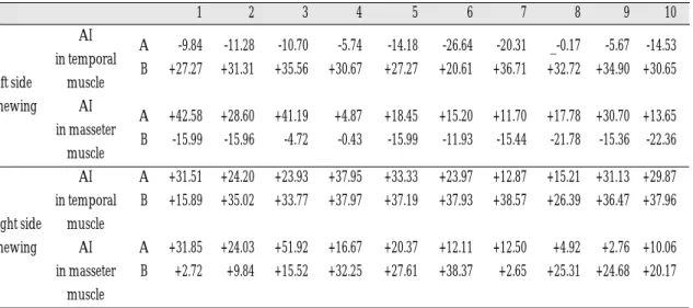
Table Ⅴ. The asymmetry index of masseter and temporal muscle electromyographic activity during chew- ing in left and right side(Unit:%)
AI A -9.84 -11.28 -10.70 -5.74 -14.18 -26.64 -20.31 _-0.17 -5.67 -14.53 in temporal
B +27.27 +31.31 +35.56 +30.67 +27.27 +20.61 +36.71 +32.72 +34.90 +30.65 left side muscle
chewing AI
A +42.58 +28.60 +41.19 +4.87 +18.45 +15.20 +11.70 +17.78 +30.70 +13.65 in masseter
B -15.99 -15.96 -4.72 -0.43 -15.99 -11.93 -15.44 -21.78 -15.36 -22.36 muscle
AI A +31.51 +24.20 +23.93 +37.95 +33.33 +23.97 +12.87 +15.21 +31.13 +29.87 in temporal B +15.89 +35.02 +33.77 +37.97 +37.19 +37.93 +38.57 +26.39 +36.47 +37.96 right side muscle
chewing AI A +31.85 +24.03 +51.92 +16.67 +20.37 +12.11 +12.50 +4.92 +2.76 +10.06 in masseter B +2.72 +9.84 +15.52 +32.25 +27.61 +38.37 +2.65 +25.31 +24.68 +20.17
muscle
(A: bilateral mastication group, B: unilateral mastication group)
1 2 3 4 5 6 7 8 9 10
right side was preferred chewing side(PCS) and left side was non-preferred chewing side(NPCS) in unilateral mastication groups. 20 subjects were asked to chew in left and right side three times and the mean of 3 recordings was illustrated.
Mean values of asymmetry index (%) of anterior temporal and masseter muscle in each group were shown in table Ⅴ.
The asymmetry index ranges from +100% (high- er activity of chewing side muscle) to -100%
(higher activity of chewing side muscle). When an index score is close to 0%, this means that muscles on the right and left sides were activated sym- metrically.
Mean values of the activity index (%) of anterior tem- poralis and masseter muscle in each group on left and right side respectively were shown in table Ⅵ.
Table Ⅵ revealed that the values of activity index in bilateral mastication group are positive substantially in same side of chewing side, that is to say, masseter muscle of chewing side is more active than temporal muscle. Therefore, the val- ues of activity index in bilateral mastication group are negative substantially in same side of chewing side, preferred chewing side, and this means temporal muscle in preferred chewing side is more active than masseter muscle.
To the difference of these values of asymmetry index and activity index during rest, clenching and chewing between bilateral and unilateral masti- cation group. Table Ⅶ represents the results of the independent sample t-test for all index values. The T- test was done in each conditions, at the p < .05 and p < .01 level of significance.
Table Ⅵ. The activity index(%) of temporal to masseter electromyographic activity during chewing in left and right side(Unit:%)
AI A -14.86 -8.63 -20.07 +6.83 -12.45 -8.95 -0.98 -1.87 -5.84 +7.59 left side in right side B +51.11 +52.45 +23.19 +11.79 +51.11 +37.25 +54.95 +52.60 +53.81 +55.56 chewing AI A +38.32 +31.04 +32.91 +17.28 +20.14 +32.44 +30.38 +15.80 +30.55 +34.50 in left side B +12.25 +9.72 +18.09 +19.17 +12.25 +6.23 +7.69 +2.37 +8.22 +8.22 AI A +6.34 +2.93 +7.13 +5.00 +6.12 +36.02 +34.22 +26.98 +48.26 +52.07 right side in right side B -8.77 -3.13 -0.74 -2.79 -2.26 -4.42 -1.93 -1.31 +0.51 -2.05 chewing AI A +5.96 +3.12 -25.41 +27.41 +19.86 +1.09 +10.11 +7.14 +17.50 +16.67 in left side B +4.51 +23.13 +18.55 +3.73 +8.43 +4.93 +9.01 +2.46 +13.46 +17.28 (A: bilateral mastication group, B: unilateral mastication group)
1 2 3 4 5 6 7 8 9 10
Table Ⅶ. The difference in asymmetry index and activity index between bilateral and unilateral mastication group
rest 0.053 0.005** 0.002** 0.211
clenching in maximum intercuspation 0.228 0.051 0.632 0.027*
chewing in preferred side(right) 0.016* 0.111 0.023* 0.147
chewing in non-preferred side(left) 0.000** 0.520 0.000** 0.383
* indicates significant difference (P< .05)
** indicates significant difference (P< .01)
Asymmetry index in Asymmetry index in Activity index Activity index temporal m. masseter m. in right side in left side
First of all, the differences between bilateral and unilateral mastication group were the most distinct in the asymmetry indices of temporal muscle and the activity indices in right side chewing(PCS) during chewing in non-preferred side(left) and these differences were significant at the p<.01 level. Also, asymmetry indices of mas- seter muscle and activity indices in right side during rest were differed between bilateral and unilateral mastication group at the p<.01 level of significance. And, asymmetry indices of tempo- ral muscle and the activity index in right side chew- ing(PCS) during chewing in preferred side(right) were differed between bilateral and unilateral mas- tication group at the p<.05 level of significance. But, except activity indices in left side, the differ- ences between bilateral and unilateral mastication group during clenching were not significant.
DISCUSSION
The stomatognathic system is consisted of occlusion, masticatory muscles and temporo- mandibular joints. The stomatognathic apparatus develops in an almost symmetric pattern, with two paired bones (the maxillae) and a single symmetric bone (the mandible) bearing two symmetric joints, several pairs of muscles, and two symmetric rows of teeth.14The inter-relationship among occlusion, masticatory muscles and temporo- mandibular joints are intricate and must be pre- cisely coordinated for functional mastication and alterations of this symmetric arrangement are most usually found in the dental arches or in the supporting bones, but the effects of such an alteration are likely to involve the apparatus as a whole.14 Nevertheless, several compensatory adaptations to the altered anatomical situations may be found, and assessment of the patient should be aimed mostly at the functional impair- ment.14For instance, continual unilateral mastication may alter the coordination of masticatory muscles
it produce strain in a single muscle unlike bilat- eral mastication. Therefore contributes to more severe damage to the stomatognathic system.
Habitual unilateral mastication is related to stom- atognathic dysfunction. The analysis of the mas- ticatory muscle activity in subjects with altered mas- ticatory functions could, therefore, provide use- ful data on the functional impact of masticatory system. These activities can be investigated using surface EMG, which allows the monitoring of some of the main masticatory muscles( mas- seter, temporal muscle), with results that do not significantly differ from those obtained with intramuscular recordings. Results from a bio- feedback study suggested that equalization of masticatory muscle activity may be important in the successful treatment of some types of the stomatognathic dysfunction.15
Since electromyography(EMG) was introduced by Moyers in 1949 for dental research, several investigation have examined jaw muscle elec- trical activity in the diagnosis of mandibular dysfunction.4,9,10,16In the present study, the influ- ence of continual unilateral mastication on mas- ticatory muscle activity was tested in a group of unilateral masticators and irregular muscular coordinations were found.
The initial recordings at rest showed very light activity in the muscles, with similar values for all subjects. The average values(1.08㎶) from all the muscles supports the presence of basal tonus.10 Present studies represent very symmetric rest position values, i. e. 1.0㎶ to 1.5㎶ for the temporal muscle and 0.5㎶ to 1.0㎶ respectively, in the bilateral mastication group.
The electromyographic analysis of this study gen- erally exhibited positive values in asymmetry indices for all recorded muscles and the larger asymmetry indices were observed in masseter mus- cle. These results reveal higher activity level in chewing side masticatory muscle than non-chew- ing side, particularly in masseter muscle.11Even
though the electromyographic analysis exhibited clear differences between the bilateral mastication group and the unilateral mastication group.
The coordination of muscle activity during mastication is thought to be a measure of dys- function. It has been suggested that the con- tralateral masseter muscle initiates activity before the ipsilateral muscle, and the ipsilateral anteri- or temporal muscle initiates activity before the con- tralateral temporal muscle.5
Our data indicate that, during chewing, the masseter acts asymmetrically, both in firing lev- el and in timing. The chewing side masseter was always more active than the non-chewing side, and EMG peaks appeared earlier at the non-chewing side than at the chewing side. Similar results were reported previously.17The latter report also showed a decrease of the chewing / non-chewing side ratio with increasingly powerful mastication.
A further increase in chewing force will probably lead to asymmetrical contraction. Such contrac- tions were found in a study where maximal effort unilateral bite forces were produced. The full contribution of both masseters is then required for the high level of force to be produced.17
The present study shows that the electromyo- graphic values during rest were clearly asymmetric in subjects with unilateral mastication group than in bilateral mastication group and muscle tonus of temporalis was higher than in bilateral mastication group and that the activity and asymmetry index during maximal clenching were slightly higher in subjects with unilateral mas- tication group than in bilateral mastication group, but no significant difference was found between the two groups.
In bilateral masticators, the AI did not equal zero.
This indicates that bilateral masticators have a dete- rioration on the symmetry of muscle activity, as reported previously.18
In the present study, the AI of the masseter muscles detected the asymmetry of muscle activ-
ity and was relative to parafunctional habit.4 The difference between the masseter and ante- rior temporal muscle activities was considered dependant on differences in the functional role of each muscle. The anterior temporal muscle activ- ity could be due in part to stabilization of the jaw and in part to the requirements of generating biting forces. The masseter muscle has a long- moment arm length favouring its biting capa- bilities.19If the symmetry of masseter muscle activity is impaired. the temporal muscles may cor- rect the imbalance. Therefore, even when there is an apparent asymmetry in the activity of temporal muscles, this asymmetry should be interpreted as a correction of the masseter muscle asymmetry and hence not abnormal by itself.
Unilateral mastication is thought to impair the symmetry of masseter muscles, leading to various disturbances of the stomatognathic system.
Therefore, an analysis of mastication habits was included in the study. Using Kendall’s ranked correlation coefficient, a significant correlation between mastication habits and stomatog- nathic dysfunction was demonstrated(p<0.01).4 Developmentally, masticatory organs are normally symmetric. Natural mastication is performed bilaterally without shifting to one side.12
It is possible that unilateral mastication affects the symmetry of muscle activity, or, conversely, it is possible that the asymmetry of muscle activ- ity induces unilateral mastication. The muscle activ- ity level during maximal clenching reflects the max- imal contractility of these muscles.13 Because even normal subjects show some degree of asym- metry of muscle activity, some difference in the contractibility between right and left muscle sand is accepted as physiological asymmetry.
The masticatory muscles usually exert their func- tion when the clenching level is about 20% of max- imum.20Therefore, the slight imbalance observed between left and right muscle activities of bilat- eral mastication subjects will not cause any imbal-
ance in the masticatory function. However, if the asymmetry index of the masseter muscle is great, the masticatory function can be adversely affected, probably leading to unilateral masti- cation. Unilateral mastication and the asymmetry index during maximal clenching are related.
That is, pain or muscle fatigue due to stomatog- nathic dysfunction syndrome can induce habitual unilateral mastication.8
The unbalance of the masticatory musculature can lead to cervical pains, headaches, earaches, clicking, discal degeneration, breaking and lateral deviations of the mandible. These symptoms can be attributed to an involuntary alteration of the muscular activity of one or more muscles producing an unbalance in the mouth opening and closing mechanism.3
CONCLUSIONS
The purpose of this study was to obtain basic data which is electromyographic activity in uni- lateral masticators.
Twenty subjects, ranging from 21 to 27 years of age without symptoms of stomatognathic system, were selected from dental students in Chosun University by the questionnaire and oral exami- nation. Among the twenty subjects, ten bilateral mastication subjects and ten unilateral mastication subjects were divided.
The electromyographic activities of anterior temporal and masseter muscle were investigated by BIO-PAK system(Bio-Research Associates Inc. USA). Each subjects were measured three times during rest, clenching in maximum intercuspation and gum chewing and the mean of 3 recordings were compared and analyzed.
The following results were obtained:
1. In electromyographic activity during rest, records of anterior part of temporal muscle in bilateral mastication group were ranged 1.0-
1.5V and superficial part of masseter muscle were ranged 0.5-1.0V and pattern of mus- cle activity of right and left side was sym- metrical. But,in unilateral mastication group, records of anterior part of temporal muscle was higher than that of bilateral mastication group(p<.01) and patterns of muscle activity of right and left side in both muscle were asymmetrical.(p<.05)
2. In electromyographic activity during clench- ing in maximum intercuspation, records of superficial part of masseter muscle were higher than anterior part of temporal muscle in both group. Muscle activity of temporal muscle in unilateral mastication group was a little higher than bilateral mastication group and asymmetry of activity pattern in temporal and masseter muscle was shown but these dif- ferences were not statistically significant.(p<.05) 3. In electromyographic activity during gum chewing, temporal muscle was activated ear- lier than masseter muscle and maximum bite force is derived from masseter muscle in both group. The order of muscle activating time in bilateral mastication group was mas- seter muscle of chewing side, contralateral mas- seter muscle, temporal muscle of chewing side and contralateral temporal muscle But, in unilateral mastication group, electromyo- graphic activity of masseter and temporal muscle of preferred chewing side, regard- less of right or left side chewing, was higher than that of bilateral mastication group and especially, difference in masseter muscle was statistically significant.(p<.01)
Based on the above results, our study sug- gested that recording of masticatory muscle activity will be helpful in the effective diagnosis and treatment of some types of the parafunc- tional habits.
Fig. 1.Electrode placed in recorded position (BIO- PAK system, Bio-Research Associates Inc. USA)
Fig. 2. The electromyographic activity during rest Fig. 3.The electromyographic activity during clench- ing
Fig. 4.The electromyographic activity during chewing
(left : during left side chewing, right : during right side chewing)
REFERENCES
1. Benito Rilo, Jose Luis da Silva, Francisco Gude, Urbano Sahara. “Myoelectric activity during uni- lateral chewing in healthy subjects: Cycle duration and order of muscle activation”J Prosthet Dent.
80:462-466,1998.
2. G. Zhang, X. Huang. SW. “Effect of unilateral bite splint on mastication in the miniature pig”J Oral Rehabil. 21:613-622, 1994.
3. C. Hagberg. “The amplitude distribution of elec- tromyographic activity of masticatory muscles during unilateral chewing”J Oral Rehabil.13:567- 574, 1986.
4. H. Abekura, H. Kotani, H. Tokuyama, T. Hamada.
“Asymmetry of masticatory muscle activity dur- ing intercuspal maximal clenching in healthy sub- jects and subjects with stomatognathic dysfunction syndrome”J Oral Rehabil. 22:699-704, 1995.
5. Khalid M. Balkhi. “Activity of anterior tempo- ralis and masseter muscles during deliberate uni- lateral mastication”J Orofacial Pain 7:89-97, 1993.
6. D. Bevilaqua-Grosso, V. Monteiro-Pedro, RR. De Jesus Guirro, F.Berzin “A physiotherapeutic ap- proach to craniomandibular disorder: a case report”
J Oral Rehabil. 29:268-273, 2002.
7. K. Nishigawa, M. Nakano, E. Bando. “Study of jaw movement and masticatory muscle activity during unilateral chewing with and without balancing side molar contacts”J Oral Rehabil. 24:691-696,1997.
8. C. Hagberg. “The amplitude distribution of el- cetromyographic activity in painful masseter mus- cles during unilateral chewing”J Oral Rehabil.
14:531-540, 1987.
9. Ambra Michenlotti, Mauro Farella, stefano Vollaro, Roberto Martina.”Mandibular rest position and elec- trical activity of the mastication”J Prosthet Dent.
78(1):48-53, 1997.
10. B. Rilo, U. Santana, MJ. Mora, CM. Cadarso.
“Myoelectrical activity of clinical rest position and jaw muscle activity in young adults”J Oral Rehabil 24:735-740, 1997
11. UC. Belser, AG. Hannam. “The contribution of the
deep fibers of the masseter muscle to selected tooth- clenching and chewing tasks”J Prosthet.
Dent. 56(5):629-635, 1986.
12. Ramfjord, SP., Ash, MM.,”Functional disturbances of temporomandibular joints and muscles”
Occlusion. 62-160, 1986.
13. Moritani, T., De vries, HA. “Neural factors versus hypertrophy in the time course of muscle strength gain”Am J Physical Med. 58:115-121, 1979.
14. VF. Ferrario, C. Sforza, G. Serrao. “The influence of crossbite on the coordinated electromyographic activity of human masticatory muscles during mastication”J Oral Rehabil. 26:575-581 1999.
15. RS. McCarroll, M. Naeije, YK. Kim, TL. Hansson.
“The immediate effect of splint-induced changes in jaw positioning on the asymmetry of submax- imal masticatory muscle activity”J Oral Rehabil.
16:163-170, 1989.
16. David M. Hickman, Richard Cramer, Morgantown.
W. Va., Hershey Pa.”The effect of different condy- lar positions on masticatory muscle electromyo- graphic activity in human”Oral Surg. Oral Med. Oral Path. 85(1):18-23, 1998.
17. T. M. G. J. Van Eijden, NG. Blanksma, P. Brugman
“Amplitude and timing of EMG activity in the hu- man masseter muscle during selected motor tasks”
J Dent Res. 72(3):599-606, 1993.
18. Buxbaum, JD., Parente, FJ., Ramsey, WO., Staling, LM. “A comparison of centric relation with max- imum intercuspation based on quantitative elec- tromyography”J Oral Rehabil. 9:45-55, 1982.
19. Jimenze, ID. “Dental stability and maximal mas- ticatory muscle activity”J Oral Rehabil. 14:591- 595, 1987.
20. Andreson, DJ. “A methods of recording masti- catory load”J Dent Res. 32:785-789, 1953.
Reprint request to:
DR. DONG-WANKANG
DEPT. OFPROSTHODONTICS,COLLEGE OFDENTISTRY,CHOSUNUNIVERSITY 421 SEOSUK-DONG,DONG-GU,GWANGJU,KOREA
E-mail : [email protected]