Laparoscopy/Robotics
Single-Surgeon Experience With Robot-Assisted
Ureteroneocystostomy for Distal Ureteral Pathologies in Adults
Ziho Lee, Shailen Sehgal, Elton Llukani1, Christopher Reilly1, Leo Doumanian1, Jack Mydlo1, David Inkoo Lee, Daniel Dong-In Eun1
Division of Urology, University of Pennsylvania School of Medicine, Philadelphia, PA, 1Department of Urology, Temple University School of Medicine, Philadelphia, PA, USA
Purpose: To demonstrate our technical approach for robot-assisted ureteroneocy- stostomy (R-UNC) for benign and malignant distal ureteral pathologies.
Materials and Methods: Between January 2009 and January 2013, a total of 10 patients underwent R-UNC in the distal ureter by a single surgeon. Indications for R-UNC were as follows: idiopathic (3), fistula (2), iatrogenic (2), malignancy (2), and chronic ves- icoureteral reflux (1).
Results: Tension-free anastomosis was attained in all 10 R-UNC procedures. A psoas hitch was performed in 6/10 cases (60%). Intravesical and extravesical reimplantations were completed in 5/10 (50%) and 5/10 cases (50%), respectively. A nonrefluxing ureter was constructed in 2/10 cases (20%). The patients’ mean age was 52.9±16.6 years, their mean body mass index was 30.8±6.3 kg/m2, the mean operative time was 211.7±69.3 minutes, mean estimated blood loss was 102.5±110.8 mL, and mean length of stay was 2.8±2.3 days. There were no intraoperative complications. There was one Clavien- Dindo grade I and one Clavien-Dindo grade II postoperative complication. The mean postoperative follow-up duration was 28.5±15.5 months. Two patients had recurrence of ureteral strictures at 3 months postoperatively and were managed successfully with balloon dilation.
Conclusions: Our technique for R-UNC demonstrates good perioperative outcomes.
However, underlying periureteral inflammation and pelvic adhesions may predispose patients for stricture recurrence after R-UNC.
Keywords: Minimally invasive surgical procedures; Reconstructive surgical procedures;
Ureter
This is an Open Access article distributed under the terms of the Creative Commons Attribution Non-Commercial License (http://creativecommons.org/licenses/by-nc/3.0) which permits unrestricted non-commercial use, distribution, and reproduction in any medium, provided the original work is properly cited.
Article History:
received 8 May, 2013 accepted 31 May, 2013
Corresponding Author:
Daniel Dong-In Eun
Department of Urology, Temple University School of Medicine, 255 S 17th Street, Suite #2101 Philadelphia, PA 19103, USA TEL: +1-215-470-2530 FAX: +1-215-875-9714 E-mail: Daniel.Eun@tuhs.
temple.edu
INTRODUCTION
Definitive surgical management of patients with distal ureteral pathologies may involve ureteroneocystostomy (UNC), which is the reimplantation of the ureter into the bladder. Traditionally, UNC is performed via an open lower abdominal incision [1]. Because a tension-free anastomosis between the ureter and the bladder is crit- ical for a successful UNC, patients with ureteral stric- tures that are extensive or involve more proximal seg- ments of the distal ureter are typically managed with a
concomitant psoas hitch or Boari flap or both during UNC [2,3].
Although robot-assisted UNC (R-UNC) has emerged as a treatment option, the literature regarding R-UNC is lim- ited to a handful of small case series with limited follow-up [4]. The purpose of the present study was two-fold: first, to demonstrate our technical approach for R-UNC for benign and malignant distal ureteral pathologies, and second, to report functional outcomes in patients with a mean fol- low-up of 28.5 months.
FIG. 1. Port placement.
MATERIALS AND METHODS
An Institutional Review Board approved retrospective re- view was performed on 10 patients (1 male and 9 female patients) who underwent R-UNC by a single surgeon be- tween January 2009 and January 2013. All procedures were performed by using the da Vinci surgical robotic plat- form (Intuitive Surgical Inc., Sunnyvale, CA, USA). There were eight left-sided and two right-sided ureteral pathologies. Indications for R-UNC were as follows: idio- pathic (3), fistula (2), iatrogenic (2), malignancy (2), and chronic vesicoureteral reflux (1). We defined recurrence as any symptomatic or imaging evidence of postoperative ure- teral stricture recurrence.
1. Presurgical evaluation
All patients were evaluated with computed tomography urography, cystoscopy, and retrograde or antegrade pye- lography to delineate the anatomy, location, and extent of ureteral injury. Mercaptoacetyltriglycine (MAG3) renal scans were performed when renal compromise was suspected. Females with a history of gynecologic malig- nancy underwent an exam under anesthesia, with possible cystoscopy, biopsy, or urinary cytology.
2. Surgical technique
1) Patient preparation and port placement
After induction of general anesthesia and administration of prophylactic intravenous antibiotics and subcutaneous heparin, the patient was placed in a modified dorsal lith- otomy position. The patient’s arms were padded and tucked in an anatomical position, and a nasogastric tube and an 18-Fr Foley catheter were placed. Pneumoperitoneum was established by using a Veress needle.
A total of 6 ports were used, and the port placement strat- egy for left-sided ureteral pathology is described (Fig. 1).
Port 1 was a 12-mm robotic camera port, which was placed
at the umbilicus. Port 2 was an 8-mm robotic instrument port, which was placed 8 cm to the patient’s left of Port 1.
Port 3 was an 8-mm robotic instrument port and was placed 8 cm to the patient’s left of Port 2. Port 4 was an 8-mm ro- botic instrument port and was placed 8 cm to the patient’s right of Port 1. Port 5 was a 12-mm assistant port and was placed 6 cm above the patient’s right iliac crest. Port 6 was a 5-mm port for suction and irrigation and was placed on the patient’s right lateral border of the rectus abdominis, 2 cm above the level of the umbilicus.
2) Ureter dissection
R-UNC was performed with a 0° camera and a Maryland bipolar grasper, monopolar hook cautery, and Cobra grasp- er (Intuitive Surgical Inc.) used in the left, right, and fourth robotic arms, respectively. Although on the left side, ure- teral exposure usually requires sigmoid colon mobi- lization, on the right side, ureteral exposure typically does not necessitate colonic mobilization. After identification of the ureter at its junction with the iliac vessels, the ureter was dissected and traced distally.
A vessel loop can be used for gentle retraction to minimize direct handling of the ureter. Ureteral dissection, however, may be particularly difficult in the presence of in- flammation or fibrosis, which often accompanies the un- derlying ureteral pathology and obliterates normal dis- section planes. The dissection of fibrotic periureteral planes may be facilitated by retraction by a skilled bedside assistant. In all portions of the procedure, the ureter was never directly grasped, and monopolar cautery was never directly applied to the ureter to preserve the ureteral blood supply and prevent excessive devascularization. The ure- ter was transected and trimmed back to healthy perfused edges by using robotic monopolar shears.
3) Bladder dissection
At this point, the bladder was dropped from the anterior abdominal wall and freed from the peritoneum, and the contralateral superior vesical artery was clipped and div- ided to fully mobilize the bladder toward the side of the af- fected ureter. The bladder was then filled with 400 mL of normal saline, and the anatomical layout was evaluated to determine if there was sufficient bladder mobilization for a tension-free anastomosis with the transected and pre- pared ureter. If a direct R-UNC did not seem optimal owing to tension, a psoas hitch was performed prior to anasto- mosis.
4) Psoas hitch
A 2-0 Vicryl suture on a SH needle (Ethicon, Somerville, NJ, USA) was placed deeply on the superior and ipsilateral wall of the bladder and pexed longitudinally through the psoas fascia. The psoas fascial stitch was placed in a longi- tudinal orientation. The needle should travel in a shallow but long fashion to allow for maximal suture traction strength while minimizing potential for genitofemoral nerve entrapment. This stitch can be redundantly placed
FIG. 2. Gastro-epiploic flap.
two to three times to prevent the stitch from tearing. Given the technical difficulties of tying this stitch while under sig- nificant traction, a Hem-o-loc Weck clip (Teleflex Medical, Research Triangle Park, NC, USA) can be clipped to a V-loc (Covidien, Mansfield, MA, USA) suture and slid along the direction of the suture’s barb to create a one-way winch mechanism. This technique avoids the need to tie knots un- der tension.
5) Intravesical reimplantation
An intravesical reimplantation was started by incising the bladder in the midline for approximately 5 cm by use of mo- nopolar cautery. A second cystotomy was created at the site of anastomosis by using a combination of sharp dissection and monopolar cautery. To create a refluxing anastomosis, the ureter was pulled through the cystostomy site and a running 5-0 Vicryl or Monocryl suture on a RB-1 needle (Ethicon) was used to approximate the spatulated edges of the ureter to the bladder. Particular care was taken to ap- proximate the mucosal edges. After completion of the pos- terior half of the anastomosis, a guidewire was advanced through the urethral council tip Foley catheter and roboti- cally guided from the catheter tip into the new ureteral orifice. A 6-Fr ureteral stent was pushed up and deployed by using the Seldinger technique. Once the stent was prop- erly deployed, the remainder of the anastomosis was completed. To create a nonrefluxing ureter, a classic 5:1 tunnel length to width ratio was utilized. The tunnel was created from the outside of the bladder by using inter- rupted 2-0 Vicryl sutures, which were imbricated over the ureter, resulting in a nonrefluxing mechanism.
6) Extravesical reimplantation
An extravesical reimplantation was begun by creating a cystotomy at the reimplantation site without making a for- mal midline incision into the bladder. To create a refluxing ureter, the ureter was spatulated and anastomosed to the bladder by using a running 5-0 Vicryl or Monocryl suture
on an RB-1 needle (Ethicon). Particular care was taken to approximate the mucosal edges. After completion of the posterior half of the anastomosis, the cystostomy site was utilized to direct a guidewire placed in a similar fashion as above. A 6-Fr ureteral stent was placed. The remainder of the anastomosis was then completed. To create a non- refluxing ureter, a classic 5:1 tunnel length to width ratio was utilized. The tunnel was closed with the ureter tucked underneath by using 2-0 Vicryl.
7) Gastro-epiploic flap technique
A gastro-epiploic flap can be developed to keep suture lines separate and improve blood flow in patients who have ques- tionable perfusion to the site of reconstruction, such as in patients with a history of radiation treatment. In our tech- nique, the flap was laparoscopically created after port placement, but before docking the robot. The omental apron was identified at the greater curvature of the stomach. A gastro-epiploic based flap was created with the help of a laparoscopic LigaSure device (Covidien). Once it was confirmed that the flap could easily reach the lowest portion of the pelvis, the distal end of the flap was clipped to the deep pelvis so that it could be accessed at a later time.
After completing the R-UNC, the gastro-epiploic flap was pexed at the site of reconstruction and interposed between critical suture lines (Fig. 2).
8) Postsurgical care
A Jackson-Pratt drain, which was left in the pelvis follow- ing R-UNC, was removed before discharge from the hospital. A Foley catheter was left for 5 days postope- ratively. Stents were left in the ureter for 6 weeks postoperatively. A retrograde ureteropyelogram was per- formed in the operating room at the time of stent removal to confirm patency of the anastomosis. Follow-up imaging consisted of a computed tomography urogram to assess kid- ney drainage at 2 weeks, 3 months, 6 months, and 1 year;
ultrasound was used to monitor the kidney thereafter. A MAG3 scan can be performed if there is any suspicion of obstruction.
RESULTS
Tension-free anastomosis was attained in all 10 R-UNC procedures; 6/10 cases (60%) required a psoas hitch. Intra- vesical and extravesical reimplantations were completed in 5/10 (50%) and 5/10 cases (50%), respectively. A non- refluxing ureter was constructed in 2/10 cases (20%). The patients’ mean age was 52.9±16.6 years, their mean body mass index (BMI) was 30.8±6.3 kg/m2, the mean operative time was 211.7±69.3 minutes, mean estimated blood loss (EBL) was 102.5±110.8 mL, and mean length of stay was 2.8±2.3 days. There were no intraoperative complications.
There was one Clavien-Dindo grade I and one Clavien- Dindo grade II postoperative complication. The mean post- operative follow-up duration was 28.5±15.5 months. Two patients (both patients at 3 months postoperatively) dem-
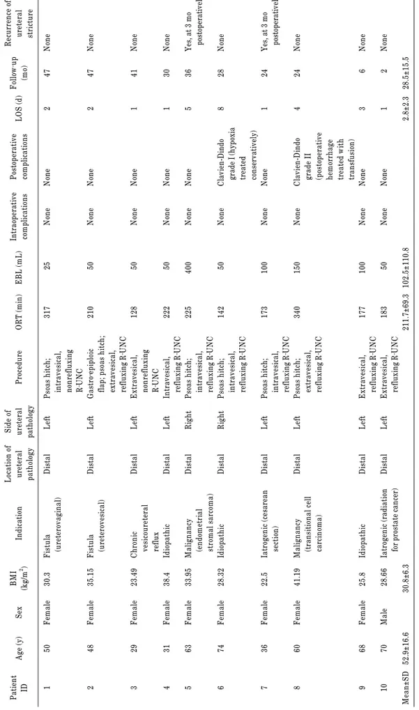
TABLE 1. Summary of perioperative variables Patient IDAge (y)SexBMI (kg/m2 )IndicationLocation of ureteral pathology Side of ureteral pathologyProcedureORT (min)EBL (mL)Intraoperative complicationsPostoperative complicationsLOS (d)Follow‐up (mo)
Recurrence of ureteral stricture 1 2 3 4 5 6 7 8 9 10 Mean±SD
50 48 29 31 63 74 36 60 68 70 52.9±16.6
Female Female Female Female Female Female Female Female Female Male
30.3 35.15 23.49 38.4 33.95 28.32 22.5 41.19 25.8 28.66 30.8±6.3
Fistula (ureterovaginal) Fistula (ureterovesical) Chronic vesicoureteral reflux Idiopathic Malignancy (endometrial stromal sarcoma) Idiopathic Iatrogenic (cesarean section) Malignancy (transitional cell carcinoma) Idiopathic Iatrogenic (radiation for prostate cancer)
Distal Distal Distal Distal Distal Distal Distal Distal Distal Distal
Left Left Left Left Right Right Left Left Left Left
Psoas hitch; intravesical, nonrefluxing R‐UNC Gastro‐epiploic flap; psoas hitch; extravesical, refluxing R‐UNC Extravesical, nonrefluxing R‐UNC Intravesical, refluxing R‐UNC Psoas hitch; intravesical, refluxing R‐UNC Psoas hitch; intravesical, refluxing R‐UNC Psoas hitch; intravesical, refluxing R‐UNC Psoas hitch; extravesical, refluxing R‐UNC Extravesical, refluxing R‐UNC Extravesical, refluxing R‐UNC
317 210 128 222 225 142 173 340 177 183 211.7±69.3
25 50 50 50 400 50 100 150 100 50 102.5±110.8
None None None None None None None None None None
None None None None None Clavien-Dindo grade I (hypoxia treated conservatively) None Clavien-Dindo grade II (postoperative hemorrhage treated with transfusion) None None
2 2 1 1 5 8 1 4 3 1 2.8±2.3
47 47 41 30 36 28 24 24 6 2 28.5±15.5
None None None None Yes, at 3 mo postoperatively None Yes, at 3 mo postoperatively None None None BMI, body mass index; ORT, operative time; EBL, estimated blood loss; LOS, length of stay; R-UNC, robot-assisted ureteroneocystostomy; SD, standard deviation.
onstrated symptomatic evidence of stricture disease. Both patients were managed successfully with balloon dilation.
Perioperative variables are summarized in Table 1.
DISCUSSION
Reddy and Evans [5] reported the first laparoscopic UNC (L-UNC) in adults in 1994. Subsequent reports confirmed the feasibility of L-UNC [6-8], and Rassweiler et al. [9]
found L-UNC to be associated with less EBL, less post- operative analgesia, and shorter length of stay, while maintaining comparable functional outcomes to open UNC. Despite this, L-UNC is not widely performed because it is a technically challenging procedure owing to the diffi- culty of visualizing the deep retropubic space [7,8], the diffi- culty of dissecting in the limited pelvic working space [8], and the difficulty of intracorporeal suturing in the pelvis [10].
Robot-assisted surgery maintains the inherent advan- tages of laparoscopic surgery, such as smaller incisions, less postoperative pain, faster recovery, and shorter hospi- tal stay, while also providing high-definition, three-dimen- sional magnified vision; tremor filtering; wristed in- strumentation; and improved ergonomics for the surgeon [11-13]. The enhanced degree of visualization, the ability to work in tight anatomical spaces, and the ability to pre- cisely suture make robot-assisted surgery particularly well suited for technically challenging procedures such as UNC. Although multiple reports have shown the feasi- bility of R-UNC [14-20], the current literature regarding R-UNC in adults is limited [4,21].
Hemal et al. [15] reported the largest series to date, in which multiple surgeons from two institutions performed 18 R-UNC procedures for distal ureteral pathology. The procedures involved extravesical or intravesical reimplan- tation of a refluxing ureter with or without a psoas hitch.
There were no intraoperative or postoperative complica- tions: no evidence of strictures was seen at a mean follow-up of 13.5 months (range, 6 to 28 months). Schimpf and Wagner [19] reported the largest single-surgeon series to date that included 11 R-UNC procedures for distal ureteral pathologies. Only extravesical, refluxing reimplantations were performed, with or without a psoas hitch or Boari flap.
There was one intraoperative vascular complication, two postoperative complications (ileus and hematuria requir- ing fulguration), and no evidence of ureteral stricture re- currence at a mean follow-up of 20.5 months (range, 6 to 28 months) [22]. Patil et al. [18] reported the largest pro- spective series, in which multiple surgeons from three in- stitutions performed 12 R-UNC procedures for distal ure- teral pathologies. Only intravesical, nonrefluxing re- implantations with psoas hitches were performed. There were no intraoperative or postoperative complications.
Likewise, there was no evidence of ureteral stricture re- currence after a mean follow-up of 15.5 months (range, 17 to 65 months).
Perioperative and functional outcomes of R-UNC have
been compared with those of open UNC. Kozinn et al. [16]
reported a retrospective comparison of 10 R-UNC proce- dures and 10 age- and BMI-controlled open UNC proce- dures for middle and distal ureteral pathology. Despite the limited number of patients, R-UNC was associated with similar operative time and success rates and lower EBL and hospital stay when compared with open UNC [16].
Our experience with R-UNC demonstrates the variety of technical approaches and considerations for benign and malignant distal ureteral pathologies. In our series, non- refluxing and refluxing ureters were extravesically and in- travesically reimplanted into the bladder, with and with- out a psoas hitch. Our experience is consistent with prior reports that demonstrate the feasibility and safety of R-UNC. The mean operative time, EBL, and complication rates from our particular study were similar to those of pre- viously reported R-UNC case series [15,16,18]. A post- operative complication was seen in 2/10 patients (20%).
One patient developed transient hypoxia and was man- aged conservatively (Clavien-Dindo grade I). One patient developed postoperative hemorrhage and was managed with 2 units of packed red blood cells (Clavien-Dindo grade II). Our report, however, was associated with a higher rate of stricture recurrence than those previously reported (20%, 2/10 patients). One potential explanation for this is that, at the time of R-UNC, both patients were noted to have severe periureteral inflammation and fibrosis and adhe- sions in the deep pelvis. Also, both patients had a history of prior abdominal and pelvic surgeries. Nevertheless, these two patients were managed successfully with balloon dilation, and all 10 patients are currently stent-free and without clinical or radiologic evidence of obstruction at a mean follow-up of 28.5 months.
Despite being one of the largest single-surgeon and sin- gle-institution R-UNC case series, the major limitation in our study was that only 10 patients were included.
Although initial reports, ours included, suggest that R-UNC is associated with shorter length of stay, less post- operative pain, less EBL, and improved cosmesis compared with open UNC, future studies incorporating larger num- bers of patients across multiple institutions will be val- uable in validating the role of R-UNC in the treatment of patients with middle and distal ureter pathologies.
CONCLUSIONS
Our technique for R-UNC demonstrates good perioper- ative outcomes. However, underlying periureteral in- flammation and pelvic adhesions may predispose patients for stricture recurrence after R-UNC.
CONFLICTS OF INTEREST
Ziho Lee, Elton Llukani, Christopher Reilly, and Leo Doumanian have no conflicts of interest or financial ties to disclose. Jack Mydlo is a consultant to Medical Diagnostic Laboratories. David Inkoo Lee receives study support from Johnson and Johnson and Pfizer and is a lecturer for
Intuitive Surgical, Ethicon Endosurgery, and Covidien.
Daniel Dong-In Eun is a proctor for Intuitive Surgical and a lecturer for Covidien.
REFERENCES
1. Paquin AJ Jr. Ureterovesical anastomosis: the description and evaluation of a technique. J Urol 1959;82:573-83.
2. Benson MC, Ring KS, Olsson CA. Ureteral reconstruction and by- pass: experience with ileal interposition, the Boari flap-psoas hitch and renal autotransplantation. J Urol 1990;143:20-3.
3. Stief CG, Jonas U, Petry KU, Sohn C, Bektas H, Klempnauer J, et al. Ureteric reconstruction. BJU Int 2003;91:138-42.
4. Phillips EA, Wang DS. Current status of robot-assisted laparo- scopic ureteral reimplantation and reconstruction. Curr Urol Rep 2012;13:190-4.
5. Reddy PK, Evans RM. Laparoscopic ureteroneocystostomy. J Urol 1994;152(6 Pt 1):2057-9.
6. Castillo OA, Litvak JP, Kerkebe M, Olivares R, Urena RD. Early experience with the laparoscopic boari flap at a single institution.
J Urol 2005;173:862-5.
7. Ehrlich RM, Gershman A, Fuchs G. Laparoscopic vesicoureter- oplasty in children: initial case reports. Urology 1994;43:255-61.
8. Lakshmanan Y, Fung LC. Laparoscopic extravesicular ureteral reimplantation for vesicoureteral reflux: recent technical advances. J Endourol 2000;14:589-93.
9. Rassweiler JJ, Gozen AS, Erdogru T, Sugiono M, Teber D.
Ureteral reimplantation for management of ureteral strictures:
a retrospective comparison of laparoscopic and open techniques.
Eur Urol 2007;51:512-22.
10. Asimakopoulos AD, Hoepffner JL, Mugnier C, Gaston R, Piechaud T. Laparoscopic extravesical ureteric re-implantation.
BJU Int 2011;108:1918-32.
11. Ahlering TE. Robotic versus laparoscopic radical prostatectomy.
Nat Clin Pract Urol 2004;1:58-9.
12. Lanfranco AR, Castellanos AE, Desai JP, Meyers WC. Robotic surgery: a current perspective. Ann Surg 2004;239:14-21.
13. Menon M, Kaul S, Bhandari A, Shrivastava A, Tewari A, Hemal A. Potency following robotic radical prostatectomy: a ques- tionnaire based analysis of outcomes after conventional nerve sparing and prostatic fascia sparing techniques. J Urol 2005;174:
2291-6.
14. Uberoi J, Harnisch B, Sethi AS, Babayan RK, Wang DS. Robot-as- sisted laparoscopic distal ureterectomy and ureteral re- implantation with psoas hitch. J Endourol 2007;21:368-73.
15. Hemal AK, Nayyar R, Gupta NP, Dorairajan LN. Experience with robot assisted laparoscopic surgery for upper and lower benign and malignant ureteral pathologies. Urology 2010;76:1387-93.
16. Kozinn SI, Canes D, Sorcini A, Moinzadeh A. Robotic versus open distal ureteral reconstruction and reimplantation for benign stricture disease. J Endourol 2012;26:147-51.
17. Laungani R, Patil N, Krane LS, Hemal AK, Raja S, Bhandari M, et al. Robotic-assisted ureterovaginal fistula repair: report of effi- cacy and feasiblity. J Laparoendosc Adv Surg Tech A 2008;18:
731-4.
18. Patil NN, Mottrie A, Sundaram B, Patel VR. Robotic-assisted lap- aroscopic ureteral reimplantation with psoas hitch: a multi-in- stitutional, multinational evaluation. Urology 2008;72:47-50.
19. Schimpf MO, Wagner JR. Robot-assisted laparoscopic Boari flap ureteral reimplantation. J Endourol 2008;22:2691-4.
20. Williams SK, Leveillee RJ. Expanding the horizons: robot-as- sisted reconstructive surgery of the distal ureter. J Endourol 2009;23:457-61.
21. Rassweiler J, Pini G, Gozen AS, Klein J, Teber D. Role of laparo- scopy in reconstructive surgery. Curr Opin Urol 2010;20:471-82.
22. Schimpf MO, Wagner JR. Robot-assisted laparoscopic distal ure- teral surgery. JSLS 2009;13:44-9.