서론
성공적인 임플란트의 주위 조직에서 일어나는 골유착 (osseointegration)은 연조직의 게재없는 골과 임플란트 표 면의 직접적인 구조적, 기능적 결합을 의미한다1). 골유착을 얻기 위해 처음 제안된 술식은 4~6개월 치유기간 후 임플 란트에 부하를 가하는 것이었으나, 즉시 부하(immediate
loading)나 초기 부하(early loading) 술식 역시 높은 예견 성이 있음이 보고되면서2,3,4) 초기 안정성에 대한 중요성이 부각되었다.
Adell 등1,5)은 임플란트의 초기 안정성이 골유착의 필수 요건이라고 하였고, 골유착의 효과적 예견 지표로 제안되었 다6,7). 또한 높은 초기 안정성은 임플란트에서 좀 더 예견성 있는 즉시 부하를 가능하게 한다3).
임플란트의 초기 안정성을 평가하는 다양한 방법이 제시 되었다. 임플란트 상부를 두드려 보는 tapping test5)와 방 사선 검사8)는 안전성을 평가하기에는 부정확한 단점이 있 고, 조직학적 분석9), 제거 토크 분석(removal torque analysis)10)은 침습적인 문제가 있기에 이러한 방법들은 장
Resonance Frequency Analysis(RFA)를 이용한 임플란트 종류간의 초기 안정성 비교
오준호, 장문택
*전북대학교 치의학전문대학원 치주과학교실
Comparison of initial implant stability measured by Resonance Frequency Analysis between different implant systems
Jun-ho Oh, Moontaek Chang
*Department of Periodontology, School of Dentistry, Chonbuk National University
ABSTRACT
Purpose: The objective of this study was to compare initial implant stability measured by RFA between different implant systems during the initial healing period.
Material and Methods: Fifty-four patients (36 males/18 females) who had been treated at the Department of Periodontology, Chonbuk National University Dental Hospital during the period between January and November in 2007 were included in the study. The mean age of the subjects was 49 years old (18 to 77). A total of 104 implants (Type A: 3i Osseotite
®, Type B:
Replace
®select, Type C: ITI implant) were placed following the manufacturer's standard surgical protocols. Implant stability quotient (ISQ) readings were obtained for each implant at the time of surgery, 2-, and 4-month postoperatively.
Result: No implant was failed during the observation period. At the baseline, the difference between mean ISQ values of 3 implant systems was statistically significant (p<0.05). However, at 2-, and 4-month following implant surgery, no significant difference was observed between ISQ values of the implant systems. In the same implant, the ISQ values of Type B and C implants increased (p<0.05), but those of Type A implants decreased during the 2-month healing period. The mean ISQ values of Type B and C implants showed a increasing tendency, while those of Type A implants were stable for the 4-month follow-up period.
Conclusion: Within limits of this study, it can be concluded that implant design and surface topography of implant might influence the ISQ value and changing pattern during the initial healing period. (J Korean Acad Periodontol 2008;38:529-534) KEY WORDS: Resonance Frequency Analysis; dental implant; implant stability; initial healing.
Correspondence: Dr. Moontaek Chang
Department of Periodontology, School of Dentistry, Chonbuk National University, Gumam-dong, Dukjin-gu, Chonju, 634-18, Korea.
E-mail: [email protected], Tel: 82-2-250-2116, Fax: 82-2-250-2259
* 이 논문은 2007년도 전북대학교병원 연구비 지원으로 수행됨.
Received: Aug 13, 2008; Accepted: Sep 11, 2008
기간 연구에 적합하지 못하다. 이런 문제점을 극복하기 위 해 Periotest가 고안되어 임플란트의 골유착을 평가하기 위 해 사용되었다11). 그러나 Periotest값은 측정 방향과 위치에 많은 영향을 받기에 생역학적 지표(biomechanical param- eter)로 사용되기에는 정확성이 의심스럽다12,13).
Meredith14)는 동물실험에서 임플란트에 변환기 (transducer)를 연결하고 resonance frequency(RF)를 측정 하여 임플란트 주변 골을 분석하는 비침습적 방법을 제시하 였다. Implant stability quotient(ISQ)로 표시되는 RFA 측 정값은 0~100까지 값을 가지며, 높은 값은 안정성이 높음 을 의미한다14,15).
RFA 측정으로 임플란트의 초기 안정성을 보고한 많은 연 구들7,14,15,19,21-23,26,27,29,30)이 있으나, 이들 연구들은 대부분 한 가지 종류의 임플란트에서 초기 안정성을 비교 분석하였
으며7,15,19,22,23,29,30), 다른 형태의 임플란트 종류간 초기 안정
성을 비교 분석한 연구19)는 드물다.
본 연구의 목적은 RFA를 이용하여 초기 골유착 기간 중 에 다른 종류의 임플란트 시스템 사이의 ISQ의 비교 및 변 화를 알아보고자 한다.
재료 및 방법
1. 연구 대상
2007년 1월부터 11월까지 전북대학교 치과병원 치주과에 서 임플란트 매식 수술을 받은 환자를 대상으로 하였다. 연 구에 포함된 환자는 모두 54명(남성: 36명, 여성: 18명)으로 평균 나이는 49세(18~77세), 이들 가운데 흡연자는 18명이 었다.
표면 처리가 다른 세 종류의 임플란트- Type A (Osseotite®, 3i-implant Innovations, West Palm Beach, Florida, USA), Type B(Replace® Select Tapered, Nobel Biocare, Göteborg, Sweden), Type C(ITI, Institut Straumann AG, Basel, Switzerland)-가 매식되었다.
Type A 임플란트는 산부식 처리가 된 OsseotiteTM 표면이 고 Type B 임플란트는 electrochemical process 방법을 이 용하여 티타늄 표면에 자연적인 산화막을 입힌 TiUniteTM 표면이며 다른 임플란트와 달리 tapered design을 가진다.
Type C 임플란트는 SLA(Sandblast Large grit Acid etch)
표면을 가지고 2.8mm shoulder가 존재하여 지대주 (abutment)와의 연결이 치조골 상방에 위치하게 된다. 매식 된 총 104개의 임플란트 가운데 Type A 임플란트는 36개, Type B는 48개, Type C는 20개이었으며, 임플란트의 길이 는 10~15 mm, 직경은 3.5~6mm로 분포되었다. 임플란트 는 전치부에 19개(18.3%), 소구치부에 14개(13.5%), 대구치 부에 71개(68.2%), 그리고 상악에 49개(47.1%), 하악에 55 개(52.9%) 식립되었다. 일회 수술법(one-stage)으로 식립 된 임플란트는 58개(55.8%), 이회 수술법으로 식립된 임플 란트는 46개(44.2%)였고 Type C 임플란트는 모두 일회 수 술법으로 식립되었다.
구강내 소독 후 2% Hcl lidocaine(1:100000 epinephrine 함유)(Xylestesin-A, 3M ESPE, Seefeld, Germany)으로 수술부위를 마취하고, 절개 후 판막을 거상한 다음 각 임플 란트 시스템 제조사에서 추천한 방법으로 식립하였다. 피질 골이 두꺼운 type I(Lekholm & Zarb classification, 1985) 에서는 bone tapping drill을 사용하였다. 식립시 주변에 골결손부가 존재한 35개(33.7%) 임플란트 주위에는 자가골 이나 이종골(Bio-Oss®, Geistlich AG, Wollhusen, Switzerland), 또는 이를 혼합하여 골증대술을 실시하였다.
2명 환자에 식립된 7개 임플란트는 잔존골이 부족하여 상악동 이식술 후 6개월째 식립하였다(모두 Type B 임플란 트). 전치부에 단독 즉시 임플란트가 식립된 오직 한 명의 환자를 제외한 나머지 환자에서는 발치 후 골이 충분히 치 유된 시기에 식립되었다.
흡수성 차폐막을 사용하여 골재생유도술을 실시한 4명의 환자에서 차폐막이 노출되는 문제를 제외하고 연구기간 중 다른 후유증은 관찰되지 않았다. 노출된 차폐막 부위는 chlorhexidine(클로헥신®, 대웅제약, 성남시, 대한민국)을 사용하여 4~6주간 치료 후 치유되었다.
2. 연구방법
각 임플란트에 맞는 magnetic peg(smartpegTM)를 연결 하고 OsstellTM Mentor(Integration Diagnostics AB, Göteborg, Sweden)을 이용하여 협측에서 2회 측정하였다.
일회법으로 시술된 임플란트는 수술시, 2, 4개월 후에 RFA 를 측정하였고, 2회법으로 시행된 임플란트에서는 수술시, 지대주 연결시(술 후 4개월)에 측정하였다.
3. 통계분석
수술시와 2개월째 측정한 임플란트 시스템간의 ISQ 값 비교는 Mann-Whitney U test, 동일 임플란트에서 측정된 수술시, 그리고 2개월째 ISQ 값의 비교는 paired t-test를 사용하였다.
결과
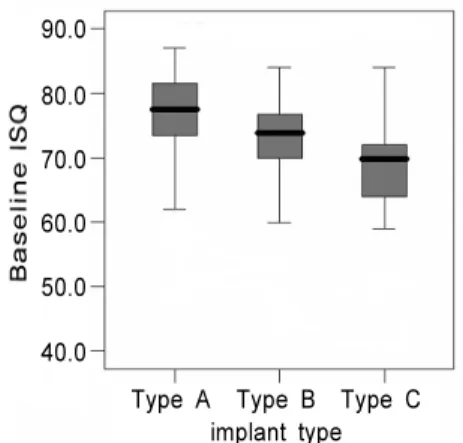
임플란트 식립시 Type A 임플란트의 평균 ISQ 값은 76.2(범위: 48.5~87.0), Type B의 평균 ISQ 값은 72.7(범 위: 45.0~84.0), Type C의 평균 ISQ 값은 67.6(범위:
41.0~84.0)으로 나타났다. 세 그룹의 ISQ 값은 통계적으로 유의한 차이를 보였다(p<0.05, Fig. 1). 그러나 임플란트 식립 후 2개월째 측정한 평균 ISQ 값은 Type A, B, C에서 각각 76.8(범위: 55.5~86.0), 77.8(범위: 67.0~85.5),
76.4(범위: 64.0~82.5)로 통계적으로 유의한 차이를 보이 지 않았다(Fig. 2).
임플란트 수술시와 2개월 후 모두 측정된 임플란트를 대 상(Type A; 14개, Type B; 14개, Type C; 16개)으로 한 ISQ 값 변화를 관찰하면 Type A에서는 감소한 반면, 나머 지 Type B, C에서는 증가하는 경향을 보였다(Fig. 3).
Type B와 Type C는 2개월 관찰기간 동안 통계적으로 유의 한 증가가 관찰되나(P<0.05), Type A에서의 감소는 통계 적 유의성이 없었다.
4개월째 ISQ값이 조사된 36개의 임플란트(Type A; 13개, Type B; 15개, Type C; 8개)를 포함하여 수술시, 2, 4개월 사이의 ISQ 값 변화를 각 임플란트 시스템 별로 관찰하였다 (Fig. 4). Type A 임플란트는 관찰기간 중 거의 변화가 없 었지만, Type B, C 임플란트는 관찰기간 중 증가하는 경향 을 보였다.
Figure 4. Change of mean ISQ values for 4-month after implant surgery in 3 implant types
90.0 84.0 78.0 72.0 66.0
60.0 Baseline 2-moth 4-moth
Type A Type B Type C
Figure 3. Comparison of mean ISQ values between implant surgery and 2-month postoperatively in 3 implant types
<0.05) 90.0
80.0 70.0 60.0 50.0 40.0
Type A Type B Type C implant type
* *
Baseline 2-month Figure 1. Baseline ISQ values of 3 implant types.
90.0 80.0 70.0 60.0 50.0 40.0
Type A Type B Type C implant type
Figure 2. 2-month ISQ values of 3 implant types.
90.0 80.0 70.0 60.0 50.0 40.0
Type A Type B Type C implant type
고찰
임플란트의 즉시 또는 초기 부하 술식이 예견성 있는 치 료로 보고되면서3,4) 임플란트의 초기 안정성의 중요성이 부 각되었다. 초기 안정성을 비침습적 방법으로 측정하기 위 하여 RFA가 개발되었다14).
이번 연구에서 세 임플란트 시스템의 수술시 ISQ 값은 Type C에서 가장 낮은 수치가 기록되었다. 이 임플란트 시 스템은 즉시 또는 초기 부하 술식에 성공적으로 많이 사용 되고 있으며16-18), 일반적으로 보철 치료 전 약 6주의 치유 기간을 추천한다16,23). 이번 실험에서도 Type C 임플란트는 골이식이 필요치 않은 부위에 모두 일회 수술법으로 식립되 었고, 대부분 3개월째 보철물이 장착되어 4개월째 RFA 값 은 측정하지 못했다.
Ersanli 등19)은 세 가지 임플란트 시스템의 RFA 비교에 서 본 연구에 포함된 Type C와 동일한 임플란트의 초기 평 균 ISQ 값이 다른 임플란트 시스템보다 낮은 값(평균 ISQ:
상악; 55, 하악; 60)을 가짐을 보고하였다. 이들의 연구에서 측정된 ISQ값은 이번 연구에서 사용한 OsstellTM Mentor (magnetic device)의 이전 모델 Osstell(electronic device) 로 측정되었다. 두 장치의 ISQ 값을 비교한 연구에서 Mentor로 측정한 값이 8~12정도 높은 값을 보였음20)을 감 안하면 이번 연구에서의 평균 ISQ 67.6과 Ersanli 등19)의 측정값이 유사함을 알 수 있다. 이렇게 낮은 값을 보이는 이 유는 Meredith 등21)이 언급한 치조골 상부 임플란트 높이 (supracrestal implant height)와 RFA 값의 강한 상관관계 에서 찾을 수 있다. 3mm의 치조골 상부 임플란트 should- er때문에 낮은 ISQ가 측정되었으며 만약 이 길이가 보정된 다면 다른 임플란트와 유사한 값을 보였을 것이다22). 또 다 른 이유로는 일부 임플란트(20개 중 4개)에서 식립시 약간 의 임플란트 자체의 골내 회전(rotation)이 존재하였기에 낮 은 ISQ 값을 보였을 수 있다.
많은 연구에서 2개월 이내의 초기 ISQ 값의 변화에 대해 서 조사되었다19,20,23). Barewal 등23)은 다양한 골질에 식립 된 SLA(Sand blast large grit acid-etched) 표면 처리된 임플란트에서의 ISQ 값을 술 후 6주까지 매주, 그리고 8주, 10주째 측정하였다. ISQ 값은 골질에 상관없이 3주째 가장 낮은 값을 보였고, 4주 이후부터는 골질 그룹간 차이가 없 었으며, 6주 이후부터는 안정기(plateau stage)에 이른다고
하였다. 이러한 변화는 초기 골접촉(primary bone contact) 에 의한 임플란트 수술 후 초기의 기계적 유지가 시간이 지 남에 따라 임플란트와 주위 골사이의 교직골(woven bone) 이 층판골(lamellar bone)로 대체되는 이차적인 고정 과정 이 반영된 것이라고 하였다23,24). 이는 Roberts 등25)의 토끼 실험에서 골에 야기된 상처의 치유과정에서 초기(0~6주)에 가골(callus)이 생기고, 6주째부터 층판골이 침착되는 것과 일치하는 내용이다. 본 연구에서 수술시와 2개월 후 값이 동시에 측정된 임플란트를 대상으로 관찰시 Type A를 제외 한 임플란트에서 ISQ 값의 증가 경향이 관찰되는 것은 이러 한 층판골 침착에 의한 결과로 생각될 수 있다.
반면 초기 평균 ISQ 값이 높은 Type A에서는 ISQ 값이 거의 변화 없이 유지되고 있었으며, 오히려 통계학적으로 유의하진 않지만 약간의 감소가 관찰되었다. 결과적으로 2 개월째에는 임플란트 시스템 사이에서 평균 ISQ 값의 유의 한 차이가 관찰되지 않았다. 이는 낮은 초기 ISQ 값을 보이 는 임플란트는 시간이 지나면서 그 값이 증가하고, 높은 초 기 ISQ 값을 보이는 임플란트는 시간에 따라 그 값이 감소 하여 결과적으로 임플란트간 ISQ 값에 차이가 줄어드는 catch up effect로 설명될 수 있다7,23,30).
수술시, 2, 4개월 후 각각의 시기에 조사된 임플란트 그 룹간의 ISQ 평균 변화값을 비교하면 Type B, C 임플란트에 서는 그 값이 지속적으로 증가하고, Type A 임플란트는 거 의 변화가 없음이 관찰된다. 이러한 시간에 따른 ISQ 값의 변화는 임플란트의 형태, 표면 처리 등에 영향을 받은 것으 로 보인다. 이런 차이는 다른 연구에서도 관찰되는데 Oʼ Sullivan 등26,27)은 동물실험과 인체해부 실험에서 tapered design의 임플란트에서 높은 RFA값이 관찰됨을 보고하였 고, 거친 표면(rough surface)을 가진 임플란트에서 평활면 (machined surface)을 가진 임플란트에 비해 높은 골-임플 란트 접촉(bone-implant contact)을 보임을 증명하였다28). Glauser 등15)은 성공한 임플란트와 실패한 임플란트의 RFA 값의 변화를 조사하였다. 81개의 Brånemark 임플란트 중 9개(11.2%)가 보철 수복 후 1년 내 실패하였는데 실패한 임플란트는 성공한 임플란트와 달리 1개월 후 현저히 RFA 값이 저하하였으며, 이들 결과에서 RFA의 임플란트 실패 예견성의 효용성을 언급했다. 반면 Huwiler 등29)은 연구에 포함된 임플란트에서 단지 한 개의 임플란트가 실패했고, 임플란트의 동요가 임상적으로 탐지된 후에 ISQ 값의 갑작 스러운 저하가 관찰되었기에 RFA의 예측능력에 의문을 가
졌으며, Nedir 등30)은 RFA가 임플란트 동요도를 재현성 있 게 진단하지 못함을 보고했다. 이번 연구에서 관찰 기간 중 실패한 임플란트는 없었으므로, RFA가 임플란트 실패를 예 견하기 위해 사용될 수 있는 효용성을 파악할 수는 없었다.
따라서 실패한 임플란트를 다수 포함하는 연구로 RFA의 진 단적 가치에 대한 평가가 필요할 것이다.
RFA를 이용하여 측정된 임플란트 형태와 표면이 다른 세 종류의 임플란트의 초기 안정성을 비교한 본 연구에서 임플 란트 수술 시에는 임플란트 종류간에 ISQ 값의 유의한 차이 가 존재하였지만, 술 후 2, 4개월째에는 통계적으로 유의한 차이가 관찰되지 않았다. 시간에 따른 ISQ 값의 변화는 임 플란트 시스템별로 다른 형태의 변화를 보였다. 그러므로 적절한 임플란트 부하 시기를 결정하기 위하여 RFA를 사용 할 경우 임플란트 시스템별로 정상적 범위의 ISQ 값을 설정 할 필요가 있다. 그리고 초기 ISQ 값이나 변화값으로 임플 란트의 성공 여부를 예측하는 RFA의 효용성에 대한 연구가 필요할 것이다.
참고문헌
1. Brånemark PI, Hanson BO, Adell R. osseointegrated im- plants in the treatment of the edentulous jaw. Experience from a 10-year period. Scand J Plast Reconst Surg 1977;11 (Suppl 16):1-32.
2. Albrektsson, T. & Isidor, F. Consensus report of session V.
IN Lang, N-P. & Karring, T., des. Proceedings of the 1st European workshop on periodontology. London: Quintessence Publ. Co. 1994;365-369.
3. Szmucler-Moncler S, Piattelli A, Favero GA, Dubruille JH.
Consideration preliminary to the application of early and immediate loading protocols in dental implantology. Clin Oral Implants Res 2000;11:12-25.
4. Degidi M, Piattelli A. Immediate functional and non func- tional loading of dental implants: a 2- to 60- month fol- low-up study of 646 titanium implants. J Periodontol 2003;74:225-241.
5. Adell, R., Lekholm, Ul, Rockler, B. & Brånemark, PI. A 15-year study of osseointegrated implants in the treatment of the edentulous jaw. International Journal of Oral Surgery 1981;10:387-416.
6. Meredith, N. Assessment of implant stability as a prog-
nostic determinant. International Journal of Prosthodontics 1998;11:491-501.
7. Friberg, B., Sennerby, L., Lin den, B., Grondahl, K. &
Lekholm, U. Stability measurements of one-stage Brånemark implants during healing in mandibles. A clinical resonance frequency analysis study. International Journal of Oral &
Maxillofacial Surgery 1999a;28:266-272.
8. Sunden S, Grondahl K, Grondahl HG. Accuracy and pre- cision in the radiographic diagnosis of clinical instability in Brånemark dental implants. Clin Oral Implants Res 1995;6:
220-226.
9. Albreksson, T.& Jacobsson, M. Bone-metal interface in osseointegration. Journal of Prosthetic Dentistry 1987;57:
597-607.
10. Carlsson, L., Rostlund, T., Albrektsson, B. & Albrektsson, T. Removal torques for polished and rough titanium implants. International journal of Oral and Maxillofacial Implants 1988;3:21-24.
11. Olivé, J. & Aparicio, C. Periotest method as a measure of osseointegrated oral implant stability. International Journal of Oral and Maxillofacial Implants 1990;5:390-400.
12. Caulier, H., Naert, I., Kalk, W. & Jansen , J.A The rela- tionship of some histologic parameters, radiographic evalua- tions, and Periotest measurements of oral implants: an ex- perimental animal study. International journal of oral and maxillofacial implants 1997;3:380-386.
13. Derhami, Kl, Wolfaardt, J.F., Faulkner, G. & Grace M.
Assessment of the periotest device in baseline mobility measurements of craniofacial implants. International journal of oral and maxillofacial implants 1995;10:221-229.
14. Meredith N, Alleyne D, Cawley P. Quantitative determi- nation of the stability of the implant-tissue interface using resonance frequency analysis. Clin Oral Implants Res 1996;7:261-267.
15. Glauser R, Sennerby L, Meredith N, et al. Resonance fre- quency analysis of implants subjected to immediate or ear- ly functional occlusal loading. Successful vs. failing implants. Clin Oral Implants Res 2004;15:428-434.
16. Cochran DL, Buser D, tenBruggenkate C, et al. The use of reduced healing times on ITI implants with a sandblasted and acid etched surface: Early results from clinical trials on ITI SLA implants. Clin Oral Implants Res 2002;13:144-153.
17. Buser D, Schenk R, Steinemann S, et al. Influence of sur- face characteristics on bone integration of titanium
implants. A histometric study in miniature pigs. J Biomed Mater Res 1991;25:889-902.
18. Romeo E, Chiapasco M, Lazza A, et al. Implant-retained mandibular overdentures with ITI implants. Clin Oral Implants Res 2002;13:495-501.
19. Ersanli S, Karabuda C, Leblebicioglu B. Resonance fre- quency analysis one-stage dental implant stability during the osseointegration period. J Periodontol 2005;76;1066-1071.
20. Valderrama P, Oates TW, Jones AA, et al. Evaluation of two different resonance frequency devices to detect implant stability: A clinical trial. J periodontol 2007;78:262-272.
21. Meredith N, Book K, Friberg B, Jemt T, Sennerby L.
Resonance frequency measurements of implant stability in vivo. A cross-sectional and longitudinal study of resonance frequency measurements on implants in the edentulous and partially dentate maxilla. Clin Oral Implants Res 1997;8:
226-233.
22. Zix J, Kessler-Liechti G, Mericske-stern R. Stability meas- urements of 1-staged implants in the maxilla by means res- onance frequency analysis: A pilot study. Int J Oral Maxillofac Implants 2005;20:747-752.
23. Barewal RM, Oates TW, Meredith N, Cochran DL.
Resonance frequency measurement of implant stability in vivo on implants with a sandblasted and acid-etched surface. Int J Oral Maxillofac Implants 2003;18:641-651.
24. Cochran KL, Schenk R, Lussi A, Higginbottom FL, Buser
D. Bone response to unloaded and loaded titanium implants with a sandblasted and acid-etched surface: A histometric study in the canine mandible. J Biomed Mater Res 1988;40:1-11.
25. Roberts W. Bone tissue interface. J Dent Educ 1988;52(12):
804-809.
26. OʼSullivan D, Sennerby L, Meredith N. Influence of im- plant taper on the primary and secondary stability of os- seointegrated titanium implants. Clin Oral Implants Res 2004;15:474-480.
27. OʼSullivan D, Sennerby L, Meredith N. Measurements com- paring the initial stability of five designs of dental im- plants: a human cadaver study. Clin Oral Implants Res 2000;2:85-92.
28. Davies JE. Mechanism of endosseous integration. Int J Prosthodont 1998;11:391-401.
29. Huwiler MA, Pjetursson BE, Bosshardt DD, Salvi GE, Lang NP. Resonance frequency analysis in relation to jaw- bone characteristics and during early healing of implant installation. Clin Oral Implants Res 2007;18:275-280.
30. Nedir R, Bischof M, Szmukler-Moncler S, Bernard JP, Samson J. Predicting osseointegration by means of implant primary stability. A resonance-frequency analysis study with delayed and immediately loaded ITI SLA implants.
Clin Oral Implants Res 2003;15:520-528.