Received:November 5, 2020, Revised:December 9, 2020, Accepted:December 14, 2020 Corresponding to:Hoon-Suk Cha http://orcid.org/0000-0001-5391-5376
Division of Rheumatology, Department of Medicine, Samsung Medical Center, Sungkyunkwan University School of Medicine, 81 Irwon-ro, Gangnam-gu, Seoul 06351, Korea. E-mail:[email protected]
Copyright ⓒ 2021 by The Korean College of Rheumatology. All rights reserved.
This is an Open Access article, which permits unrestricted non-commerical use, distribution, and reproduction in any medium, provided the original work is properly cited.
The Effects of Sex and Estrogen on Radiographic Progression of Ankylosing Spondylitis in Korean Patients
Hyemin Jeong, M.D., Ph.D.1, Eun-Kyung Bae, M.S.2, Jiwon Hwang, M.D., Ph.D.3, Eun-Jung Park, M.D.4,
Jaejoon Lee, M.D., Ph.D.5, Chan Hong Jeon, M.D., Ph.D.1, Eun-Mi Koh, M.D., Ph.D.5, Hoon-Suk Cha, M.D., Ph.D.5
1Division of Rheumatology, Department of Internal Medicine, Soonchunhyang University Bucheon Hospital, Bucheon, 2Samsung Biomedical Research Institute, Seoul, 3Department of Internal Medicine, Samsung Changwon Hospital, Changwon, 4Department of Internal Medicine, National Medical Center, 5Department of Medicine, Samsung Medical Center, Sungkyunkwan University School of Medicine, Seoul, Korea
Objective. Ankylosing spondylitis (AS) is a chronic inflammatory disease with obvious male preponderance. Males show more severe radiographic manifestations compared with females. This study aimed to evaluate the effects of sex and estrogen on the radiographic progression of AS. Methods. A total of 101 patients with AS were included in this study. All of the radiographs were scored using the modified Stoke AS Spine Score (mSASSS). Serum levels of 17β-estradiol (E2), dickkopf-1 (Dkk1), and leptin were detected by enzyme-linked immunosorbent assay. The generalized estimating equations model was used to evaluate fac- tors associated with spinal radiographic progression. Results. The mean age at disease onset was 27.3±10.7 years, and 16 pa- tients (15.8%) were female. In the multivariable analysis, body mass index (β-coefficient=0.12; p=0.047) and levels of Dkk1 (β-coefficient=−0.11; p<0.001), and female (β-coefficient=−1.40; p=0.001) were associated with radiographic progression. Among male patients with AS, baseline C-reactive protein (β=0.11; p=0.005) and mSASSS (β=0.21; p=0.030) were also associated with radiographic progression. E2 and leptin levels were not significantly related to the radiographic progression. Conclusion. Although female patients were associated with less radiographic progression in AS, there was no sig- nificant relationship between serum estrogen level and radiographic progression. Results of current study suggests that genetic factors or other environmental factors associated with female may influence radiographic progression in patients with AS. (J Rheum Dis 2021;28:76-84)
Key Words. Ankylosing spondylitis, Estrogen, Female
INTRODUCTION
Ankylosing spondylitis (AS) is a chronic inflammatory disease that causes inflammation in the sacroiliac joints and spine. AS predominantly affects young males. The male-to-female ratio ranges from approximately 2:1 to 5:1 in patients with AS [1-3]. Male patients are also asso- ciated with more severe radiographic progression [1].
The pathogenesis of sexual difference in AS is uncertain.
Sexual difference in AS could be due to genetics, environ- mental factors, or hormones. Kobak et al. [4] reported a male case of coexisting AS and Klinefelter’s syndrome. In
that case, the disease status of AS was milder both clin- ically and radiologically. Furthermore, radiologic findings of this case are reported to be similar to radiologic images of female AS cases, and X chromosomes observed in 47XXY may have an important role in the expression of AS disease [5]. Baseline radiographic damage, elevated levels of C-reactive protein, and cigarette smoking are as- sociated with radiographic progression [6]. Although male patients with AS are more often smokers than fe- males with AS, the effect of smoking on the radiographic difference between males and females is uncertain.
Jimenez-Balderas et al. [7] reported that 17β-estradiol
levels was lower in premenopausal female patients with active AS than in inactive AS, and the clinical activity of AS was improved after oral estrogen therapy. On the oth- er hand, Giltay et al. [8] reported that serum testosterone level was not elevated in male patients with AS compared with controls. Both serum testosterone level and the ratio of 17β-estradiol/testosterone were normal in patients with AS [9].
Estrogen receptors are expressed on most immune cells, and estrogen is involved in the immune response [10].
Estrogen may have an immune-modulatory effect on the disease activity of AS. We previously evaluated the estro- gen levels in patients with AS, and we found that high es- trogen levels were associated with radiographic pro- gression in AS [11]. This study has limitations due to the small sample size. Therefore, the aim of this study was to investigate the effects of sex and estrogen on the spinal radiographic progression in a large number of patients with AS.
MATERIALS AND METHODS
Study population
Subjects were selected from a pool of patients who vis- ited the rheumatologic clinic of Samsung Medical Center in Seoul, South Korea from May 2012 to May 2015. All pa- tients were diagnosed with AS by a rheumatologist and fulfilled the 1984 modified New York criteria for AS [12].
The inclusion criteria were defined as follows: over 18 years of age with the presence of baseline and paired fol- low-up radiographic data at a minimum interval of two years. Patients who were using anti-tumor necrosis factor (TNF) drugs due to the insufficient effect of non-steroidal anti-inflammatory drugs were also included. Patients whose baseline and recent X-rays of both the cervical and lumbar spine were not available were excluded. Patients with advanced AS whose baseline modified Stoke AS Spine Score (mSASSS) score was 72 (maximal score) were also excluded. Pregnant women were excluded.
Forty-seven patients from our previous study were in- cluded in this study [11]. Fifty-four patients were newly included for a total of 101 patients analyzed in the current study. The study was approved by the Institutional Review Board (IRB) of Samsung Medical Center, and written informed consent was obtained from all subjects in the study (IRB no. 2012-04-107).
Parameters collected
The baseline clinical characteristics were collected, in- cluding patient symptoms and history, body mass index (BMI), C-reactive protein (CRP), erythrocyte sed- imentation ratio (ESR), HLA-B27 test using PCR amplifi- cation with sequence-specific primers (BioSewoom Inc., Seoul, Korea), and X-rays of the cervical and lumbar spine. The ESR and CRP levels were measured at the first visit of outpatient clinic. The serum estradiol (E2), Dickkopf-1 (Dkk1), and leptin levels were measured in all patients using a human estrogen (E) enzyme-linked im- munosorbent assay (ELISA) kit (CUSABIO, College Park, MD, USA), Dkk1 ELISA kit (R&D Systems, Minneapolis, MN, USA), and leptin ELISA kit (R&D Systems). Blood samples were obtained once at a routine visit at the rheu- matologic clinic during the enrollment period. For fe- males, menstrual cycle information was inquired about in an interview before the blood test, which was performed only in the follicular phase. Additional information re- garding the presence or history of uveitis or enthesitis, psoriasis, inflammatory bowel disease, and peripheral ar- thritis was obtained from medical records.
Radiographic scoring
Paired recent radiographs of the cervical and lumbar spine and those at baseline were scored in all patients. All of the images collected during the follow-up periods were scored by two trained rheumatologists using the mSASSS [13]. Readers were blinded to the patient’s clinical char- acteristics but knew the chronology. The mean scores of the two readers were calculated. The X-ray follow-up du- ration was defined as the time interval between the base- line and the collection date of the most recent images.
The change in the mSASSS unit between baseline and the most recent image was divided by the X-ray follow-up duration. Patients with an mSASSS progression ≥1 unit/year were defined as progressors, and patients with a rate <1 unit/year were defined as non-progressors. The sacroiliitis grade was defined as the sum of the right and left sacroiliitis grades according to the modified New York criteria [12]. The intra-class correlation coefficient of the score for the two readers was 0.88 (95% confidence inter- val 0.66∼0.97).
Statistical analysis
Descriptive statistics were used to identify the charac- teristics of the study population. Clinical comparisons were performed using t-tests, and the Mann–Whitney
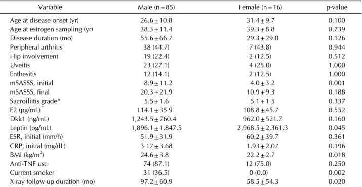
Table 1. Baseline clinical characteristics
Variable Male (n=85) Female (n=16) p-value
Age at disease onset (yr) 26.6±10.8 31.4±9.7 0.100
Age at estrogen sampling (yr) 38.3±11.4 39.3±8.8 0.739
Disease duration (mo) 55.6±66.7 29.3±29.0 0.126
Peripheral arthritis 38 (44.7) 7 (43.8) 0.944
Hip involvement 19 (22.4) 2 (12.5) 0.512
Uveitis 23 (27.1) 4 (25.0) 1.000
Enthesitis 12 (14.1) 2 (12.5) 1.000
mSASSS, initial 8.9±11.2 4.0±3.2 0.001
mSASSS, final 20.3±21.9 10.9±9.3 0.188
Sacroiliitis grade* 5.5±1.6 5.1±1.5 0.337
E2 (pg/mL)† 114.1±35.9 108.8±45.7 0.552
Dkk1 (ng/mL) 1,243.5±760.4 962.0±521.7 0.160
Leptin (pg/mL) 1,896.1±1,847.5 2,968.5±2,361.3 0.045
ESR, initial (mm/h) 51.9±31.9 60.2±39.7 0.361
CRP, initial (mg/dL) 3.17±3.68 1.93±2.07 0.196
BMI (kg/m2) 24.6±3.8 22.2±2.7 0.018
Anti-TNF use 74 (87.1) 12 (75.0) 0.250
Current smoker 31 (36.5) 0 (0.0) 0.002
X-ray follow-up duration (mo) 97.2±60.9 58.5±54.3 0.020
Data are expressed as mean±standard deviation or number (%). mSASSS: modified Stoke Ankylosing Spondylitis Spine Score, E2:
17β-estradiol, Dkk1: dickkopf-1, ESR: erythrocyte sedimentation rate, CRP: C-reactive protein, BMI: body mass index, TNF: tumor necrosis factor. *Sacroiliitis grade refers to the sum of sacroiliitis grades of each side according to the modified New York criteria.
†Reference range for E2 are 0∼400 pg/mL.
U-test was performed for continuous variables, as appropriate. Chi-square tests were used for categorical variables. The Spearman correlation analysis was used to investigate the relationship between disease duration and initial mSASSS. Radiographic progression over time was investigated using generalized estimating equations (GEE). The variables associated with p-value ≤0.20 were included in the multivariate analysis. A simple correla- tion analysis was used to analyze the correlation between two continuous parameters. Statistical analysis was exe- cuted using SAS version 9.4 (SAS Institute, Cary, NC, USA) and R version 3.0.3 (The R Foundation for Statistical Computing, Vienna, Austria).
RESULTS
A total of 101 patients were included and analyzed. The clinical characteristics of the patients are shown in Table 1. The mean age at disease onset was 27.3±10.7 years, and 16 patients (15.8%) were female. Mean E2 level was 113.2±37.4 pg/mL. Eighty-six patients (85.2%) used an- ti-TNF agents. Female patients showed lower initial mSASSS score than male patients. Leptin level was higher
and BMI was lower in female patients than in male patients. The X-ray follow-up duration was significantly higher in male patients than in female patients (97.2±60.9 vs. 58.5±54.3 months; p=0.020). Among the 101 patients, 35 (34.7%) were categorized as pro- gressors (ΔmSASSS/year ≥1 unit/year), and 66 (65.3%) were categorized as non-progressors (ΔmSASSS/year <1 unit/year). Table 2 shows a comparison of the character- istics of non-progressors and progressors. Disease dura- tion before the initial rheumatology department visit was significantly longer in progressors than in non-progressors.
The proportion of females was significantly higher in the non-progressor group than the progressor group. E2 level was significantly higher in the progressor group com- pared with the non-progressor group. Initial CRP level and BMI were significantly higher in the progressor group. The Dkk1 level was significantly lower in the pro- gressor group, and leptin levels were not significantly dif- ferent between the two groups. Figure 1A shows the nat- ural course of radiographic progression in patients with AS. Variable progression rates were seen within and across patients. The mean X-ray follow-up time was 91.2±61.2 months, and the mean ΔmSASSS/year was
Table 2. Comparison of characteristics between non-progressors (ΔmSASSS <1 unit/year) and progressors (ΔmSASSS ≥1 unit/year)
Variable Non-progressor (n=66) Progressor (n=35) p-value
Age at disease onset (yr) 26.8±11.5 28.3±9.2 0.528
Age at estrogen sampling (yr) 36.3±11.6 42.6±8.5 0.006
Disease duration (mo) 37.1±38.4 78.4±87.5 0.011
Female 14 (21.2) 2 (5.7) 0.048
Peripheral arthritis 32 (48.5) 13 (37.1) 0.275
Hip involvement 17 (25.8) 4 (11.4) 0.123
Uveitis 18 (27.3) 9 (25.7) 0.866
Enthesitis 9 (13.6) 5 (14.3) 0.928
mSASSS, initial 3.8±4.5 16.3±13.4 <0.001
mSASSS, final 17.9±20.2 21.6±22.7 0.445
Sacroiliitis grade* 4.94±1.37 6.31±1.62 <0.001
Estrogen (pg/mL)† 107.7±37.5 123.6±35.6 0.042
Dkk1 (ng/mL) 1,303.7±778.9 1,001.2±598.7 0.048
Leptin (pg/mL) 1,939.0±1,838.8 2,305.4±2,189.4 0.375
ESR, initial (mm/h) 49.1±35.4 61.0±27.3 0.087
CRP, initial (mg/dL) 2.31±2.75 4.21±4.37 0.024
BMI (kg/m2) 23.1±3.2 26.3±3.8 <0.001
Anti-TNF use 53 (80.3) 33 (94.3) 0.079
Current smoker 17 (25.8) 14 (40.0) 0.140
X-ray follow-up duration (mo) 93.3±58.3 119.1±66.9 0.080
Data are expressed as mean±standard deviation or number (%). mSASSS: modified Stoke Ankylosing Spondylitis Spine Score, Dkk1: dickkopf-1, ESR: erythrocyte sedimentation rate, CRP: C-reactive protein, BMI: body mass index, TNF: tumor necrosis factor, E2: 17β-estradiol. *Sacroiliitis grade refers to the sum of sacroiliitis grades of the each sides according to the modified New York criteria. †Reference ranges for E2 are 0∼400 pg/mL.
Figure 1. (A) The natural course of radiographic progression. The black line indicates the mean radiographic progression of the en- tire patient cohort. (B) Predicted value of radiographic progression over time in multivariable analysis according to sex. mSASSS:
modified Stoke Ankylosing Spondylitis Spine Score.
1.03±1.36 units/year. The X-ray follow-up duration was not significantly different between non-progressors and progressors (93.3±58.3 vs. 119.1±66.9 months; p=0.080).
Among the male patients, 33 (38.8%) were categorized as progressors, and 52 (61.2%) were categorized as non-
progressors (Table 3). Initial mSASSS, Dkk1, initial CRP and BMI levels were significantly different between non-progressors and progressors among male patients.
Interactions with clinical characteristics on spinal radio- graphs with the GEE model are shown in Table 4. In uni-
Table 3. Comparison of characteristics between non-progressors (ΔmSASSS <1 unit/year) and progressors (ΔmSASSS ≥1 unit/year) among male patients with ankylosing spondylitis (n=85)
Variable Non-progressor (n=52) Progressor (n=33) p-value
Age at disease onset (yr) 25.9±11.8 27.5±8.9 0.507
Age at estrogen sampling (yr) 35.6±12.1 42.6±8.7 0.005
Disease duration (mo) 38.5±40.4 82.4±88.6 0.003
Peripheral arthritis 26 (50.0) 12 (36.4) 0.218
Hip involvement 15 (28.8) 4 (12.1) 0.108
Uveitis 14 (26.9) 9 (27.3) 0.972
Enthesitis 7 (13.5) 5 (15.2) 0.827
mSASSS, initial 3.9±4.8 16.8±13.7 <0.001
mSASSS, final 19.2±21.5 22.0±23.0 0.601
Sacroiliitis grade* 4.90±1.33 6.39±1.61 <0.001
Estrogen (pg/mL)† 109.6±36.7 121.1±34.1 0.153
Dkk1 (ng/mL) 1,384.0±823.6 1,022.1±595.0 0.032
Leptin (pg/mL) 1,591.7±1,497.3 2,375.7±2,235.8 0.082
ESR, initial (mm/h) 47.2±34.3 59.3±26.6 0.088
CRP, initial (mg/dL) 2.51±2.96 4.19±4.44 0.039
BMI (kg/m2) 23.4±3.3 26.5±3.7 <0.001
Anti-TNF use 43 (82.7) 31 (93.9) 0.132
Current smoker 17 (32.7) 14 (42.4) 0.364
X-ray follow-up duration (mo) 88.7±58.3 110.6±63.4 0.106
Data are expressed as mean±standard deviation or number (%). mSASSS: modified Stoke Ankylosing Spondylitis Spine Score, Dkk1: dickkopf-1, ESR: erythrocyte sedimentation rate, CRP: C-reactive protein, BMI: body mass index, TNF: tumor necrosis factor, E2: 17β-estradiol. *Sacroiliitis grade refers to the sum of sacroiliitis grades of the each sides according to the modified New York criteria. †Reference ranges for E2 are 0∼400 pg/mL.
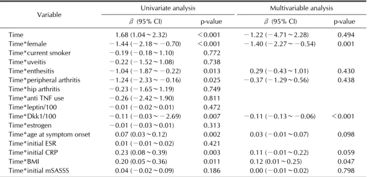
Table 4. Interactions with baseline characteristics and spinal radiographic progression with generalized estimating equations model (n=101)
Variable Univariate analysis Multivariable analysis
β (95% CI) p-value β (95% CI) p-value
Time 1.68 (1.04∼2.32) <0.001 −1.22 (−4.71∼2.28) 0.494
Time*female −1.44 (−2.18∼−0.70) <0.001 −1.40 (−2.27∼−0.54) 0.001
Time*current smoker −0.19 (−0.18∼1.10) 0.772
Time*uveitis −0.22 (−1.52∼1.08) 0.738
Time*enthesitis −1.04 (−1.87∼−0.22) 0.013 0.29 (−0.43∼1.01) 0.430
Time*peripheral arthritis −1.24 (−2.33∼−0.16) 0.025 −0.37 (−1.29∼0.56) 0.438
Time*hip arthritis −0.23 (−1.65∼1.19) 0.749
Time*anti TNF use −0.26 (−2.42∼1.90) 0.811
Time*leptin/100 −0.01 (−0.02∼0.01) 0.472
Time*Dkk1/100 −0.11 (−0.03∼−2.69) 0.007 −0.11 (−0.13∼−0.06) <0.001
Time*estrogen −0.01 (−0.03∼0.01) 0.313
Time*age at symptom onset 0.07 (0.03∼0.12) 0.002 0.03 (−0.01∼0.07) 0.098
Time*initial ESR 0.01 (−0.01∼0.02) 0.421
Time*initial CRP 0.23 (0.08∼0.39) 0.003 0.11 (−0.01∼0.22) 0.059
Time*BMI 0.20 (0.05∼0.36) 0.011 0.12 (0.01∼0.25) 0.047
Time*initial mSASSS 0.04 (−0.02∼0.09) 0.186 0.00 (−0.01∼0.02) 0.798
TNF: tumor necrosis factor, Dkk1: dickkopf-1, ESR: erythrocyte sedimentation rate, CRP: C-reactive protein, BMI: body mass index, mSASSS: modified Stoke Ankylosing Spondylitis Spine Score, CI: confidence interval.
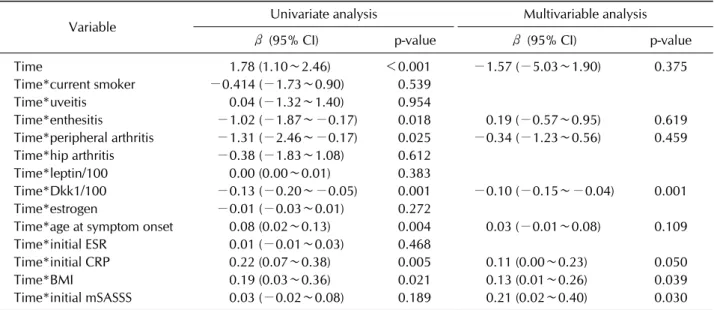
Table 5. Interactions with baseline characteristics and spinal radiographic progression with generalized estimating equations model among male patients with ankylosing spondylitis (n=85)
Variable Univariate analysis Multivariable analysis
β (95% CI) p-value β (95% CI) p-value
Time 1.78 (1.10∼2.46) <0.001 −1.57 (−5.03∼1.90) 0.375
Time*current smoker −0.414 (−1.73∼0.90) 0.539
Time*uveitis 0.04 (−1.32∼1.40) 0.954
Time*enthesitis −1.02 (−1.87∼−0.17) 0.018 0.19 (−0.57∼0.95) 0.619
Time*peripheral arthritis −1.31 (−2.46∼−0.17) 0.025 −0.34 (−1.23∼0.56) 0.459 Time*hip arthritis −0.38 (−1.83∼1.08) 0.612
Time*leptin/100 0.00 (0.00∼0.01) 0.383
Time*Dkk1/100 −0.13 (−0.20∼−0.05) 0.001 −0.10 (−0.15∼−0.04) 0.001
Time*estrogen −0.01 (−0.03∼0.01) 0.272
Time*age at symptom onset 0.08 (0.02∼0.13) 0.004 0.03 (−0.01∼0.08) 0.109
Time*initial ESR 0.01 (−0.01∼0.03) 0.468
Time*initial CRP 0.22 (0.07∼0.38) 0.005 0.11 (0.00∼0.23) 0.050
Time*BMI 0.19 (0.03∼0.36) 0.021 0.13 (0.01∼0.26) 0.039
Time*initial mSASSS 0.03 (−0.02∼0.08) 0.189 0.21 (0.02∼0.40) 0.030
Dkk1: dickkopf-1, ESR: erythrocyte sedimentation rate, CRP: C-reactive protein, BMI: body mass index, mSASSS: modified Stoke Ankylosing Spondylitis Spine Score, CI: confidence interval.
variable analysis, age at symptom onset, CRP, and BMI were positively associated with radiographic progression.
Female, presence of enthesitis, peripheral arthritis, and Dkk1 level were negatively associated with radiographic progression. In multivariable analysis, Dkk1 was sig- nificantly associated with less radiographic progression, and BMI was associated with increased risk of radio- graphic progression. Female patients were associated with less radiographic progression. However, estrogen level and leptin level were not significantly associated with radiographic progression. Figure 1B shows the pre- dicted value of radiographic progression according to sex in multivariable analysis. The slope of the male patients was higher than that of the females over the follow-up time. In the subgroup analysis of male patients with AS (n=85), CRP level, BMI, and initial mSASSS were sig- nificantly associated with radiographic progression in a multivariable analysis (Table 5). Dkk1 level was asso- ciated with decreased risk of radiographic progression. In addition, serum estrogen level and BMI showed a weak positive correlation (Pearson correlation coefficient [r]=0.225; p=0.023).
DISCUSSION
We investigated the effects of sex and estrogen on the ra- diographic progression of spondyloarthritis (SpA).
Although female was associated with less radiographic progression, estrogen level was not significantly asso- ciated with radiographic progression in patients with AS.
Several studies have investigated the differences between men and women in patients with AS. These studies corre- spond well with a previous study in which adrenal and go- nadal hormones were not altered in patients with AS [14], and women had less radiographic changes com- pared with men with AS [15].
The sex hormone estrogen is involved in the immune re- sponse and affects lymphoid and myeloid cell differ- entiation, cytokine production, and Th cell polarization.
Estrogen decreased the production of Th1 cells and in- creased the production of Th2 cells [16]. Estrogen in- creased IL-10 and IL-4 expression, whereas it decreased the expression of TNFα [17]. Estrogen also inhibits Th17 cell differentiation through downregulation of ROR γT expression [18]. Thus, in a Th1-mediated auto- immune disease such as rheumatoid arthritis (RA), dis- ease activity often decreases during pregnancy ,and Th2-mediated diseases such as systemic lupus eryth- ematosus often flare or initially develop in the post- partum period [19]. In contrast to RA, it seems that preg- nancy does not improve the symptoms of AS [20].
Ostensen and Ostensen [21] reported that disease activ- ity during pregnancy was unchanged in 33.2%, improved in 30.9%, and worsened in 32.5% of patients with AS.
However, postpartum flares were observed in 60% of the patients. Postpartum flares can be explained by an in- crease in soluble TNF receptor during pregnancy com- pared to non-pregnant women and a decrease in the TNF receptor during the postpartum period in patients with rheumatic disease [22].
We previously reported that BMI and estrogen level pre- dict radiographic progression in patients with AS [11].
The results of this study were in contrast to the expect- ation that estrogen would have an anti-inflammatory effect. Because the number of patients included in the previous study was small, the number of patients enrolled in the current study was twice that of previous studies, and the GEE method was used to analyze factors asso- ciated with radiographic progression to adjust for time.
Serum levels of leptin and Dkk1 were analyzed as well as other traditional risk factors. Although estrogen level was higher in the progressors than non-progressors, we could not find a statistically significant association between es- trogen level and radiographic progression in the GEE analysis in the current study. Interestingly, BMI was sig- nificantly associated with radiographic progression in both previous and current studies. Moreover, estrogen level was positively related to BMI in both our previous and current studies, which corresponds well with other reports [23].
Sex hormones can influence body weight and body fat and also affect estrogen level [24]. In males, estrogen is derived from the tissue aromatization of testosterone [23]. However, the serum estradiol level does not de- crease with age as much as the serum total testosterone level because aromatase activity increases with age, and there is an age-associated increase in fat mass. Adipokines, which are a secreted form of adipocytes in fat tissue, are bioactive substances having an immunomodulatory ef- fect [25]. Serum levels of adipokines, such as leptin, TNFα, IL-6, and IL-1β, positively correlate with BMI, and the concentrations of adipokines are increased in patients with metabolic syndrome [26]. In patients with AS, adi- pokines are related to spinal radiographic progression.
Among adipokines, serum levels of resistin and visfatin are elevated in patients with AS, and visfatin was a pre- dictive factor of radiographic progression [27]. High es- trogen levels in patients with radiographic progression might result from the secondary elevation of estrogen caused by fat aromatization. Although leptin level was not significantly associated with radiographic pro- gression in the current study, several adipokines might be
associated with a high inflammatory burden in obese pa- tients with AS and ultimately result in more rapid radio- graphic progression. Obesity is related to a poor response to tumor necrosis factor inhibitor (TNFi) in patients with AS [28]. Obesity is also associated with TNFi dis- continuation related with anti-drug antibody formation [29]. We previously investigated the effect of estrogen on the disease activity of SpA using a zymosan-induced SKG mice model, and we found that estrogen significantly re- duced the disease activity of SpA [30]. Considering that BMI was also associated with radiographic progression in AS, the anti-inflammatory effect of estrogen might be overcome by the pro-inflammatory cytokines produced by fat tissue.
Estrogen may aggravate SpA considering the direct modulation of osteoblastic activity by estrogen [31].
Estrogen deficiency causes osteoporosis in postmenopausal women. Pro-inflammatory cytokines such as IL-1, IL-6, and TNFα increase bone resorption and are down- regulated by estrogen [32]. Other studies reported that estrogen upregulates TGFβ, an inhibitor of bone re- sorption that acts on osteoclasts to decrease osteoclast activity and increase apoptosis [33]. Estrogen also sup- presses the receptor activator of nuclear factor kappa-B li- gand-induced osteoclast differentiation. These reports suggest that estrogen inhibits bone resorption and in- creases bone formation.
The baseline CRP level and initial mSASSS were also sig- nificantly associated with radiographic progression espe- cially in male patients, which is consistent with the re- sults of German Spondyloarthritis Inception Cohort (GESPIC) patients [6]. We also evaluated the role of Wnt inhibitors in the disease activity of SpA. Dkk1 is a potent inhibitor of the Wnt signaling pathway and has been re- ported to be involved in the etiology of AS. Although Dkk1 has been reported to be involved in the etiology of AS, data on the Dkk1 serum level is conflicting. Daoussis et al. [34] reported that Dkk1 level was increased in pa- tients with AS compared with the control group.
However, another study showed that Dkk1 level was low- er in patients with AS than the control group [35,36]. A recent meta-analysis reported that serum Dkk1 level was higher in AS patients than in the control group [37]. The current study showed that the Dkk1 level was negatively associated with radiographic progression in patients with SpA. This corresponds well with the previous findings of the GESPIC study, which reported that patients with no syndesmophyte formation show significantly higher
Dkk1 level compared with those with syndesmophyte formation [38]. In accordance with our results, a recent study showed that Dkk1 level was significantly higher in AS patients with no syndesmophytes [39]. Blockade of Dkk1 significantly reduced bone erosions and osteoclast count in the TNF tg mice model [40]. The Dkk1 blockade enhanced collagen type “X” expression, which results in the formation of hypertrophic chondrocytes and anky- loses of the sacroiliac joint in the TNF tg mice. Dkk1 level was increased in patients with AS who received NSAID or TNFi [34,36]. Although Dkk1 might be dysfunctional in AS [34], the results of the current study suggests that Dkk1 might inhibit radiographic progression in patients with SpA.
There are several limitations in this study. First, most of the patients had established AS and had been treated with appropriate medications for a long time. Although we could not analyze the disease activity of AS such as the Ankylosing Spondylitis Disease Activity Score or Bath Ankylosing Spondylitis Disease Activity Index, the dis- ease activity of most of the included patients was low. It is not suitable to evaluate the effect of estrogen on the early phase of AS. We focused on the effect of estrogen on ra- diographic progression rather than disease activity.
Second, estrogen level was assessed just one time at a routine visit at the rheumatologic clinic during the study enrollment period. Furthermore, estrogen level in the hu- man body is affected by various factors such as sex, age, and BMI. Other unknown confounding factors could not fully be adjusted. A hormone analysis of the human body has limitations, and interpretation must be performed with caution. This is a major limitation to evaluating the effect of estrogen on radiographic progression. Third, es- trogen levels were not compared with a normal control group. Forth, the possibility of future radiographic pro- gression in the non-progressor group could not be com- pletely excluded due to variation in the timing of fol- low-up X-ray. Although patients with a follow-up X-ray for a period of 2 years or longer were included, there is a concern about selection bias. However, we investigated the effects of sex and estrogen on the spinal radiographic progression in SpA patients with relatively long-term X-ray follow-up data including other traditional risk factors.
CONCLUSION
Although female patients were associated with less ra-
diographic progression in AS, there was no significant re- lationship between serum estrogen level and radio- graphic progression. In addition, high BMI and lower Dkk1 were associated with spinal radiographic progression.
Genetic factors or other environmental factors associated with female may influence radiographic progression in patients with AS rather than female sex hormones.
Further study is warranted to evaluate the effects of estro- gen on the disease activity of AS and reveal the patho- physiology of estrogen in patients with AS.
CONFLICT OF INTEREST
No potential conflict of interest relevant to this article was reported.
AUTHOR CONTRIBUTIONS
H.J. conceptualized the study, collected the data, ana- lyzed data, and drafted the manuscript. E.K.B., E.J.P., and J.H. collected data and analyzed data. J.L., C.H.J., and E.M.K. collected the data and reviewed the article. H.S.C.
conceptualized the study, collected the data, and critically reviewed the article.
REFERENCES
1. Lee W, Reveille JD, Davis JC Jr, Learch TJ, Ward MM, Weisman MH. Are there gender differences in severity of ankylosing spondylitis? Results from the PSOAS cohort.
Ann Rheum Dis 2007;66:633-8.
2. Will R, Edmunds L, Elswood J, Calin A. Is there sexual in- equality in ankylosing spondylitis? A study of 498 women and 1202 men. J Rheumatol 1990;17:1649-52.
3. Jeong H, Yoon JY, Park EJ, Hwang J, Kim H, Ahn JK, et al.
Clinical characteristics of nonradiographic axial spondy- loarthritis in Korea: a comparison with ankylosing spondylitis.
Int J Rheum Dis 2015;18:661-8.
4. Kobak S, Yalçin M, Karadeniz M, Oncel G. Coexistence of ankylosing spondylitis and Klinefelter's syndrome. Case Rep Rheumatol 2013;2013:543953.
5. Armstrong RD, Macfarlane DG, Panayi GS. Ankylosing spondylitis and Klinefelter's syndrome: does the X chromo- some modify disease expression? Br J Rheumatol 1985;24:
277-81.
6. Poddubnyy D, Haibel H, Listing J, Märker-Hermann E, Zeidler H, Braun J, et al. Baseline radiographic damage, ele- vated acute-phase reactant levels, and cigarette smoking status predict spinal radiographic progression in early axial spondylarthritis. Arthritis Rheum 2012;64:1388-98.
7. Jimenez-Balderas FJ, Tapia-Serrano R, Madero-Cervera JI, Murrieta S, Mintz G. Ovarian function studies in active an- kylosing spondylitis in women. Clinical response to estro-
gen therapy. J Rheumatol 1990;17:497-502.
8. Giltay EJ, Popp-Snijders C, van Schaardenburg D, Dekker-Saeys BJ, Gooren LJ, Dijkmans BA. Serum testosterone levels are not elevated in patients with ankylosing spondylitis. J Rheumatol 1998;25:2389-94.
9. Arniaud D, Mattei JP, Boyer J, Roux H. Sex hormones in spondylarthropathies. A study in 57 patients. Rev Rhum Engl Ed 1998;65:21-6.
10. Cunningham M, Gilkeson G. Estrogen receptors in im- munity and autoimmunity. Clin Rev Allergy Immunol 2011;
40:66-73.
11. Jeong H, Bea EK, Lee J, Koh EM, Cha HS. Body mass index and estrogen predict radiographic progression in the spine in ankylosing spondylitis. Joint Bone Spine 2015;82:473-4.
12. van der Linden S, Valkenburg HA, Cats A. Evaluation of di- agnostic criteria for ankylosing spondylitis. A proposal for modification of the New York criteria. Arthritis Rheum 1984;27:361-8.
13. Creemers MC, Franssen MJ, van't Hof MA, Gribnau FW, van de Putte LB, van Riel PL. Assessment of outcome in ankylos- ing spondylitis: an extended radiographic scoring system.
Ann Rheum Dis 2005;64:127-9.
14. Straub RH, Struhárová S, Schölmerich J, Härle P. No alter- ations of serum levels of adrenal and gonadal hormones in patients with ankylosing spondylitis. Clin Exp Rheumatol 2002;20(6 Suppl 28):S52-9.
15. Baraliakos X, Listing J, von der Recke A, Braun J. The natural course of radiographic progression in ankylosing spondyli- tis: differences between genders and appearance of charac- teristic radiographic features. Curr Rheumatol Rep 2011;
13:383-7.
16. Giltay EJ, van Schaardenburg D, Gooren LJ, Popp-Snijders C, Dijkmans BA. Androgens and ankylosing spondylitis: a role in the pathogenesis? Ann N Y Acad Sci 1999;876:
340-64; discussion 365.
17. Javadian A, Salehi E, Bidad K, Sahraian MA, Izad M. Effect of estrogen on Th1, Th2 and Th17 cytokines production by proteolipid protein and PHA activated peripheral blood mononuclear cells isolated from multiple sclerosis patients.
Arch Med Res 2014;45:177-82.
18. Chen RY, Fan YM, Zhang Q, Liu S, Li Q, Ke GL, et al.
Estradiol inhibits Th17 cell differentiation through in- hibition of RORγT transcription by recruiting the ERα/REA complex to estrogen response elements of the RORγT promoter. J Immunol 2015;194:4019-28.
19. Wilder RL. Hormones, pregnancy, and autoimmune diseases.
Ann N Y Acad Sci 1998;840:45-50.
20. Ostensen M, Husby G. Ankylosing spondylitis and pregnancy.
Rheum Dis Clin North Am 1989;15:241-54.
21. Ostensen M, Ostensen H. Ankylosing spondylitis--the fe- male aspect. J Rheumatol 1998;25:120-4.
22. Østensen M, Förger F, Nelson JL, Schuhmacher A, Hebisch G, Villiger PM. Pregnancy in patients with rheumatic dis- ease: anti-inflammatory cytokines increase in pregnancy and decrease post partum. Ann Rheum Dis 2005;64:839-44.
23. Vermeulen A, Kaufman JM, Goemaere S, van Pottelberg I.
Estradiol in elderly men. Aging Male 2002;5:98-102.
24. O'Sullivan AJ, Hoffman DM, Ho KK. Estrogen, lipid oxida- tion, and body fat. N Engl J Med 1995;333:669-70.
25. Ouchi N, Parker JL, Lugus JJ, Walsh K. Adipokines in in-
flammation and metabolic disease. Nat Rev Immunol 2011;11:85-97.
26. Deng Y, Scherer PE. Adipokines as novel biomarkers and regulators of the metabolic syndrome. Ann N Y Acad Sci 2010;1212:E1-19.
27. Syrbe U, Callhoff J, Conrad K, Poddubnyy D, Haibel H, Junker S, et al. Serum adipokine levels in patients with anky- losing spondylitis and their relationship to clinical parame- ters and radiographic spinal progression. Arthritis Rheumatol 2015;67:678-85.
28. Micheroli R, Hebeisen M, Wildi LM, Exer P, Tamborrini G, Bernhard J, et al. Impact of obesity on the response to tumor necrosis factor inhibitors in axial spondyloarthritis.
Arthritis Res Ther 2017;19:164.
29. Hwang J, Kim HM, Jeong H, Lee J, Ahn JK, Koh EM, et al.
Higher body mass index and anti-drug antibodies predict the discontinuation of anti-TNF agents in Korean patients with axial spondyloarthritis. Rev Bras Reumatol Engl Ed 2017;57:311-9.
30. Jeong H, Bae EK, Kim H, Eun YH, Kim IY, Kim H, et al.
Estrogen attenuates the spondyloarthritis manifestations of the SKG arthritis model. Arthritis Res Ther 2017;19:198.
31. Majeska RJ, Ryaby JT, Einhorn TA. Direct modulation of os- teoblastic activity with estrogen. J Bone Joint Surg Am 1994;76:713-21.
32. Pacifici R. Estrogen, cytokines, and pathogenesis of post- menopausal osteoporosis. J Bone Miner Res 1996;11:
1043-51.
33. Manolagas SC. Birth and death of bone cells: basic regu- latory mechanisms and implications for the pathogenesis and treatment of osteoporosis. Endocr Rev 2000;21:115-37.
34. Daoussis D, Liossis SN, Solomou EE, Tsanaktsi A, Bounia K, Karampetsou M, et al. Evidence that Dkk-1 is dysfunc- tional in ankylosing spondylitis. Arthritis Rheum 2010;62:
150-8.
35. Kwon SR, Lim MJ, Suh CH, Park SG, Hong YS, Yoon BY, et al. Dickkopf-1 level is lower in patients with ankylosing spondylitis than in healthy people and is not influenced by anti-tumor necrosis factor therapy. Rheumatol Int 2012;32:
2523-7.
36. Liao HT, Lin YF, Tsai CY, Chou TC. Bone morphogenetic proteins and Dickkopf-1 in ankylosing spondylitis. Scand J Rheumatol 2018;47:56-61.
37. Zhang L, Ouyang H, Xie Z, Liang ZH, Wu XW. Serum DKK-1 level in the development of ankylosing spondylitis and rheumatic arthritis: a meta-analysis. Exp Mol Med 2016;48:e228.
38. Heiland GR, Appel H, Poddubnyy D, Zwerina J, Hueber A, Haibel H, et al. High level of functional dickkopf-1 predicts protection from syndesmophyte formation in patients with ankylosing spondylitis. Ann Rheum Dis 2012;71:572-4.
39. Sakellariou GT, Iliopoulos A, Konsta M, Kenanidis E, Potoupnis M, Tsiridis E, et al. Serum levels of Dkk-1, sclero- stin and VEGF in patients with ankylosing spondylitis and their association with smoking, and clinical, inflammatory and radiographic parameters. Joint Bone Spine 2017;84:
309-15.
40. Uderhardt S, Diarra D, Katzenbeisser J, David JP, Zwerina J, Richards W, et al. Blockade of Dickkopf (DKK)-1 induces fusion of sacroiliac joints. Ann Rheum Dis 2010;69:592-7.