Therapeutic Hypothermia after Decompressive Craniectomy in Malignant Cerebral Infarction
Jun Young Chang, M.D., Jeong-Ho Hong, M.D.*, Jin-Heon Jeong, M.D., Sung-Jin Nam, M.D., Ji-Hwan Jang, M.D.†, Jae Seung Bang, M.D.†, and Moon-Ku Han, M.D.‡
Department of Interdisciplinary Care Medicine, Seoul National University Bundang Hospital, Seongnam; *Department of Neurology, Keimyung University Dongsan Medical Center, Daegu; Departments of †Neurosurgery, and ‡Neurology, Seoul National University Bundang Hospital, Seongnam, Korea
Decompressive hemicraniectomy followed by subsequent therapeutic hypothermia can reduce mortality in patients with malignant cerebral infarction without significantly increasing risk. We report three cases of malignant cerebral infarction treated with hemi- craniectomy followed by hypothermia. Case 1 received elective decompressive surgery and hypothermia. Case 2 developed subsequent cerebral infarction with uncal herniation. Therefore, emergent decompressive surgery and hypothermia was performed in this case.
Despite surgery and hyperosmolar therapy, case 3 received hypothermia treatment for refractory increased intracranial pressure. All pa- tients survived with a score of 4 or 5 on the modified Rankin scale. Therefore, we suggest that application of hypothermia after hemi- craniectomy is safe and feasible. Several possible modifications can be made to improve the management strategy in order to increase the benefits of hypothermia treatment.
Key Words: brain edema; cerebral infarction; decompressive craniectomy; hypothermia.
Received on February 4, 2014 Revised on April 2, 2014 Accepted on April 16, 2014
Correspondence to: Moon-Ku Han, Department of Neurology, Seoul National University Bundang Hospital, 82 Gumi-ro 173beon-gil, Bundang-gu, Seongnam 463-707, Korea
Tel: +81-31-787-7464, Fax: +82-31-787-4059 E-mail: [email protected]
93
This is an Open Access article distributed under the terms of the Creative Commons Attribution Non-Commercial License (http://creativecommons.org/
licenses/by-nc/3.0/) which permits unrestricted non-commercial use, distribution, and reproduction in any medium, provided the original work is properly cited.
ⓒ 2014 Korean Society of Critical Care Medicine
Malignant cerebral infarction consists of 3% to 10% of supra- tentorial cerebral infarctions and 80% of the patients die without proper treatment.[1,2] Early decompressive surgery prior to her- niation is known to reduce 1- and 6-month mortality to 4.8 and 19.1%, respectively.[3] Moderate hypothermia is also a useful therapeutic option for space-occupying cerebral edema, sig- nificantly lowering elevated intracranial pressure (ICP) and pro- tecting from further neuronal damage.[4] Moreover, therapeutic hypothermia after the hemicraniectomy has an additional effect on decreasing mortality and functional disability without in- creasing the occurrence of adverse events.[5] We have studied three patients with malignant cerebral infarction who underwent
decompressive hemicraniectomy and subsequent hypothermia without serious complications.
CASE REPORT
1) Case 1
A 26-year-old man presented to the emergency room (ER) with right hemiparesis, global aphasia, and drowsiness with a history of pulmonary embolism and exertional dyspnea for a month. He was brought to a nearby tertiary hospital and initial brain magnetic resonance imaging (MRI) revealed large acute infarction in the left middle cerebral artery (MCA) with left proximal internal carotid artery (ICA) occlusion. After recanali- zation therapy had failed, the patient was transferred to the neuroICU in our hospital. Upon arrival at the neuroICU, initial National Institutes of Health Stroke Scale (NIHSS) score of the patient was 22. As a development of malignant cerebral edema was predicted, the patient received an elective hemicraniectomy 14 hours after symptom onset and therapeutic hypothermia was
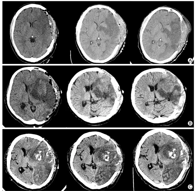
A
B
C
Fig. 1. Serial brain CT of the patients. (A) Case 1: Brain CT was checked at postoperative day 0, 4, 7. Midline shifting was minimal and the cerebral edema ceased to progress between day 4 and day 7. (B) Case 2: Brain CT was performed at postoperative day 1, 4 and 7. Midline shifting and obstructive hydrocephalus were evident at day 1. Cerebral edema was gradually decreased during hypo- thermia treatment. (C) Case 3: Brain CT was evaluated at postoperative day 0, 4 and 6. Large hemispheric infarction was accom- panied by hemorrhagic transformation at day 0. Despite the infarct area became more evident, the degree of midline shifting did not increase till day 8.
subsequently applied. During that period, the progression of cer- ebral edema and midline shifting was minimal on serial brain computed tomography (CT) images (Fig. 1a). After verifying that cerebral edema ceased to progress, rewarming was initiated 6 days following hypothermia. Due to hypotension caused by hy- pothermia and anti-shivering medications, inotropic agents were administered for 7 days. Sinus bradycardia (with a heart rate of 60 beats/min or less) occurred during the first 3 days after apply- ing hypothermia but recovered without specific management.
Mechanical ventilation was maintained for 9 days and discontinued after pneumonia was resolved. The patient was transferred to the recovery ward at 21 days after admission. The patient survived and was discharged with mild improvement of NIHSS total score of 16. Modified Rankin scale (MRS) score at discharge was the same as the initial score of 5. The laboratory results for hypercoagulable states including anti-phospholipid antibodies and prothrombin gene mutation were all negative.
2) Case 2
A 50-year-old woman with history of paroxysmal atrial fi- brillation and hypertension was found after having fallen in her bathroom with an altered mental status. In the ER, she presented with drowsiness, was unable to speak and had decreased with- drawal response to pain stimuli on the right side implying right hemiplegia or hypesthesia. Initial NIHSS was 22. Diffusion- weighted image revealed a superior division of the left MCA, anterior cerebral artery (ACA) territorial infarction with mas- sive hemorrhagic transformation and midline shifting (4.3 mm).
Based on the decision of the attending neurologist, hyperosmolar therapy was initiated instead of surgery. The midline shifting was increased to 8.2 mm on the fourth day and ceased to progress on the seventh day without obvious neurologic deterioration. However, at day 10, her mental status became stuporous, left pupil was di- lated and nonreactive to light. Brain CT revealed a newly devel- oped inferior division of the left MCA infarction with uncal herniation. Emergent decompressive hemicraniectomy was per- formed and moderate hypothermia was induced after the operation.
Midline shifting and obstructive hydrocephalus gradually de- creased during the treatment (Fig. 1b). Hypothermia was main- tained for 2 days and slowly rewarmed for 16 h. Normothermia was continued for 2 days. Due to hypotension at the beginning of the hypothermia therapy, continuous inotropic agents were infused for 4 days to maintain mean arterial pressure of 90 to 110 mmHg. Transient slow ventricular response of atrial fi- brillation with a heart rate less than 40 beats/min was observed, but recovered immediately without medications. As the mental status of the patient improved to near alert, pupillary reflex was restored and midline shifting decreased to 2 mm in the brain CT image, we decided to move her to the general ward on the elev- enth day after neuroICU readmission. The NIHSS score at dis- charge was 22 and MRS score was 5. The MRS score 3 months later did not improve.
3) Case 3
A 76-year-old man with history of atrial fibrillation and Parkinsonism developed an acute right-sided hemiparesis and global aphasia (NIHSS score of 23). In the ER, brain MRI re- vealed a left MCA infarction with distal ICA occlusion. The oc- cluded vessel was completely recanalized after thrombolysis.
However, the patient became stuporous and brain CT revealed aggravated cerebral edema after the procedure. We decided to perform a decompressive hemicraniectomy at 17 hours after symptom onset. Despite the surgery and hyperosmolar therapy, the left pupil became dilated, nonreactive and midline shift pro-
gressed to 7 mm at follow-up CT imaging (Fig. 1c). Moderate hypothermia was induced at 24 hours after symptom onset, maintained for 7 days and slowly rewarmed to normothermia for 24 hours. After maintaining normothermia for 30 hours, hy- pothermia was discontinued. Bradycardia with a rate below 40 beats/min was sustained for 3 days and was resolved without treatment. The methicillin-resistant Streptococcus aureus asso- ciated cellulitis at the tracheostomy site was treated with an in- travenous vancomycin. Cerebral edema gradually improved and the patient was able to open his eyes in response to painful stimuli. The patient stayed at the neuroICU for 23 days. The NIHSS and MRS scores at discharge were 21 and 4, respectively.
The characteristics of the patients are summarized in Table 1.
Decompressive craniectomy
A question mark-shaped skin incision was made 1 cm ahead of a tragus. A large craniectomy including frontal, temporal and parietal bones was performed. Dura was opened in semicircular and radial shapes to retain a sufficient space for the swollen brain. Duroplasty was done using lyophilized bovine pericar- dium (Lyoplant®, B. Braun). The infarcted area of the brain was not removed. The bone flap was stored in 100% ethanol until performing cranioplasty, usually at least 6 weeks after craniectomy.
Therapeutic hypothermia
Moderate hypothermia (33.5°C) achieved by a surface cool- ing device (Arctic Sun®, Medivance, USA) was started after the operation. Surface cooling pads were attached at the trunk, back, and both thighs and core temperature was monitored via an esoph- ageal probe. One liter of cold saline (4°C) was administered for 30 min to reach the target temperature as soon as possible. To mini- mize shivering, warming blankets were routinely used for all hypothermic patients. In case shivering was not well controlled with surface warming, intravenous meperidine (50-100 mg bo- lus followed by 12.5-50 mg/h), dexmedetomidine (loading dose 1 µg/kg over 10 min followed by an infusion of 0.3-1.5 µg/kg-1/h-1), fentanyl (25-75 µg bolus dose followed by 50-200 µg/h), and magnesium (1 g/h and titrate to serum magnesium of 3 mmol/L) were used in that sequence. The duration of hypo- thermia maintenance was determined by clinical decision of the physician. Due to a risk of rebound cerebral edema, rewarming was slowly conducted at a rate of 0.1°C/h. The latter two cases retained normothermia for a certain period after rewarming was completed. During the treatment, the occurrence of possible side effects such as hypo- or hypertension, bradycardia, arrhythmia,
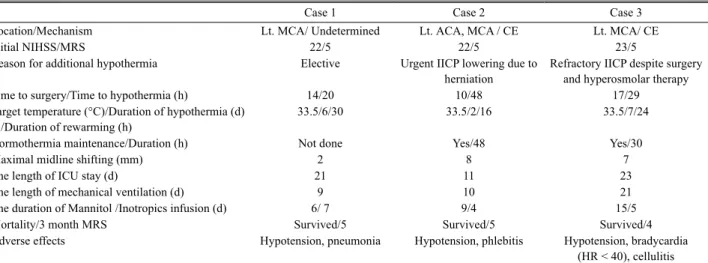
Table 1. Characteristics and outcomes of the three patients underwent decompressive hemicraniectomy followed by moderate hypothermia
Case 1 Case 2 Case 3
Location/Mechanism Lt. MCA/ Undetermined Lt. ACA, MCA / CE Lt. MCA/ CE
Initial NIHSS/MRS 22/5 22/5 23/5
Reason for additional hypothermia Elective Urgent IICP lowering due to
herniation
Refractory IICP despite surgery and hyperosmolar therapy
Time to surgery/Time to hypothermia (h) 14/20 10/48 17/29
Target temperature (°C)/Duration of hypothermia (d) /Duration of rewarming (h)
33.5/6/30 33.5/2/16 33.5/7/24
Normothermia maintenance/Duration (h) Not done Yes/48 Yes/30
Maximal midline shifting (mm) 2 8 7
The length of ICU stay (d) 21 11 23
The length of mechanical ventilation (d) 9 10 21
The duration of Mannitol /Inotropics infusion (d) 6/ 7 9/4 15/5
Mortality/3 month MRS Survived/5 Survived/5 Survived/4
Adverse effects Hypotension, pneumonia Hypotension, phlebitis Hypotension, bradycardia
(HR < 40), cellulitis Lt: left; MCA: middle cerebral artery; ACA: anterior cerebral artery; CE: cardioembolism; NIHSS: National Institutes of Health Stroke Scale; MRS:
Modified Rankin scale; IICP: increased intracranial pressure; HR: heart rate.
electrolyte imbalance, coagulopathy, and infection were closely monitored and managed.
The protocol of intensive care management during therapeutic hypothermia
All patients who underwent decompressive hemicraniectomy were sedated using midazolam, remifentanyl, and cisatracurium aimed at a Ramsay score of 4 or 5. We monitored arterial blood pressure directly through the radial artery. Fluid infusion and in- otropic medication were adjusted to maintain mean arterial pres- sure (MAP) in a range of 90-110 mmHg, cerebral perfusion pressure (CPP) over 70 mmHg, and central venous pressure from 8-12 cmH2O. The position of the patients was maintained with their head elevated about 30°. Hyperosmolar therapy was indicated when ICP exceeded 20 mmHg. Blood glucose level was kept between 120-150 mg/dl, and arterial blood CO2 be- tween 36 and 40 mmHg. Therapeutic hypothermia was termi- nated if severe bleeding, intractable hypotension (MAP < 50 mmHg despite fluid resuscitation and inotropic medications), or multi-system organ failure occurred.
DISCUSSION
Based on several mechanisms of neuroprotection,[6-8] the additional effect of hypothermia on MCA after hemi- craniectomy has been explored and revealed improved func- tional outcome without significant side effects.[5] Nevertheless, the sample size of the previous study was too small to generalize the results.[5] The DEcompressive surgery Plus hypoTHermia for Space-Occupying Stroke (DEPTH-SOS) study planned as a
multicenter, randomized controlled trial has just commenced with the primary outcome of mortality at day 14.[9] Though none of the patients died, the MRS of the patients at 3 months was 4 or 5, indicating severe functional disability remained after 3 months.
The addition of therapeutic hypothermia might be helpful for preventing death from the disease, but it is insufficient to im- prove functional outcome and thereby quality of life.
The time interval between symptom onset and starting hypo- thermia ranged from 20 to 48 hours in our cases. Due to a con- cern that adverse events such as hypotension or coagulopathy might be problematic on applying hypothermia before or during the surgery, hypothermia was applied as soon as possible after the surgery. However, intraoperative hypothermia has been used during intracranial aneurysm clipping and thoraco-abdominal aortic aneurysm surgery, which have been proven safe for re- ducing an ischemic insult of the brain and spinal cord induced by transient occlusion of the vessel.[10,11] According to a pre- vious animal study, focal ischemic injury progresses more rap- idly and the time window for preventing neurologic injury is nar- row compared with a traumatic brain injury or global ischemia.[12]
Once hypothermic therapy is chosen, the initiation should be prompt even before surgery and maintained during the procedure.
The latency to reach the target temperature should also be mini- mized by cold saline infusion and appropriate shivering management.
A consensus on optimal cooling duration has not yet been reached for malignant cerebral infarction. In our cases, the dura- tion of maintaining target temperature was 2, 6, and 7 days, respectively. Usually, infarct tissue swelling was maximal be- tween the second and fifth day after stroke onset[13] and we maintained the treatment for at least 48 hours. In the patient who
had maintained hypothermia for 2 days, the degree of maximal midline shifting was greatest, but the length of ICU stay was the least among the three. In the patient with the longest duration of hypothermia for 7 days, the length of ICU stay and mechanical ventilation was also the longest. The duration of hypothermia should be individualized by weighing the risks and benefits and be maintained at least until the cerebral edema stops increasing.
The treating physician should also carefully evaluate any occur- rence of neurologic deterioration after discontinuing the hypo- thermia treatment.
Rewarming was slowly performed after ascertaining that cer- ebral edema ceased to progress. We adopted a slower rate of 0.05 to 0.1°C/h, which took 16 to 30 hours. ICP elevation and CPP decrement was slow with more gradual and controlled re- warming after hypothermia.[14] When rewarming rate is not cau- tiously regulated, the risk of developing rebound edema increases.[15,16] However, in our cases, rebound edema did not occur. Owing to a detrimental effect of fever in cerebral ische- mia,[17] we decided to retain normothermia for 30 and 48 hours in the latter two patients at risk of developing fever caused by infection.
The most frequent side effect was hypotension that occurred in all of the cases and required intravenous inotropic agents to maintain target mean arterial pressure. Whether the low blood pressure was attributed to the hypothermia, sedatives or an- ti-shivering medications is unclear. Severe, sustained brady- cardia without intractable hypotension occurred in one patient but this patient regained normal heart rate spontaneously despite ongoing hypothermia. The slowing of metabolism, decreased left ventricular contractility and cardiac output induced by hypo- thermia causes hypotension and bradycardia, although sympto- matic bradycardia or arrhythmia do not occur frequently at body temperatures above 30°C.[18] As the myocardium becomes re- fractory to antiarrhythmic medications or defibrillation,[18] termi- nation of the hypothermia should be considered in the presence of symptomatic bradycardia or arrhythmia. Hospital-acquired pneu- monia, venous access site phlebitis, and cellulitis were reported in the cases, all of which were cured by proper management. Regardless of the treatment duration, no significant infection occurred.
Recent meta-analyses confined to studies of cerebral ischemia or traumatic brain injury found that the occurrence of infection was not significantly different between hypothermia and control groups.[19] With a close monitoring in the neuroICU, we could easily detect and safely manage the treatment-related complications.
In summary, we report the treatment of three cases of malig- nant cerebral infarction with decompressive craniectomy and
therapeutic hypothermia. Application of hypothermia after hemi- craniectomy was safe and feasible. Several modifications of management strategy could possibly maximize the benefit of hypothermia. We should strive to reduce time from symptom on- set to treatment initiation. Duration of cooling should be main- tained until progression of edema is halted, followed by slow, controlled rewarming. Maintenance of normothermia in patients with combined infection should also be considered. Given our present findings, we are planning to conduct a randomized con- trolled trial comparing the beneficial effect of hemicraniectomy alone and hemicraniectomy with therapeutic hypothermia in the malignant cerebral infarction.
REFERENCES
1) Hacke W, Schwab S, Horn M, Spranger M, De Georgia M, von Kummer R: 'Malignant' middle cerebral artery territory infarction: clinical course and prognostic signs. Arch Neurol 1996; 53: 309-15.
2) Vahedi K, Hofmeijer J, Juettler E, Vicaut E, George B, Algra A, et al: Early decompressive surgery in malignant infarction of the middle cerebral artery: a pooled analysis of three rand- omised controlled trials. Lancet Neurol 2007; 6: 215-22.
3) Mori K, Nakao Y, Yamamoto T, Maeda M: Early external de- compressive craniectomy with duroplasty improves func- tional recovery in patients with massive hemispheric em- bolic infarction: timing and indication of decompressive sur- gery for malignant cerebral infarction. Surg Neurol 2004; 62:
420-9.
4) Schwab S, Schwarz S, Spranger M, Keller E, Bertram M, Hacke W: Moderate hypothermia in the treatment of patients with severe middle cerebral artery infarction. Stroke 1998;
29: 2461-6.
5) Els T, Oehm E, Voigt S, Klisch J, Hetzel A, Kassubek J:
Safety and therapeutical benefit of hemicraniectomy com- bined with mild hypothermia in comparison with hemi- craniectomy alone in patients with malignant ischemic stroke. Cerebrovasc Dis 2006; 21: 79-85.
6) Chi OZ, Liu X, Weiss HR: Effects of mild hypothermia on blood-brain barrier disruption during isoflurane or pento- barbital anesthesia. Anesthesiology 2001; 95: 933-8.
7) Xu L, Yenari MA, Steinberg GK, Giffard RG: Mild hypo- thermia reduces apoptosis of mouse neurons in vitro early in the cascade. J Cereb Blood Flow Metab 2002; 22: 21-8.
8) Kimura A, Sakurada S, Ohkuni H, Todome Y, Kurata K:
Moderate hypothermia delays proinflammatory cytokine
production of human peripheral blood mononuclear cells.
Crit Care Med 2002; 30: 1499-502.
9) Neugebauer H, Kollmar R, Niesen WD, Bosel J, Schneider H, Hobohm C, et al: DEcompressive surgery Plus hypo- THermia for Space-Occupying Stroke (DEPTH-SOS): a pro- tocol of a multicenter randomized controlled clinical trial and a literature review. Int J Stroke 2013; 8: 383-7.
10) Frank SM, Parker SD, Rock P, Gorman RB, Kelly S, Beattie C, et al: Moderate hypothermia, with partial bypass and seg- mental sequential repair for thoracoabdominal aortic aneurysm. J Vasc Surg 1994; 19: 687-97.
11) Hindman BJ, Todd MM, Gelb AW, Loftus CM, Craen RA, Schubert A, et al: Mild hypothermia as a protective therapy during intracranial aneurysm surgery: a randomized pro- spective pilot trial. Neurosurgery 1999; 44: 23-32.
12) Polderman KH: Application of therapeutic hypothermia in the ICU: opportunities and pitfalls of a promising treatment modality. Part 1: Indications and evidence. Intens Care Med 2004; 30: 556-75.
13) Cheng-Mei S, Alvord EC, Jr, Berry RG: Swelling of the brain following ischemic infarction with arterial occlusion.
Arch Neurol 1959; 1: 161-77.
14) Steiner T, Friede T, Aschoff A, Schellinger PD, Schwab S,
Hacke W: Effect and feasibility of controlled rewarming af- ter moderate hypothermia in stroke patients with malignant infarction of the middle cerebral artery. Stroke 2001; 32:
2833-5.
15) Schwab S, Georgiadis D, Berrouschot J, Schellinger PD, Graffagnino C, Mayer SA: Feasibility and safety of moderate hypothermia after massive hemispheric infarction. Stroke 2001; 32: 2033-5.
16) Hong JH, Jeong JH, Chang JY, Yeo MJ, Jeong HY, Bae HJ, et al: Repeated hypothermia for rebound cerebral edema after therapeutic hypothermia in malignant cerebral infarction.
Korean J Crit Care Med 2013; 28: 221-4.
17) Hajat C, Hajat S, Sharma P: Effects of poststroke pyrexia on stroke outcome : a meta-analysis of studies in patients. Stroke 2000; 31:410-4.
18) Polderman KH: Application of therapeutic hypothermia in the ICU: opportunities and pitfalls of a promising treatment modality. Part 1: Indications and evidence. Intens Care Med 2004; 30: 556-75.
19) Geurts M, Macleod MR, Kollmar R, Kremer PH, van der Worp HB: Therapeutic hypothermia and the risk of infection:
a systematic review and meta-analysis. Crit Care Med 2014;
42: 231-42.