• 검색 결과가 없습니다.

Predisposing Factors and Clinical Impact of Linear Intracrania l Calcification Following External Ventricular Drainage

N/A
N/A
Protected

Academic year: 2021

Share "Predisposing Factors and Clinical Impact of Linear Intracrania l Calcification Following External Ventricular Drainage"

Copied!
7
0
0

로드 중.... (전체 텍스트 보기)

전체 글

(1)

© Copyright

Keimyung University School of Medicine 2017

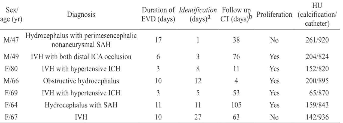
External ventricular drainage (EVD) is a common procedure performed in neurosurgical field. The purpose of this study was to introduce the linear intracranial calcification formed along EVD tract and to investigate its incidence, predisposing factors, and clinical impact. A total of 59 patients who underwent EVD insertion over a 1-year period were included in this study. The clinical factors and radiographic features between the occurrence and the non-occurrence groups were analyzed to investigate the predisposing factors and clinical impact related to the linear intracranial calcification in EVD tract. The linear intracranial calcification following EVD insertion occurred in 7 patients (11.9%). Among various risk factors assessed, only usage of bone dust ( p =0.003) had contributed to linear intracranial calcification with statistical significance in univariate logistic regression analysis. Housefield unit (HU) scale was different between calcification (872.57 ± 46.15 HU) and EVD catheter (169.00 ± 61.35 HU). This study indicates that using bone dust for sealing a burr hole is the only predisposing factor for linear intracranial calcification formed in EVD tract.

Keywords: Bone dust, External ventricular drainage, Linear intracranial calcification

Introduction

External ventricular drainage (EVD) is one of the most widely performed neurosurgical procedures. Although it is useful to control increased intracranial pressure or for drainage of intraventricular hemorrhage, there are several complications related to procedure such as infection, hemorrhage and intracranial calcification, rarely. In our

Received: March 27, 2017 Revised: April 26, 2017 Accepted: May 31, 2017

Corresponding Author: Chang Young Lee, M.D., Department of Neurosurgery, Keimyung University School of Medicine,

56 Dalseong-ro, Jung-gu, Daegu 41931, Korea Tel: +82-53-250-7730

E-mail: [email protected]

• The authors report no conflict of interest in this work.

Department of Neurosurgery, Keimyung University School of Medicine, Daegu, Korea

Yoon Gyo Jung, M.D., Chang Young Lee, M.D.

Predisposing Factors and Clinical Impact of Linear Intracranial

Calcification Following External Ventricular Drainage

(2)

center, by accident, one computed tomography (CT) scan taken after removal of the EVD catheter demonstrated longitudinal radiopaque lesions along EVD catheter tract, which could be considered as torn EVD silicon catheter (Fig. 1). However, the undamaged tip of EVD catheter confirmed at the time of its removal by attending doctor and its different housefield unit (HU) scale from that of the catheter made the lesion diagnosed as not torn catheter, but the linear calcification. Thus, the purpose of this study was to introduce the linear intracranial calcification in EVD tract and to investigate predisposing factors and clinical impact of the calcification.

Materials and Methods

Patients’ population

The authors retrospectively reviewed the m e d i c a l r e c o r d s a s w e l l a s t h e p r e a n d postoperative CT scans of the 59 patients who underwent EVD from September 2013 to August 2014. There were 29 male (49%) and 30 (51%) female in this study, ranging in age from 24 to 94 years old (mean ± SD: 61.07 ± 13.67). The patients were divided into two groups according to the occurrence of linear intracranial calcification after EVD removal. The clinical and radiological factors were compared between the occurrence group and the non-occurrence group.

Clinical and radiological evaluations

T h e o c c u r r e n c e o f l i n e a r i n t r a c r a n i a l calcification was defined as longitudinal radiopaque lesions along EVD catheter tract in CT scan and lower HU scale of the lesion than that of EVD catheter. Blood calcium level was categorized into three groups: low (<8.7), normal (8.7-10.4), high (>10.4). Clinical symptoms were quoted from Fahr’s syndrome which is characterized by abnormal deposition of calcium in areas of the brain that control movements including basal ganglia, thalamus, dentate nucleus, cerebral cortex, cerebellum, subcortical white matter, and hippocampus [1,2].

Surgical procedure and management

All ventriculostomy were performed by two neurosurgeons in operation room with surgical drape, clearly. Burr hole was made at Kocher’s point by using electronic high-speed surgical drill.

Bone dust on drilling the bone was collected. Then Fig. 1. Follow-up computed tomography

demonstrates linear intracranial calcification following external ventricular drainge on right side.

Radiopaque lesion on left side was exteranl

ventricular drainge catheter. It is difficult to

recognize linear calcification along catheter tract

via visual inspection on the CT. It may erroneously

lead one to consider torn tip of EVD catheter

remained in brain parenchyma.

(3)

dura mater and arachnoid membrane were cut by No.15 blade and coagulated by bipolar coagulation.

EVD catheter tip was placed into anterior horn of lateral ventricle and cortex was covered with Surgicel

(Ethicon, North Ryde, NSW, Australia).

Neurosurgeon A used the bone dust collected to fill a burr hole for the purpose of preventing sunken d e f o r m i t y o f s c a l p o v e r b u r r h o l e s i t e.

Neurosurgeon B didn’t use the bone dust, but absorbable gelatin sponge (Cutanplast sponge;

Cure Sys Ltd, Bucheon-si, South Korea) to fill a burr hole. Any other skills and the type of equipment the two neurosurgeons used were not different from each other. All the patients had pre- operative CT, post-operative CT, post-removal CT and follow up CT, serially. Therefore, those serial CT examinations were compared to identify formation, location and proliferation of linear intracranial calcification in EVD tract.

Statistical analysis

A univariate logistic regression analysis was performed with Fisher’s exact test and student’s t-test to assess the relationship between each factors and the occurrence of linear intracranial calcification. The statistical significance was set at p

< 0.05.

Results

Patients’ characteristics are summarized in Table 1. Linear intracranial calcification in EVD tract after removal of EVD catheter was found in 7 patients (11.9%). T h e d e m o g r a p h i c d a t a w a s n o t significantly associated with linear intracranial calcification in EVD tract. Comorbidities, antithrombotics usage and smoking or alcohol consumption were also not related to the

occurrence of linear intracranial calcification in EVD tract. Causes of performing EVD were various, but all of them did not have the statistical significance. In operation, the rate of using bone dust in patients with linear intracranial calcification (100%) was higher than that in those without linear intracranial calcification (38.5%) with statistical significance ( p <0.01). However, any other operative factors were not related to the occurrence of linear intracranial calcification. Information of 7 patients with linear intracranial calcification was demonstrated in Table 2. Mean duration of EVD was 8.57 ± 5.00 days. Duration of identification of calcification from operation (days) was varied from 1 to 27 (mean: 9.57 ± 8.68 days). Mean HU scale of EVD catheter (872.57 ± 46.15 HU) was higher than that of the calcification (169.00 ± 61.35 HU). It was most important means to differentiate the calcification from EVD catheter. Clinical symptoms associated with linear intracranial calcifications were listed in Table 3. Neurological symptoms like seizure, coma and spasticity were not presented.

Dementia was manifested on two patients.

However, dementia did not be considered as newly onset clinical symptom associated with linear intracranial calcifications, because it is a common symptom related with stroke attack. Movement disorder like involuntary movement was not presented. Neuropsychiatric features like psychosis were found in one patient, which was just temporary, mild and gradually subsided after several days.

Discussion

External ventricular drainage was first

performed as early as 1744 by Claude-Nicholas

LeCat [3,4]. Nowaday, it is a well-established

procedure in the management of hydrocephalus,

(4)

intraventricular hemorrhage, intracranial hypertension, and central nervous system bacterial

infections [5-7]. Since its introduction into the clinical field, there have been numerous changes in Table 1. Characteristics in 59 patients with external ventricular drainage

OG (n=7) NOG (n=52) Total (n=59) p- value

Sex Male 3(42.9) 26(50) 29

Female 4(57.1) 26(50) 30

Age (years)

Mean age 63.14 ± 11.57 60.79 ± 14.00 61.07 ± 13.67 0.673

Comorbidity

Hypertension 3(42.9) 29(55.8) 32(54.2)) 0.692

Diabetes mellitus 4(57.1) 12(23.1) 16(27.1) 0.078

Cerebrovascular attack 1(14.3) 12(23.1) 13(22)

Meningitis 5(71.4) 19(36.5) 24(40.7) 0.109

Blood Calcium 0.109

Low (<8.7) 5(71.4) 19(36.5) 24(40.7)

Normal (8.7-10.4) 2(28.6) 33(63.5) 35(59.3)

High (>10.4) 0(0) 0(0) 0(0)

Cause of EVD

Hypertensive ICH 2(28.6) 26(50) 28(47.5) 0.428

Subarachnoid hemorrhage 2(28.6) 14(26.9) 16(27.1)

Cerebellar infarction 0(0) 5(9.6) 5(8.5)

MCA occlusion 1(14.3) 3(5.8) 4(6.8) 0.405

Intraventricular hemorrhage 1(14.3) 2(3.8) 3(5.1) 0.320

Posttraumatic hydrocephalus 1(14.3) 0(0) 1(1.7) 0.119

Arteriovenous malformation 0(0) 1(1.9) 1(1.7)

Tuberculosis meningitis 0(0) 1(1.9) 1(1.7)

Medication

Antiplatelet 2(28.6) 12(23.1) 14(23.7) 0.666

Anticoagulants 0(0) 5(9.6) 5(8.5)

Calcium channel blocker 3(42.9) 38(73.1) 41(69.5) 0.184

Alcohol 1(14.3) 24(46.2) 25(42.4) 0.221

Smoking 2(28.6) 13(25) 15(25.4)

Operation

Bone dust 7(100) 20(38.5) 27(45.8) 0.003

EVD duration (days) 8.57 ± 5.00 10.38 ± 9.24 9.57± 8.68 0.614

EVD side 0.521

Right 4(57.1) 20(38.5) 24(40.7)

Left 0(0) 5(9.6) 5(8.5)

Both 3(42.9) 27(51.9) 30(50.8)

OG: occurrence group, NOG: non-occurrence group, EVD: external ventricular drainage, ICH: intracerebral

hemorrhage, MCA: middle cerebral artery.

(5)

technique, materials used, indication for the procedure [3]. In Korea, because national medical insurance policy does not allow for using a plate for covering a burr hole site, an neurosurgeon in our center used bone dust for covering a burr hole to prevent sunken deformity of scalp over burr hole site by stimulating ossification. However, as one of rare complications, the authors experienced several cases of linear intracranial calcifications along external ventricular drainage tract mimicking torn EVD silicon catheter remained. In this study,

its incidence rate is about 11.9%. The authors introduced the linear intracranial calcification in EVD tract and investigated predisposing factors and clinical impact of the calcification. Alorainy [5]

suggested that small bone pieces pushed into the brain parenchyma from the calvarium during insertion of the drainage tube might cause small bone fragment in the tract of drain. Findings in one patient of the present study are consistent with the suggestion of Alorainy [5]. The linear calcification adjacent to EVD catheter in the patient was Table 2. Summary of 7 patients with linear intracranial calcification

Sex/

age (yr) Diagnosis Duration of

EVD (days) Identification

(days)a Follow up

CT (days)b Proliferation

(calcification/ HU catheter) M/47 Hydrocephalus with perimesencephalic nonaneurysmal SAH 17 1 38 No 261/920

M/49 IVH with both distal ICA occlusion 6 3 76 Yes 204/824

F/80 IVH with hypertensive ICH 3 8 11 Yes 152/820

M/66 Obstructive hydrocephalus 10 12 4 Yes 200/895

F/69 IVH with hypertensive ICH 3 5 53 Yes 65/870

F/64 Hydrocephalus with SAH 11 11 105 Yes 159/843

F/67 IVH 10 27 63 No 142/936

SAH: subarachnoid hemorrhage, IVH: intraventricular hemorrhage, ICH: intracerebral hemorrhage, HU: housfield unit (calcification/catheter),

a

Interval from operation to identification of calcification,

b

Interval from identification to last follow up CT.

Table 3. List of clinical symptoms of patients with linear intracranial calcifications

No Seizure Spasticity Coma Dementia Involuntary

movement Psychosis

1 - - - - - -

2 - - - - +

3 - - - - - -

4 - - - - - -

5 - - - + - -

6 - - - - - -

7 - - - + - -

(6)

identified on immediate postoperative CT and the size and shape were not changed on the follow-up CT scan. However, this hypothesis is not a convincing explanation for the other 6 cases and no development of the calcification in the patients disusing bone dust. Linear calcification of these 6 cases were identified on CT scan after 48 hours from operation and proliferated in size and shape.

The other pathogenesis of linear intracranial calcification in EVD tract is that bone dust covering a burr-hole after EVD insertion can be pushed into the brain parenchyma and proliferated after EVD catheter removal. According to the report of Thomson et al [8], bone dust contains viable osteoblast that are capable of dividing and laying down bone when implanted into vascular tissues.

Consistent with the report of Thomson et al [8], all 7 cases of linear intracranial calcifications were found in the patients using bone dust. The rate of using bone dust in patients with linear intracranial calcification (100%) was higher than that in those without linear intracranial calcification (38.5%) with statistical significance ( p =0.003). In 5 of 7 patients, the calcifications were identified on CT scan examined after removal of EVD catheter. In 1 of remained 2 patients, the calcification was seen on CT scan taken after removal of contralateral EVD catheter and in the other, it was found immediate after ipsilateral EVD catheter insertion. Therefore, the authors suggest that change of pressure and spaces might develop between lateral ventricle and brain cortex after the EVD insertion or at the time the EVD was removed. The pressure change was likely to have bone dust with viable osteoblast sucked into the EVD tract. Calcification was enlarged over time because of viable osteoblast.

On the other hand, none of calcification was observed when bond defect was covered with surgicel alone followed by a watertight galeal closure in our study. It is difficult to recognize

linear calcification along catheter tract via visual inspection on the CT. It may erroneously lead one to consider torn tip of EVD catheter remained in brain parenchyme. The way to differentiate this lesion from EVD silicon tube is measuring HU scale of the lesion on CT scan. HU scale of the calcification was lower (169.00 ± 61.35 HU) than that of EVD catheter, whereas HU scale of EVD catheter was high (872.57 ± 46.15 HU). All of 7 patients have had no newly onset, long lasting clinical symptoms or signs associated with linear intracranial calcification. Although neuropsychiatric features like psychosis were found in one patient, it was not considered as being related to calcification due to its temporary development.

Surgical removal or particular medication is unnecessary so far. However, it is uncertain because of short follow-up period. Therefore, the clinical and radiological long term surveillance may be needed for the patients who have linear intracranial calcification in EVD tract, continuously.

Conclusion

The present study suggested that the cause of

linear intracranial calcification mimic torn EVD

catheter in EVD tract is bone dust covering a burr-

hole after EVD insertion. Therefore, to prevent

these complications, bone dust should be disused

and national health insurance service should

consider covering price of plate for sealing a burr

hole. It is important to differentiate linear

calcification from EVD tube silicon and long term

follow-up is necessary to assess a clinical impact of

linear intracranial calcification in EVD tract.

(7)

References

1. Saleem S, Aslam HM, Anwar M, Anwar S, Saleem M, Saleem A, et al . Fahr’s syndrome: literature review of current evidence. Orphanet J Rare Dis 2013;8:156.

2. Calabrò RS, Spadaro L, Marra A, Bramanti P. Fahr's disease presenting with dementia at onset: a case report a n d l i t e r a t u r e r e v i e w . B e h a v N e u r o l 2014;2014:750975.

3. Visish M, Brent R, Diana J, Donald M, Michael Y. The history of external ventricular drainage. J Neurosurg 2014;120:228-36.

4. Kompanje EJ, Delwel EJ. The first description of a device for repeated external ventricular drainage in the treatment of congenital hydrocephalus, invented in

1744 by Claude-Nicolas LeCat. Pediatr Neurosurg 2003;39:10-3.

5. Alorainy IA. Introduction of skull bone fragments into the brain during external ventricular drain placement.

Eur J Radiol 2001;40:218-23.

6. Stangle AP, Meyer B, Zentner J, Schramm J.

Continuous external CSF drainage-a perpetual problem in neurosurgery. Surg Neurol 1998;50:77-82.

7. Rohit K, Mark L, Jack P, Ghaus M. Prolonged external ventricular drainage with percutaneous long-tunnel ventriculostomies. J Neurosurg 1995;83:791-4.

8. Thomson S, Tyagi AK, Chumas PD. Intracranial

h y p e r t r o p h i c c a l c i f i c a t i o n c o m p l i c a t i n g

neuroendoscopy. Report of three cases. J Neurosurg

2003;98:186-9.

수치

Table 3. List of clinical symptoms of patients with linear intracranial calcifications

참조

관련 문서