Introduction
Endometriosis affects the intestinal tract in 15%
to 37% of patients with pelvic endometriosis [1].
but is difficult to distinguish from a benign or malignant neoplasm clinically [2-6]. Histologically,
endometriosis shows the endometrial glands and the stroma, and may be accompanied by smooth m u s c l e h y p e r p l a s i a i n 24% o f i n t e s t i n a l endometriosis patients [7]. Endometriosis in the intestinal wall can be misdiagnosed as a benign or m a l i g n a n t n e o p l a s m [3], a n d e s p e c i a l l y
Abstract
It is difficult to distinguish colonic endometriosis with smooth muscle metaplasia from a colonic neoplasm.
Especially, in a small biopsy specimen, marked smooth muscle proliferation can be misdiagnosed as a spindle cell lesion such as leiomyoma or as a gastrointestinal stromal tumor. We present the case of a woman aged 48 years who had a colonic polyp, raising suspicion of a submucosal tumor. In a endoscopic polypectomy specimen, the lesion showed marked smooth muscle proliferation but in the resection specimen, a nodular mass, composed of endometrial glands, stroma, and a smooth muscle component, was identified. By immunohistochemical staining, smooth muscle cells were positive for estrogen receptor, progesterone receptor, smooth muscle actin, and smooth muscle myosin heavy chain. Endometriosis in the colon can be misinterpreted as a benign or malignant neoplasm. When examining small biopsy specimens, clinicians and pathologists should be aware of the potential of this condition to mimic other intestinal diseases.
Key Words :
Colon, Endometriosis, LeiomyomaCorresponding Author: Ilseon Hwang, M.D., Department of Pathology, Keimyung University School of Medicine 1095 Dalgubeol-daero, Dalseo-gu, Daegu 704-701, Korea
Tel : +82-53-580-3809 E-mail : [email protected], [email protected]
Department of Pathology, Keimyung University School of Medicine, Daegu, Korea
Ilseon Hwang, M.D.
Colonic Endometriosis with Extensive Smooth Muscle Metaplasia
Simulating Leiomyoma
endometriosis with marked smooth muscle proliferation (metaplasia) can be misidentified as a spindle cell neoplasm of the intestinal wall. Here, we present a case with a polypoid lesion of the colon, initially interpreted as a spindle cell neoplasm, that was finally diagnosed as colonic endometriosis.
Case Report
A woman aged 48 years visited our hospital for periodical health checks. In a colonoscopic examination, a 1.2 cm-sized submucosal tumor-like lesion was found 20 cm from the anal verge (Fig.
1A). Endoscopic snare polypectomy was performed. On histological examination, the submucosal tumor-like lesion was found to be a spindle cell lesion. The lesion consisted of smooth muscle cells and the resection margin was involved by smooth muscle component. On abdominal CT, a 1.5 cm-sized hyperattenuating lesion was found to remain present in the rectosigmoid area (Fig. 1B).
The uterus showed slightly enlarged but no
a b n o r m a l s h a p e, a n d b o t h o v a r i e s w e r e unremarkable. A segmental resection was performed for removal of the smooth muscle proliferative lesion.
On gross examination, an ill-demarcated polypoid lesion (2.1 × 1.5 × 1.0 cm) was present in the sigmoid colon (Fig. 2). The cut surface showed a lobulating mass in a mucosal extension to the subserosa. The lesion was grayish tan, solid, and fibrotic, with a focal hemorrhagic area.
Necrosis was not identified in the lesion.
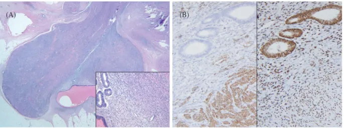
Microscopically, the lesion was snowman-like shape and had a clear boundary. The lesion consisted of smooth muscle cells with a few endometrial glands and stroma. The smooth muscle component was more than 80% of total volume and endometrial glands and stroma were centrally scattered in smooth muscle component (Fig. 3A). By immunohistochemistry, staining for the estrogen receptor (ER) and progesterone receptor (PR) was positive for endometrial glands, stroma, and the smooth muscle proliferative lesion (Fig. 3B), but negative for colonic mucosa and normal muscle. The smooth muscle proliferative
Fig. 1. (A) A 1.2 cm-sized submucosal tumor-like lesion was present in colonoscopic examination. (B) A 1.5 cm sized hyperattenuatting lesion is remained in rectosigmoid area (Abdominal CT examination).
(A) (B)
lesion stained positive for smooth muscle actin (SMA), desmin, and smooth muscle myosin heavy chain (SMMHC) (Fig. 2B). Immunohistochemical staining for C-kit, CD34, and S-100 proteins was negative, and staining for CD10 was positive on the e n d o m e t r i a l s t r o m a. T h e s m o o t h m u s c l e
proliferative lesion was distinguished from i n d i g e n o u s n o r m a l m u s c l e, a n d s h o w e d hypercellularity, moderate nuclear atypia, and occasional mitoses.
The patient has had no evidence of post- operative complication and no evidence of recurrence for 6 months after surgery.
Discussion
Endometriosis with predominant smooth muscle proliferation or smooth muscle metaplasia can be misinterpreted as submucosal tumors on endoscopic examination [2,5,7-10]. On radiologic examination, endometriosis with smooth muscle metaplasia can appear as a submucosal spindle cell tumor of the intestinal wall, including a gastrointestinal stromal tumor or leiomyoma [5,7].
In the present case, the smooth muscle component was predominant, representing more than 80% of the endometrial mass. Only the
Fig. 3. (A) Some scattered endometrial gland and stromal tissue was identified in the indicated smooth muscle proliferative lesion (H&E, × 40 and × 200). (B) Endometrial glands, stroma, and smooth muscle cells were positive for estrogen receptor (left, Immunohistochemical stain for estrogen receptor, × 200). The smooth muscle cells were positive, but endometrial gland and stromal cells were negative for smooth muscle myosin heavy chain (right, Immunohistochemical stain for smooth muscle actin, × 200).
(A) (B)
Fig. 2. A 1.5 cm-sized polypoid lesion was present in the colonic mucosa.
smooth muscle component was present in a small fragment obtained by endoscopic biopsy and resulted in misinterpretation of the condition as a spindle cell neoplasia that might be either a gastrointestinal stromal tumor or leiomyoma. If a few fragments of endometrial glands consisting relatively short columnar epithelium with prominent nucleoli or no mucinous components were identified, the biopsy lesion could be considered endometriosis with smooth muscle metaplasia of large intestine.
On immunohistochemistry, endometriosis c o m p o n e n t s, s u c h a s e n d o m e t r i u m a n d myometrium, are positive for ER and PR [8,9].
SMA, SMMHC, and desmin are expressed in the smooth muscle component, and ER and PR are also present [7].
Differential diagnoses include intravenous leiomyomatosis, intestinal leiomyoma with endometriosis, leiomyosarcoma and synovial sarcoma. In intravenous leiomyomatosis, the margin is irregular, and lacks an endometrial c o m p o n e n t s. I n t e s t i n a l l e i o m y o m a w i t h endometriosis does not show expression of either ER or PR in the smooth muscle component. PR expression can be present in leiomyoma with specific site, such as sinonasal area. However, both of ER and PR expression was not identified in l e i o m y o m a e x c e p t u t e r i n e o r i g i n [11].
Leiomyosarcoma have hypercelluar lesion with marked cellular atypia and also lacks an endometrial components. Synovial sarcoma may have similar histologic features. Especially in biphasic type, epithelial and spindle cell components are similar to endometrial glands and smooth muscle component. However, synovial sarcoma does not show expression of SMA, SMMHC and desmin. In the present case, the endometrial glands and stroma were scattered in a nodular mass of smooth muscle, which could be distinguished from normal
indigenous muscle. Therefore, the smooth muscle fraction was an endometriosis component and the condition was consistent with smooth muscle metaplasia.
Endometriosis is commonly misinterpreted as a benign or malignant neoplasia because the condition can present with variable proportions of components [12]. Especially, endometriosis with marked smooth muscle proliferation or metaplasia can be misdiagnosed as spindle cell neoplasia in a small biopsy specimen. Such misdiagnosis can lead to overtreatment such as wide resection and chemotherapy. Therefore, a tumor-like lesion in the female pelvic or abdominal cavity requires careful examination and treatment.
References
1. Forsgren H, Lindhagen J, Melander S, Wagermark J.
Colorectal endometriosis. Acta Chir Scand 1983;149:431-5.
2. Borsellino G, Buonaguidi A, Veneziano S, Borsellino V, Mariscalco G, Minnici G. Endometriosis of the large intestine. A report of 2 clinical cases. Minerva Ginecol 1993;45:443-7.
3. Kelly P, McCluggage WG, Gardiner KR, Loughrey MB. Intestinal endometriosis morphologically mimicking colonic adenocarcinoma. Histopathology 2008;52:510-4.
4. Matarese G, De Placido G, Nikas Y, Alviggi C.
Pathogenesis of endometriosis: natural immunity dysfunction or autoimmune disease? Trends Mol Med 2003;9:223-8.
5. Pickhardt PJ, Kim DH, Menias CO, Gopal DV, Arluk GM, Heise CP. Evaluation of submucosal lesions of the large intestine: part 2. Nonneoplastic causes.
Radiographics 2007;27:1693-703.
6. Zacharia TT, O'Neill MJ. Prevalence and distribution of adnexal findings suggesting endometriosis in patients
with MR diagnosis of adenomyosis. Br J Radiol 2006;79:303-7.
7. Yantiss RK, Clement PB, Young RH. Endometriosis of the intestinal tract: a study of 44 cases of a disease that may cause diverse challenges in clinical and pathologic evaluation. Am J Surg Pathol 2001;25:445-54.
8. Itoga T, Matsumoto T, Takeuchi H, Yamasaki S, Sasahara N, Hoshi T, et al. Fibrosis and smooth muscle metaplasia in rectovaginal endometriosis. Pathol Int 2003;53:371-5.
9. Kitano T, Matsumoto T, Takeuchi H, Kikuchi I, Itoga T, Sasahara N, et al. Expression of estrogen and progesterone receptors in smooth muscle metaplasia of rectovaginal endometriosis. Int J Gynecol Pathol
2007;26:124-9.
10. Yoshida M, Watanabe Y, Horiuchi A, Yamamoto Y, Sugishita H, Kawachi K. Sigmoid colon endometriosis treated with laparoscopy-assisted sigmoidectomy:
significance of preoperative diagnosis. World J Gastroenterol 2007;13:5400-2.
11. Tseng PY, Lai YS, Chen MK, Shen KH. Progesterone receptor expression in sinonasal leiomyoma: a case report and review of the literature. Int J Clin Exp Pathol 2014;7:1224-8.
12. Clement PB. The pathology of endometriosis: a survey of the many faces of a common disease emphasizing diagnostic pitfalls and unusual and newly appreciated aspects. Adv Anat Pathol 2007;14:241-60.