Long-term Management of a Gingival Fibromatosis Patient with the Primary Dentition
Chungmin Kang, Jaeho Lee, Hyungjun Choi, Jeseon Song, Seongoh Kim
Department of Pediatric Dentistry, College of Dentistry, Yonsei University
Gingival fibromatosis is a rare oral condition that is characterized by proliferative fibrous overgrowth of the attached gingiva, the marginal gingiva, and the interdental papilla, typically presenting in the growth period.
A case of a 27-month-old girl with a generalized severe gingival overgrowth is described herein. The patient had no known systemic disease, but enlarged gingival tissue had gradually covered her teeth. The excess gingival tissue was removed by conventional gingivectomy, which involved extraction of the retentive primary teeth under general anesthesia when she was 5 years old. Post surgical follow-up at 18 months after the surgery demonstrated no recurrence.
Resectional surgery of the enlarged gingival tissue is the treatment choice for gingival fibromatosis, although there is a high risk of recurrence. More frequent professional follow-ups and oral hygiene instruction might be required. A delay in the surgical treatment may have significant consequences for the patient, such as primary dentition retention and consequent delay in the eruption of the permanent teeth, difficulties in mastication and phonation, malpositioning of the teeth, and psychological problems. Early surgical treatment should be performed according to the severity of enlargement.
Key words :Gingival fibromatosis, Gingival enlargement, Idiopathic, Gingivectomy, Surgical treatment Abstract
Ⅰ. Introduction
Gingival enlargement is defined as overgrowth of the gingiva associated with an accumulation and expansion of the connective tissue1-3). The disease has a multifacto- rial etiology, including inflammatory, drug, and heredi- tary factors. However, the underlying pathologic mecha- nisms remain unknown, and thus the condition is gener- ally labeled as being explained as idiopathic4). Idiopathic gingival enlargement is also known as gingivostomatosis, idiopathic gingival fibromatosis, hereditary gingival fi- bromatosis, and congenital familial fibromatosis5,6).
Gingival fibromatosis, which was first reported in 1856 by Gross7), is characterized by non-inflammatory, non- hemorrhagic benign lesions. The hyperplastic gingiva has a normal color and a firm consistency, with abundant stippling on the adjacent gingiva8,9). It occurs throughout the gingiva in both the maxilla and mandible, as well as in other areas such as the maxillary tuberosity and the mandibular posterior buccal gingiva10,11). The gingival en- largement usually begins at the time of eruption of the permanent teeth but, less frequently, may also occur with eruption of the primary teeth11-13).
Gingival fibromatosis results in both functional and es-
Corresponding author : Seongoh Kim
Department of Pediatric Dentistry, College of Dentistry, Yonsei University, 50-1 Yonsei-ro, Seodaemun-gu, Seoul, 120-752, Korea Tel: +82-2-2228-3171 / Fax: +82-2-392-7420 / E-mail: [email protected]
Received April 22, 2014 / Revised September 1, 2014 / Accepted September 4, 2014
thetic problems in affected patients. The common out- comes of the lesions are diastema, malposition of the teeth, prolonged retention of the primary teeth, open bite, prominent lips, and open lip closure9,12,14,15)
. The suggested treatment depends upon the severity of the condition. Mild enlargement can be reduced through scaling and home care to maintain a good appearance.
However, the overgrown tissue needs to be surgically re- moved since it causes both functional and esthetic im- pairment12,16,17).
This report presents a rare case of a young patient with gingival fibromatosis who had no special medical history and was treated with resective surgery under general anesthesia. This case emphasizes the clinical significance of early management of gingival fibromatosis with respect to long- term prognosis.
Ⅱ. Case Report
A 27-month-old girl presented at the Department of Pediatric Dentistry, Yonsei University Dental Hospital with a chief complaint of non-visibility of the primary teeth due to overall gingival swelling. Neither her family nor medical histories were significant for disease trans-
mission, which made it difficult to identify the exact cause of the gingival enlargement. She was referred to the Department of Pediatrics, but the finding of blood tests and genetic screening were unremarkable for both drug- and hormone-induced gingival hyperplasia.
An intraoral examination of the patient at first dental visit revealed generalized gingival overgrowth involving attached gingiva, marginal gingiva, and interdental papilla. The posterior areas at both arches were particu- larly severely deformed by a large amount of gingival tis- sue that was sufficient to inhibit tooth eruption and im- pair mastication. Extraorally, the patient was not able to close her lips because of protrusion of the enlarged gingival tissue (Fig. 1).
The patient’s tooth eruption pattern was followed in periodic check-ups, and panoramic radiographs revealed delayed development of the maxillary primary incisor roots at the age of 3 years (Fig. 2A). Clinical examina- tion also revealed partial eruption of the teeth in the mandible, with the exception of the second primary mo- lars, and only cusp exposure of the first primary molar in the maxilla (Fig. 2B, C). Conventional gingivectomy was performed, localized to the maxillary anterior area, to facilitate eruption of the primary incisors.
Fig. 1. Initial photographs of the 27-month-old female (A) Intraoral photograph showing generalized gingival enlargement involving both the maxillary and mandibular arches. (B) Extraoral photograph showing incompetent lips.
Fig. 2. (A) Panoramic radiograph taken at the age of 3 years showing delayed root development in the maxillary primary incisors (arrows), and floating permanent tooth germ. (B) Exposure of the cusp tip of the first maxillary primary molar. (C) The hyperplastic gingival tissues covered at least one-third of the clinical crowns of almost all of the teeth in mandible.
At the age of 4 years, the patient experienced early loss of the mandibular primary incisors, which was pre- disposed by the previously reported developmental root defect. Overall gingivectomy was planned for esthetic improvement so that her appearance did not hinder her ability to form relationships with her peers at kinder- garten.
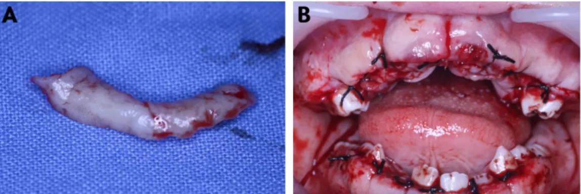
At the age of 5 years, the patient underwent gingivec- tomy of both arches under general anesthesia. Incisions were made for the gingival resection from the distal as- pect of the posterior teeth to the midline on both the fa- cial and lingual surfaces, which avoided interference with the field of view. The gingiva was incised in the apical direction between the periodontal pocket and the alveolar bone at an angle of approximately 45�. This contributed to the formation of a physiologic gingival shape (Fig. 3). A wedge-shaped slice of the gingival overgrowth was removed via external bevel gingivectomy (Fig. 4A). During the procedure, maxillary left primary incisors were extracted, the posterior crowns were ex- posed, and the wounds then sutured (Fig. 4B). No com- plications were experienced and the patient was in- structed not to brush or to chew on the surgical site. She was provided with a 0.2% chlorhexidine rinse to use af- ter brushing, twice daily, to reduce plaque accumulation.
The removed gingival tissue was assessed histological- ly and a diagnosis of gingival fibromatosis was made.
The epithelium had a normal structure, with rete pegs that penetrated deep into the connective tissue. There appeared to be more collagen fiber bundles than in nor- mal tissue, but there were no other unusual histologic features (Fig. 5).
Fig. 3. Diagrams of gingivectomy treatment for gingival fibromatosis. (A) Initial reverse bevel incision ; the dashed lines indicate where incisions were made. (B) Removal of the enlarged gingival tissue after flap elevation. (C) The flap is placed on top of the alveolar bone and sutured.
Fig. 4. (A) A slice of gingival tissue resected surgically (B) Post-operative photograph. Multiple primary and permanent teeth are visible after removing the excess gingival tissue.
Fig. 5. Histologic examination of the gingival fibromatosis tissues. (A) Hyperplastic dense connective tissue predominantly consisting of thick and irregularly arranged collagen fibers (H-E, ×40). (B) Squamous epithelium exhibited acanthosis and elongated irregular down growth of rete pegs (H-E, ×100).
The patient was recalled for follow-up observation at 1, 3, 6, 12, and 18 months postsurgically. She exhibited improvements in both esthetics and mastication func- tion, and no recurrence of gingival fibromatosis (Fig. 6).
However, she required orthodontic treatment to correct an open bite. A combination of surgical and fixed ortho- dontics was thus planned. Professional oral hygiene management through regular check-ups was emphasized to prevent a recurrence of gingival enlargement.
Ⅲ. Discussion
Gingival fibromatosis is a rare oral disease occurring in children that requires an accurate diagnosis and appro- priate treatment by a pediatric dentist18). The exact pathogenesis of the disease is unknown. However, it seems to be associated with a genetic predisposition, al- though the genes involved have yet to be identified19,20) Therefore, all cases of gingival enlargement without de- finitive causes are labeled idiopathic, even if the condi- tion is seen in other members of the same family without any other symptoms5,6).
In the case described here, the patient was diagnosed with idiopathic gingival fibromatosis because her family and medical history were not distinct; there was no evi- dence of genetic transmission. Moreover, no abnormali- ties were detected in either blood or gingival karyotypes.
The histologic characteristics of the excised gingiva are dominated by changes to connective tissue. The enlarged gingival tissue possesses a hyperplastic, parakeratinized, stratified squamous epithelium covered with a dense overgrowth of fibrous connective tissue6,21). In addition, the connective tissue contains a remarkable amount of collagen fiber bundles with few fibroblasts and a reduced
vascular supply22). The histopathologic features are not specific, and so a definitive diagnosis should be made based on the clinical findings and medical history.
According to several authors, the ideal time for surgi- cal intervention is when all of the permanent teeth have erupted, because of the associated low recurrence rate8,16,23). In the present case, however, early surgical treatment was performed to improve mastication func- tion and resolve esthetic problems at a time when she was beginning to form social relationships even though there was a risk of recurrence. Baptista14) considered that without treatment, patients can experience signifi- cant problems with mastication and pronunciation after the age of 4-5 years. Moreover, Coletta and Graner6)and Gregory et al.24) demonstrated that the psychological benefits of esthetic improvement should not be underes- timated and may outweigh the risk of recurrence. At the first visit her body weight and height were 11 kg and 80 cm, respectively, which are both less than the 20th per- centiles on the standard growth curve for children in Korea. However, improvement of her eating ability re- sulted in a positive effect on her growth and develop- ment, which subsequently increased to the 90th per- centiles on the growth curve after 6 months.
Treatment of gingival fibromatosis is approached from the viewpoint of symptom control rather than funda- mental healing, and its success rate depends upon the severity of the gingival enlargement. When it is mild, home care and scaling may be sufficient to maintain good oral health14). On the other hand, the excess tissue should be surgically removed for both esthetic and func- tional reasons. In the past, tooth extraction and reduc- tion of the underlying alveolar bone were favored treat- ments16). However, various techniques have since been Fig. 6. (A) Intraoral appearance 1 month after the surgical treatment, showing obvious improvement of the gingival condition, but malocclusion has been revealed. (B) Intraoral appearance 12 months after the surgical treatment, with decreased open bite. (C) Intraoral appearance 18 months after the surgical treatment, showing satisfactory gingival condition with no evidence of recurrence.
developed for excising the enlarged gingiva, including in- ternal or external gingivectomy accompanied by gingivo- plasty, an apically positioned flap, electrocautery, and carbon-dioxide-laser12,16,17,25)
. The present patient received conventional, external gingivectomy to remove a large amount of gingival tissue under general anesthesia.
External gingivectomy is a first choice to make physio- logic gingival contour and to remove tissue tags attached to tooth surface. Indications for this treatment are no at- tachment loss and periodontal pocket formation. If gingi- val enlargement is associated with deep pockets and se- vere loss of alveolar bone, an internal bevel gingivectomy with open-flap debridement can be advocated26). Orthodontic treatment to resolve an open bite was rec- ommended for this patient, and included a combination of both surgical and fixed orthodontics for her permanent dentition.
Recurrence can occur within several months after surgery, and so the monthly examination of the gingival condition and oral hygiene is recommended to reduce the recurrence risk. Several studies have found that recur- rence occurs faster in areas with plaque accumulation27,28). However, Emerson23)demonstrated that correcting phys- iologic contouring of the marginal gingiva is more impor- tant than good oral hygiene and the amount of calculus.
Therefore, in the present case the aim of the surgery was to produce physiologic gingival contour. We made the facial and lingual surface contours with gradual cur- vatures in all directions to facilitate the rubbing and cleaning function of the lips, cheeks, and tongue. The in- terproximal contour of adjacent teeth, of the tooth con- tact areas, and of the teeth in relation to the gingival papilla also had such a curvature that the patient can perform oral hygiene easily. Professional cleaning, oral hygiene instructions, and regular follow-up appoint- ments were also planned to minimize or delay the recur- rence in this patient.
Many recent studies have attempted to identify the bi- ological mechanism underlying gingival overgrowth.
However, more evidence is needed to support the corre- lation between gene mutation, molecular change, and the clinical features. Such evidence will provide pediatric dentists with methods not only for disease diagnosis, but also for disease prevention and treatment.
Ⅳ. Summary
Gingival fibromatosis is a rare oral condition that oc-
curs in children, and for which the pediatric dentist has important roles in both determining and providing the correct diagnosis and treatment. The diagnosis was con- firmed by the typical presentation, medical history, and histopathological features. This clinical report highlights a nonsyndromic incidence of gingival fibromatosis man- aged with surgical treatment in primary dentition and long-term follow-up. Early intervention is sometimes needed to avoid malocclusion and esthetic and functional complications. It will also help to improve the patient’s quality of life, and in particular the child’s emotional and social stability.
References
1. Takagi M, Yamamoto H, Mega H, et al. : Heterogeneity in the gingival fibromatoses. Cancer, 68:2202-2212, 1991.
2. Butler RT, Kalkwarf KL, Kaldahl WB : Drug- induced gingival hyperplasia: phenytoin, cyclosporine, and nifedipine. J Am Dent Assoc, 114:
56-60, 1987.
3. Pearlman BA : An oral contraceptive drug and gin- gival enlargement; the relationship between local and systemic factors. J Clin Periodontol, 1:47-51, 1974.
4. Sakamoto R, Nitta T, Kamikawa Y, et al.:
Histochemical, immunohistochemical, and ultra- structural studies of gingival fibromatosis: a case report. Med Electron Microsc, 35:248-254, 2002.
5. Jorgenson RJ : Gingival fibromatosis. Birth Defects Orig Artic Ser, 7:278-280, 1971.
6. Coletta RD, Graner E : Hereditary gingival fibro- matosis: a systematic review. J Periodontol, 77:753- 764, 2006.
7. Gross SD : Case of hypertrophy of gums. Louisville review:1232, 1856.
8. Bittencourt LP, Campos V, Moliterno LF, et al. : Hereditary gingival fibromatosis: review of the liter- ature and a case report. Quintessence Int, 31:415- 418, 2000.
9. Bozzo L, de Almedia OP, Scully C, Aldred MJ : Hereditary gingival fibromatosis. Report of an exten- sive four-generation pedigree. Oral Surg Oral Med Oral Pathol, 78:452-454, 1994.
10. Kelekis-Cholakis A, Wiltshire WA, Birek C : Treatment and long-term follow-up of a patient with hereditary gingival fibromatosis: a case report. J
Can Dent Assoc, 68:290-294, 2002.
11. Singer SL, Goldblatt J, Hallam LA, Winters JC : Hereditary gingival fibromatosis with a recessive mode of inheritance. Case reports. Aust Dent J, 38:427-432, 1993.
12. Bozzo L, Machado MA, de Almeida OP, et al. : Hereditary gingival fibromatosis: report of three cas- es. J Clin Pediatr Dent, 25:41-46, 2000.
13. Anderson J, Cunliffe WJ, Roberts DF, Close H : Hereditary gingival fibromatosis. Br Med J, 3:218- 219, 1969.
14. Baptista IP : Hereditary gingival fibromatosis: a case report. J Clin Periodontol, 29:871-874, 2002.
15. Goldblatt J, Singer SL : Autosomal recessive gingi- val fibromatosis with distinctive facies. Clin Genet, 42:306-308, 1992.
16. Cuestas-Carnero R, Bornancini CA : Hereditary generalized gingival fibromatosis associated with hypertrichosis: report of five cases in one family. J Oral Maxillofac Surg, 46:415-420, 1988.
17. Zackin SJ, Weisberger D : Hereditary gingival fibro- matosis. Report of a family. Oral Surg Oral Med Oral Pathol, 14:828-836, 1961.
18. Newman MG, Takei HH, Carranza FunA : Carranza's clinical periodontology. 10th ed.
Saunders/Elsevier, St. Louis, Mo., 373-390. 2006.
19. Gorlin RJ, Cohen MM, Levin LS : Syndromes of the head and neck. 3rd ed. Oxford University Press, New York, 847-852. 1990.
20. Wood NH, Anagnostopoulos C, Meyerov R, et al. : Idiopathic gingival fibromatosis: a review of the lit-
erature and a case report. Journal of the South African Dental Association, 63:298-300, 2008.
21. Nevin NC, Scally BG, Kernohan DC, Dodge JA : Hereditary gingival fibromatosis. J Ment Defic Res, 15:130-135, 1971.
22. Barros SP, Merzel J, de Araujo VC, et al. : Ultrastructural aspects of connective tissue in hered- itary gingival fibromatosis. Oral Surg Oral Med Oral Pathol, 92:78-82, 2001.
23. Emerson TG : Hereditary gingival hyperplasia. A family pedigree of four generations. Oral Surg Oral Med Oral Pathol, 19:1-9, 1965.
24. Gregory MH, Jon GF, Bruce GF : Gingival fibro- matosis with hypertrichosis. J Periodontol, 56:344- 347, 1985.
25. Kharbanda P, Sidhu SS, Panda SK, Deshmukh R : Gingival fibromatosis: study of three generations with consanguinity. Quintessence Int, 24:161-164, 1993.
26. Ramnarayan BK, Sowmya K, Rema J : Management of idiopathic gingival fibromatosis:
report of a case and literature review. Pediatr Dent, 33:431-436, 2011.
27. Kavvadia K, Pepelassi E, Alexandridis C, et al.:
Gingival fibromatosis and significant tooth eruption delay in an 11-year-old male: a 30-month follow-up.
Int J Paediatr Dent, 15:294-302, 2005.
28. Ramer M, Marrone J, Stahl B, Burakoff R : Hereditary gingival fibromatosis: identification, treatment, control. J Am Dent Assoc, 127:493-495, 1996.
유치열기에서 나타난 치은섬유종증 환자의 장기간 관리
강정민∙이제호∙최형준∙송제선∙김성오 연세대학교 치과대학 소아치과학교실
치은 섬유종증은 흔하지 않은 성장기 구강질환으로, 치은 변연과 치간 유두뿐만 아니라 부착치은의 전반에 걸친 섬유성 증 식을 특징으로 한다.
본 증례의 환아는 전반적인 치은비대를 보이는 27개월 여아로 특별한 의학적 전신병력은 없으나 오빠에게서 같은 증상을 보이는 가족력이 존재하였다. 심미적 요구와 영구치 맹출 시기를 고려하여 만 5세경에 전신마취 하에 치은절제술 및 잔존 유 치의 발치를 시행하였고 이후 1년 6개월의 추적검사 기간 동안 재발 양상은 관찰되지 않았다.
치은 섬유종증의 치료법으로 과증식된 치은 조직의 외과적 절제술을 고려할 수 있으며, 수 년 내에 재발하려는 경향이 있 으므로 전문적이고 지속적인 구강 위생 관리가 필요하다. 외과적 치료를 지연하면 영구치의 맹출 지연으로 인한 유치의 잔 존, 저작과 발음의 어려움, 부정교합 및 환자의 심리적인 문제 등을 초래하므로 치은 비대의 정도에 따라 유치열에서 외과적 치료를 할 수 있다.
주요어: 치은 섬유종증, 치은 비대, 유전성, 치은 절제술, 외과적 치료 국문초록