—1—
INTRODUCTION
Temperature is a major environmental factor that affects growth, survival, reproduction, and immune function in teleost fishes (Schreck et al., 1989; Bly and Clem, 1992;
Bowden, 2008). In particular, the acute changes of water temperature can cause stress in fish (Abele et al., 1998), combined with an increase in oxygen consumption to maintain homeostasis (Hochachka and Somero, 1984;
Bagnyukova et al., 2007). Generally, when acute oxygen consumption increases, the influx of oxygen in aerobic cells is converted to reactive oxygen species (ROS), oxy- gen radicals (O
2-), or hydrogen peroxide (H
2O
2) (Chance et al., 1979), and increased tissue oxygen consumption entails elevated rates of ROS production in mitochondria (Boveris et al., 1976). Therefore, water temperature changes, especially to high temperatures, potentially enhance ROS release (Halliwell and Gutteridge, 1989) and increased lipid peroxidation, and may affect cell viability by causing membrane damage and enzyme
inactivity. Subsequently, cell senescence, apoptosis, and the oxidation of nucleic acids and proteins may be accel- erated. The resultant DNA damage may provoke a vari- ety of physiological disorders such as accelerated aging, reduced disease resistance, and reduced reproductive ability (Kim and Phyllis, 1998; Pandey et al., 2003).
However, complex antioxidant defense systems operate to maintain homeostasis in changing environments and protect the aerobic organisms against ROS and subse- quent oxidative stress damage (Bagnyukova et al., 2007).
Antioxidant function may include ROS-scavenging or detoxifying enzymes such as superoxide dismutase (SOD), catalase (CAT), glutathione peroxidase (GPX), and glu- tathione S-transferase (GST) as well as protein (e.g., metallothionein, MT) or non-protein (e.g., vitamin C and E) compounds (McFarland et al., 1999). Antioxidant defense systems are found in the livers and kidneys of marine organisms (Basha and Usha, 2003), and they have antioxidant functions as follows. As phase 1 enzymes, SOD and CAT directly scavenge ROS; SOD removes O
2-through the process of dismutation to O
2and H
2O
2(2O
2-++H
++→H
2O
2++O
2), and then H
2O
2produced by SOD is sequentially reduced to H
2O and O
2by CAT, in
Antioxidant Defenses and Physiological Changes in Olive Flounder (Paralichthys olivaceus) in Response to Oxidative Stress Induced by Elevated Water Temperature
By Hyun Suk Shin, Kwang Wook An, Na Na Kim and Cheol Young Choi*
Division of Marine Environment & Bioscience, Korea Maritime University, Busan 606-791, Korea
ABSTRACT We determined oxidative stress caused by thermal stress in olive flounder Paralichthys olivaceus based on the altered-mRNA expression and enzymatic activity of two key antioxidant en- zymes, superoxide dismutase (SOD) and catalase (CAT), along with monitoring of several other bio- markers. When the fish were exposed to acute thermal change (from 20� � C to 25� � C and 30� � C), the ex- pression and activity of both enzymes were significantly higher at elevated temperatures (25 � � C and 30� � C) than at 20� � C. Lipid peroxidation (LPO) was also higher at 25� � C and 30� � C than at 20� � C. In addi- tion, the plasma H
2O
2concentration was significantly increased by thermal stress. Furthermore, we investigated changes due to thermal stress by measuring levels of plasma alanine aminotransferase (AlaAT) and aspartate aminotrasferase (AspAT). Both were significantly increased by thermal stress.
As an immune indicator, the lysozyme concentration was lower at 30� � C than at 20� � C, indicating that thermal stress decreases immune function. Therefore, thermal stress could induce oxidative stress and suppress immune function and can cause physiological stress.
Key words : Thermal stress, SOD, CAT, H
2O
2, LPO, lysozyme
*Corresponding author: Cheol Young Choi Tel: 82-51-410-4756, Fax: 82-51-404-4750, E-mail: [email protected]
Accepted : March 4, 2010
http://www.fishkorea.or.kr
the end CAT is an oxidoreductase that breaks down two molecules of H
2O
2into two molecules of H
2O and O
2(2H
2O
2→2H
2O
++O
2), thereby counteracting the toxicity of H
2O
2(Kashiwagi et al., 1997).
Acute water temperature changes also affect the imm- une function and capacity of fish. For example, Wang et al. (2008) and Cheng et al. (2009) reported that lysozyme activity was reduced in sea cucumbers and orange-spot- ted grouper (Epinephelus coioides) exposed to high tem- perature.
Generally, increase of water temperature during sum- mer season may often cause acute stress and even out- break of mass mortality in marine organisms (Collazos et al., 1995). In this study, we investigated the mRNA expression and/or enzymatic activities of the antioxidant enzymes SOD and CAT as well as the plasma H
2O
2con- centration and lipid peroxidation (LPO) in order to exam- ine the oxidative stress in olive flounder (Paralichthys olivaceus) exposed to high temperatures. We also exam- ined potential change of immune function during thermal stress based on the analysis of plasma lysozyme concen- tration, and determine plasma alanine aminotransferase (AlaAT) and aspartate aminotransferase (AspAT) concen- trations to measure general stress levels arisen from the thermal changes.
MATERIALS AND METHODS
1. Experimental fish and conditions
Olive flounder (n= =60; length, 10±0.5 cm; weight, 19.9±1.3 g) were obtained from a commercial fish farm (Hwanam fishery, Gijanggun, Busan, Korea) and allowed to acclimate to the experimental conditions for 2 weeks in three 300-L flow-through tank system. The water tem- perature and photoperiod were maintained at 20±1� C, and 12-h light : 12-h dark, respectively. And pH was 7.8~7.9 and ammonia was no detected (0 ppm) in water.
The fish were fed a commercial feed formed as extruded pellet (jeilfeed company, kyoungnam, Korea) twice daily (09:00 and 17:00).
2. Treatment of high water temperature
The water temperature was increased by 1� C every day from 20� C to 30� C (1� C/day) using automatic tem- perature regulation system (Johnsam Co., Boocheon, Korea), we sampled at 25� C and 30� C after 5 days and 10 days started elevating temperature, respectively. The thermal experiment was performed with three replica- tions per group, and five fish from each group [control group (20� C), experimental groups (25� C and 30� C)] were randomly selected for blood and tissue sampling. The fish were anesthetized with 200 mg/L tricaine methane- sulfonate (MS-222; Sigma, St. Louis, MO, USA) prior to blood collection. Blood was collected from the caudal
vasculature using a 3-mL syringe coated with heparin.
Plasma samples were separated by centrifugation (4� C, 10,000 rpm, 5 min) and stored at -80� C until analysis.
To obtain liver samples, the fish were euthanized by spinal transection. Immediately after collection, the tis- sue samples were frozen in liquid nitrogen and stored at -80� C until used for total RNA extraction.
3. Quantitative real-time PCR (QPCR)
QPCR was conducted to determine the relative expres- sion of SOD and CAT mRNA in the total RNA extracted from the liver using the Trizol method according to the manufacturer’s instructions (Gibco/BRL, Grand Island, NY, USA). The concentration and purity of the RNA samples was determined by UV spectroscopy at 260 and 280 nm. Total RNA (2.5 μg) was reverse transcribed in a total volume of 20 μL using an oligo-d (T)
15anchor prim- er and M-MLV reverse transcriptase (Bioneer, Korea) according to the manufacturer’s instructions. Primers for QPCR were designed with reference to the known sequences of olive flounder as follows [GenBank acces- sion no. EF681883 (SOD); GQ229479 (CAT); EU090804 ( β-actin)]: SOD forward primer (5′-CGT TGG AGA CCT GGG GAA TGT G-3 ′ ), SOD reverse primer (5 ′- ATC GTC AGC CTT CTC GTG GAT C-3 ′ ), CAT for- ward primer (5′-GGC TGA GAA GTT CCA GTT CAA TCC-3 ′ ), CAT reverse primer (5 ′-CTC CAC CTC TGC AAA GTA GTT GAC-3 ′ ), β-actin forward primer (5′- GCA AGA GAG GTA TCC TGA CC-3′) and β-actin reverse primer (5 ′-CTC AGC TCG TTG TAG AAG G- 3 ′ ). PCR amplification was conducted using an iCycler iQ Multicolor Real-Time PCR Detection System (Bio- Rad, Hercules, CA, USA) and iQ
TMSYBR Green Super- mix (Bio-Rad) according to the manufacturer’s instruc- tions. QPCR was performed as follows: denaturation at 95� C for 5 min, 35 cycles of denaturation at 95� C for 20 s, and annealing at 55� C for 20 s, and extension at 72� C for 20 s. As an internal control, experiments were dupli- cated with β-actin. All data were expressed as change with respect to the corresponding β-actin calculated cycle threshold ( ΔCt) levels. The calibrated ΔCt value (ΔΔCt) for each sample and internal control(β-actin) was calculated [ ΔΔCt= =2^- ( ΔCt
sample- ΔCt
internal control)]. Also, to ensure that the primers amplified a specific product, we performed a melting curve, melting at only one tem- perature.
4. SOD and CAT activity analysis
The tissues were homogenized in ice-cold 0.1 M phos- phate-buffered saline (PBS, pH 7.4) containing 1 mM ethylenediaminetetraacetic acid (EDTA). The homoge- nates were centrifuged at 10,000 rpm for 15 min at 4� C;
the supernatant was removed and then the remaining
sample was used for analysis. SOD and CAT activities
were determined using commercial kits supplied by
Cayman Chemical (Ann Arbor, MI, USA).
SOD activity was assessed by using a tetrazolium salt for detecting superoxide radicals generated by xanthine oxydase and hypoxanthine. One unit of SOD is defined as the amount of enzyme needed to exhibit 50% dismuta- tion of the superoxide radical according to the manufac- turer’s instructions (Cayman Chemical, USA). Absorb- ance was read at 450 nm (Victor X3, PerkinElmer, USA).
Each assay was performed in duplicate, and enzyme units were recorded as U/mL.
For CAT activity, the assay is based on the reaction of the enzyme with methanol in the presence of an optimal concentration of H
2O
2. The formaldehyde produced is measured spectrophotometrically with 4-amino-3-hydrazi- no-5-mercapto-1,2,4-triazole (Purpald
®) as the chromogen (Wheeler et al., 1990). Purpald
®specifically forms a bicyclic heterocycle with aldehydes, which upon oxida- tion changes from colorless to purple. Absorbance was read at 540 nm (Victor X3, PerkinElmer, USA). Each assay was performed in duplicate, and CAT activity was expressed as nmole/min/mL.
5. LPO assay
LPO is quantified by measuring malondialdehyde (MDA) and 4-hydroxynonenal (4-HNE), the degradation products of polyunsaturated fatty acids (PUFAs) hydro- peroxides (Esterbauer et al., 1991). A lipid hydroperoxide assay kit (Cayman Chemical, USA) was used to mea- sure hydroperoxides directly, utilizing the redox reac- tion with ferrous ion. Hydroperoxides were extracted into chloroform and reacted with ferrous ions to produce ferric ions. The resulting ferric ions were detected using thiocyanate ion as the chromagen. The hydroperoxide concentration was determined based on absorption at 500 nm (Victor X3, PerkinElmer, USA).
6. H
2O
2assay
H
2O
2concentrations were measured using the modified methods of Nouroozzadeh et al. (1994) and a Peroxidetect Kit (Sigma). Twenty microliters of olive flounder serum was added per well to flat-bottom 96-well microtiter plates. Plates were left at room temperature for 20 min to allow the serum to settle and adhere to the plate. A working color reagent was prepared by mixing 100 mL distilled water containing 100 mM sorbitol and 125 μM xylenol orange (Sigma) with 1 mL of 25 mM ferrous ammonium sulfate prepared in 2.5 M sulfuric acid (Sig- ma). Two hundred microliters of this reagent was then added to each well and allowed to incubate at room tem- perature for 1 h. Absorbance was read at 560 nm (Victor X3, PerkinElmer, USA) and the concentration of H
2O
2was interpolated from a standard curve. Concentrations are expressed as nM/mL.
7. Plasma parameters analysis
Plasma AlaAT and AspAT were measured with Bio- chemistry Autoanalyzer (model 7180; Hitachi, Tokyo, Japan). To determine the lysozyme activity of olive flo- under, plasma (50 μL) was added to 950 μL of a suspen- sion of Micrococcus lysodeikticus (0.2 mg/mL) in a 0.05 M sodium phosphate buffer (pH 6.2). The reactions were carried out at 25� C and absorbance at 530 nm was mea- sured between 0.5 and 4.5 min with a spectrophotometer.
A lysozyme activity unit was defined as the amount of enzyme producing a 0.001/min decrease in absorbance.
8. Statistical analysis
All data were analyzed using the SPSS statistical pack- age (version 10.0; SPSS Inc., USA). One way ANOVA followed by Dunnett’s post hoc test was used to compare the differences to 20� C group in the data (P ⁄ 0.05).
RESULTS
1. Expression and activity of antioxidant enzymes SOD mRNA expression was significantly increased
*
*
0 1 2 3 4
20 25 30
*
*
0 5 10 15
20 25 30
Normalized fold expressionNormalized fold expression
(A)
(B)
Temperature change (�C)
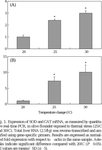
Fig. 1. Expression of SOD and CAT mRNA, as measured by quantita- tive real-time PCR, in olive flounder exposed to thermal stress (25�C and 30�C). Total liver RNA (2.5μg) was reverse-transcribed and am- plified using gene-specific primers. Results are expressed as normal- ized fold expression with respect to β-actin in the same sample. Aste- risks indicate significant difference compared with 20�C (P⁄0.05).
All values are means±SD (n==5).
about 2.4- and 3-fold in fish from the 25� C and 30� C groups respectively as relative to those from 20� C group, and CAT mRNA expression was significantly increased about 7.3- and 10.1-fold at 25� C and 30� C respectively than at 20� C (Fig. 1). SOD activity was significantly increased (P ⁄ 0.05) about 1.9- and 2.3- fold at 25� C and 30� C respectively than at 20� C. CAT activity was signif- icantly increased (P ⁄ 0.05) about 1.6-fold at 25� C and 30� C than at 20� C (Fig. 2).
2. H
2O
2concentration
The plasma H
2O
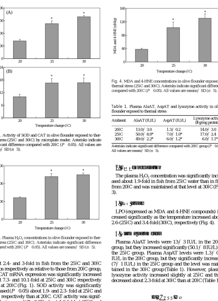
2concentration was significantly incre- ased about 1.9-fold in fish from 25� C water than in fish from 20� C and was maintained at that level at 30� C (Fig.
3).
3. LPO assay
LPO (expressed as MDA and 4-HNE compounds) in- creased significantly as the temperature increased about 2.6-(25� C) and 3.4-fold (30� C), respectively (Fig. 4).
4. Plasma parameters
Plasma AlaAT levels were 13±3 IU/L in the 20� C group, but they increased significantly (50.1±8 IU/L) in the 25� C group. Plasma AspAT levels were 1.3±0.2 IU/L in the 20� C group, but they significantly increased (7±1 IU/L) in the 25� C group and the level was main- tained in the 30� C group (Table 1). However, plasma lysozyme activity increased slightly at 25� C and then decreased about 2.3-fold at 30� C than at 20� C (Table 1).
DISCUSSION
In this study, we measured plasma H
2O
2and LPO lev-
Table 1. Plasma AlaAT, AspAT and lysozyme activity in olive flounder exposed to thermal stress
Ambient AlaAT (IU/L) AspAT (IU/L) Lysozyme activity (μg/mg protein)
20�C 13.0±3.0 1.3±0.2 14.0±3.0
25�C 50.0±8.0* 7.0±1.0* 17.0±2.4
30�C 49.0±2.2* 6.0±1.2* 6.0±1.1*
Asterisks indicate significant difference compared with 20�C group (P⁄0.05).
All values are means±SD (n==5).
*
*
0 200 400 600 800
20 25 30
* *
0 6 12 18 24
20 25 30
SOD activity(U/mL)CAT activity(nmole/min/mL)
Temperature change (�C)
(A)
(B)
Fig. 2. Activity of SOD and CAT in olive flounder exposed to ther- mal stress (25�C and 30�C) by microplate reader. Asterisks indicate significant difference compared with 20�C (P⁄0.05). All values are means±SD (n==5).
* *
0 100 200 300
20 25 30
H2O2 level(nmole peroxide/mL)
Temperature change (�C)
Fig. 3. Plasma H2O2concentrations in olive flounder exposed to ther- mal stress (25�C and 30�C). Asterisks indicate significant difference compared with 20�C (P⁄0.05). All values are means±SD (n==5).
*
*
0 40 80 120 160
20 25 30
MDA and 4-HNE(nM/g)
Temperature change (�C)
Fig. 4. MDA and 4-HNE concentrations in olive flounder exposed to thermal stress (25�C and 30�C). Asterisks indicate significant difference compared with 20�C (P⁄0.05). All values are means±SD (n==5).
els, as well as the expression and activity of the antioxi- dant enzymes SOD and CAT to understand the oxidative stress and mechanism in olive flounder exposed to the high temperature environment. We also examined lysozy- me-related immune function, AlaAT and AspAT to inves- tigate physiological changes induced by oxidative stress.
The expression and activity of antioxidant enzymes increased in olive flounder exposed to high temperature of 25� C and 30� C (Figs. 1 and 2). These results are in agreement with a previous report that found increased SOD, GPX, and GST activity in goldfish exposed to high temperatures from 3� C to 23� C (Bagnyukova et al., 2007). Also, SOD activity increased when goldfish were exposed to 35� C water (Lushchak and Bagnyukova, 2006a), as well as in mice exposed to a high temperature (38� C; Djordjevi´c et al., 2004), indicating possibly its key role in protection against ROS produced at high tem- perature (Lushchak and Bagnyukova, 2006a). Further- more, in this study, to examine oxidative stress induced by thermal stress, we investigated H
2O
2in plasma, and found that the plasma H
2O
2concentration was significant- ly increased in fish from 25� C and 30� C water than in fish from 20� C water (Fig. 3). When the Antarctic inter- tidal limpet (Nacella concinna) is exposed to a tempera- ture change of more than 10� C, its plasma H
2O
2concen- tration increases (Abele et al., 1998). An acute water temperature change induces stress (Abele et al., 1998) and increases oxygen consumption, which leads to an influx of oxygen into cells. Oxygen then is converted to ROS, O
2-, and H
2O
2, and increased oxygen consumption occurs in tissues (Boveris et al., 1976). ROS increase the risk of oxidative damage (Halliwell and Gutteridge, 1989). In addition, Harari et al. (1989) demonstrated that thermal stress accelerates the oxidation of polyamine in cells to generate ROS, so oxidative stress induced by ROS is related to the antioxidant response (Abele et al., 1998; Liu et al., 2007). Therefore, stimulated mRNA expression and enzymatic activity of the antioxidant enzymes with elevated water temperature would be tight- ly associated with the enhanced oxidative stress in olive flounder.
Moreover ROS production enhances LPO through lip- id damage, as well as increasing the expression and activ- ity of antioxidant enzymes (Bagnyukova et al., 2007). In this study, we found the LPO increased as the temperature increased (Fig. 4). This result is in agreement with Lush- chak and Bagnyukova (2006a, b), who found that prod- ucts of LPO, lipid peroxides (LOOH) and thiobarbituric acid-reactive substances (TBARS), increase quickly in goldfish tissues due to oxidative stress induced by ther- mal stress (35� C), and An and Choi (2010) reported that LPO increased in ark shell (Scapharca broughtonii) exposed to high temperature, indicating oxidative stress induced tissue damage. In addition, Chien and Hwang (2001) reported that malondialdehyde (MDA) increased
in thornfish (Terapon jarbua) exposed to 36� C water.
Also, Parihar et al. (1996) showed increased levels of LPO which, taken together with the SOD changes indi- cate increased ROS generation and oxidative stress in the liver of catfish (Heteropneustes fossilis) exposed to high temperature, and the results indicate an increase in oxidative stress of catfish exposed to high temperature.
Generally, AlaAT and AspAT are amino transfer enzy- mes and their blood concentrations are a general index of liver function in vertebrates. These enzymes can be used to evaluate the stress response caused by tempera- ture change, low oxygen, pH, ammonia, or heavy metals (Pan et al., 2003), and many studies have measured AlaAT and AspAT to examine stress levels (Vaglio and Landriscina, 1999; Choi et al., 2007, 2008a, b). In this study, plasma AlaAT levels increased during experimen- tal period (Table 1), suggesting that hepatocytes are damaged and increase stress levels due to the temperature increase, leading to a decrease in liver function. Also, lysozyme, a lysosomal enzyme implicated in the inflam- matory process, is a nonspecific humoral factor that acts under stressful conditions such as acute temperature change (Eo and Lee, 2008). It is released by leukocytes and plays an important role in antimicrobial activity (Eo and Lee, 2008). However, stress induced by acute envi- ronmental change suppresses the immune system’s ability to inhibit lysozyme activity (Wang et al., 2008). Lysozy- me activity decreased when sea cucumbers were trans- ferred from 12� C to 32� C (Wang et al., 2008), and when orange-spotted grouper (Epinephelus coioides) were transferred from 27� C to 35� C (Cheng et al., 2009).
Therefore, the stressor causes secretion of cortisol by the interrenal gland (Pickering and Pottinger, 1989; Har- ris and Bird, 2000), cortisol suppresses phagocytosis by decreasing phagocyte production (Harris and Bird, 2000), and resistance against bacterial pathogens is reduced (Maule et al., 1989). In this study, as an indicator of im- mune function, plasma lysozyme activity was measured, plasma lysozyme activity in was significantly decreased at 30� C than at 20� C (Table 1). This result is in agreement with An and Choi (2010), who found that lysozyme activ- ity decreased in ark shell exposed to high temperature, suggesting that acute temperature changes suppress im- mune function and reduce resistance against infection.
In particular, lysozyme activity increased slightly at 25� C and then decreased at 30� C. We suggest that lysozyme activity increased to enhance immune function against temperature change stress up to 25� C, but lysozyme activity was inhibited by the 30� C environment (Wang et al., 2008). So the increasing temperature in short peri- od induced acute stress and affects immune function in olive flounder by reduced lysozyme activity.
In conclusion, in the present study, the expression and
activity of the antioxidant enzymes (SOD and CAT) in
olive flounder exposed to high temperature environments
were increased, and induced oxidative stress. These re- sults indicate that antioxidant enzymes operated against thermal stress by increasing plasma H
2O
2concentrations and LPO levels in olive flounder exposed to high tem- perature environments. Additionally, thermal stress decre- ased lysozyme activity and then led to suppressed im- mune ability. Furthermore, these results, along with the changes of antioxidant enzymes (expression and activity of SOD and CAT) and H
2O
2, LPO and lysozyme gener- ated by thermal stress can provide basic data on antioxi- dant mechanism by thermal stress in fish.
ACKNOWLEDGMENTS
This research was supported in part by a project grant from Yeongnam Sea Grant, Korea and in part by the MKE (The Ministry of Knowledge Economy), Korea, under the ITRC (Information Technology Research Cen- ter) support program supervised by the NIPA (National IT Industry Promotion Agency (NIPA-2010-C1090-1021- 0015).
REFERENCES
Abele, D., B. Burlando, A. Viarengo and H.O. Pörtner. 1998.
Exposure to elevated temperatures and hydrogen per- oxide elicits oxidative stress and antioxidant response in the Antarctic intertidal limpet Nacella concinna.
Comp. Biochem. Physiol., B 120: 425-435.
An, M.I. and C.Y. Choi. 2010. Activity of antioxidant enzym- es and physiological responses in ark shell, Scapharca broughtonii, exposed to thermal and osmotic stress:
Effects on hemolymph and biochemical parameters.
Comp. Biochem. Physiol., B 155: 34-42.
Bagnyukova, T.V., V.I. Lushchak, K.B. Storey and V.I.
Lushchak. 2007. Oxidative stress and antioxidant defense responses by goldfish tissues to acute change of temperature from 3 to 23� C. J. Therm. Biol., 32:
227-234.
Basha, S.P. and A.R. Usha. 2003. Cadmium-induced antiox- idant defense mechanism in freshwater teleost Ore- ochromis mossambicus (Tilapia). Ecotoxical. Environ.
Saf., 56: 218-221.
Bly, J.E. and W. Clem. 1992. Temperature and teleost im- mune functions. Fish Shellfish Immunol., 2: 159-171.
Boveris, A., E. Cadenas and A.O.M. Stoppani. 1976. Role of ubiquinone in the mitochondrial generation of hydrogen peroxide. Biochem. J., 156: 435-444.
Bowden, T.J. 2008. Modulation of the immune system of fish by their environment. Fish Shellfish Immunol., 25:
373-383.
Chance, B., H. Sies and A. Boveris. 1979. Hydroperoxide
metabolism in mammalian organs. Physiol. Rev., 59:
527-605.
Cheng, A.C., S.A. Cheng, Y.Y. Chen and J.C. Chen. 2009.
Effects of temperature change on the innate cellular and humoral immune responses of orange-spotted grouper Epinephelus coioides and its susceptibility to Vibrio alginolyticus. Fish Shellfish Immunol., 26:
768-772.
Chien, L.T. and D.F. Hwang. 2001. Effects of thermal stress and vitamin C on lipid peroxidation and fatty acid composition in the liver of thornfish Terapon jarbua.
Comp. Biochem. Physiol., B 128: 91-97.
Choi, C.Y., B.H. Min, P.G. Jo and Y.J. Chang. 2007. Mol- ecular cloning of PEPCK and stress response of black porgy (Acanthopagrus schlegeli) to increased tempera- ture in freshwater and seawater. Gen. Comp. Endocri- nol., 152: 47-53.
Choi, C.Y., K.W. An, H.S. Shin, M.I. An and P.G. Jo. 2008a.
Changes of cytochrome P4501A mRNA expression and physiology responses in the olive flounder, Par- alichthys olivaceus, exposed to benzo[a]pyrene. Mar.
Biol. Res., 4: 470-476.
Choi, C.Y., K.W. An, Y.K. Choi, P.G. Jo and B.H. Min.
2008b. Expression of warm temperature acclimation- related protein 65-kDa (Wap65) mRNA, and physio- logical changes with increasing water temperature in black porgy, Acanthopagrus schlegeli. J. Exp. Zool., A 309: 206-214.
Collazos, M.E., C. Barriga and E. Ortega. 1995. Effect of high summer temperatures upon granulocyte phago- cytic function of the tench (Tinca tinca, L.). Comp.
Immun. Microbiol. Infect. Dis., 18: 115-121.
Djordjevi´c, J., G. Cviji´c, T. Vuˇckovi´c and V. Davidovi´c.
2004. Effect of heat and cold exposure on the rat brain monoamine oxidase and antioxidative enzyme activities. J. Therm. Biol., 29: 861-864.
Eo, J. and K.J. Lee. 2008. Effect of dietary ascorbic acid on growth and non-specific immune responses of tiger puffer, Takifugu rubripes. Fish Shellfish Immunol., 25: 611-616.
Esterbauer, H., R.J. Schaur and H. Zoliner. 1991. Chemistry and biochemistry of 4-hydroxynonenal, malonalde- hyde and related aldehydes. Free Radic. Biol. Med., 11: 81-128.
Halliwell, B. and J.M.C. Gutteridge. 1989. Free radicals in Biology and Medicine. Clarendon Press, Oxford, pp.
96-98.
Harari, P.M., D.J. Fuller and E.W. Gerner. 1989. Heat shock stimulates polyamine oxidation by two distinct mech- anisms in mammalian cell cultures. Int. J. Radiat.
Oncol. Biol. Phys., 16: 451-457.
Harris, J. and D.V. Bird. 2000. Modulation of the fish immune
system by hormones. Vet. Immunol. Immunopathol., 77: 163-176.
Hochachka, P.W. and G.N. Somero. 1984. Biochemical Adaptation. Princeton University Press, Princeton, New Jersey, 538pp.
Kashiwagi, A., K. Kashiwagi, M. Takase, H. Hanada and M.
Nakamura. 1997. Comparison of catalase in diploid and haploid Rana rugosa using heat and chemical inactivation techniques. Comp. Biochem. Physiol., B 118: 499-503.
Kim, M.O. and E.B. Phyllis. 1998. Oxidative stress in criti- cal care: is antioxidant supplementation beneficial?
J. Am. Diet. Assoc., 98: 1001-1008.
Liu, Y., W.N. Wang, A.L. Wang, J.M. Wang and R.Y. Sun.
2007. Effects of dietary vitamin E supplementation on antioxidant enzyme activities in Litopenaeus van- namei (Boone, 1931) exposed to acute salinity changes.
Aquaculture, 265: 351-358.
Lushchak, V.I. and T.V. Bagnyukova. 2006a. Temperature increase results in oxidative stress in goldfish tissues:
1. Indices of oxidative stress. Comp. Biochem. Physi- ol., C 143: 30-35.
Lushchak, V.I. and T.V. Bagnyukova. 2006b. Temperature increase results in oxidative stress in goldfish tissues:
2. Antioxidant and associated enzymes. Comp. Bio- chem. Physiol., C 143: 36-41.
Maule, A.G., R.A. Tripp, S.L. Kaattari and C.B. Schreck.
1989. Stress alters immune function and disease resis- tance in Chinook salmon (Oncorhynchus tshawytscha).
J. Endocrinol., 120: 135-142.
McFarland, V.A., L.S. Inouye, C.H. Lutz, A.S. Jarvis, J.U.
Clarke and D.D. McCant. 1999. Biomarkers of oxida- tive stress and genotoxicity in livers of field-collected brown bullhead, Ameiurus nebulosus. Arch. Environ.
Contam. Toxicol., 37: 236-241.
Nouroozzadeh, J., J. Tajaddinisarmadi and S.P. Wolff. 1994.
Measurement of plasma hydroperoxide concentrations
by ferrous oxidation-xylenol orange assay in conjunc- tion with triphenylphosphine. Anal. Biochem., 200:
403-409.
Pan, C.H., Y.H. Chien and B. Hunter. 2003. The resistance to ammonia stress of Penaeus monodon Fabricius juvenile fed diets supplemented with astaxanthin. J.
Exp. Mar. Biol. Ecol., 297: 107-118.
Pandey, S., S. Parvez, I. Sayeed, R. Haques, B. Bin-Hafeez and S. Raisuddin. 2003. Biomarkers of oxidative stress: a comparative study of river Yamuna fish Wal- lago attu (BI & Schn.). Sci. Total Environ., 309: 105- 115.
Parihar, M.S., A.K. Dubey, T. Javeri and P. Prakash. 1996.
Changes in lipid peroxidation, superoxide dismutase activity, ascorbic acid and phospholipid content in liver of freshwater catfish Heteropneustes fossilis exposed to elevated temperature. J. Therm. Biol., 21:
323-330.
Pickering, A.D. and T.G. Pottinger. 1989. Stress response and disease resistance in salmonid fish: effects of chronic elevation of plasma cortisol. Fish Physiol.
Biochem., 7: 253-258.
Schreck, C.B., C.S. Bradford, M.S. Fitzpatrick and R. Patino.
1989. Regulation of the interrenal of fishes: non-clas- sical control mechanism. Fish Physiol. Biochem., 7:
259-265.
Vaglio, A. and C. Landriscina. 1999. Changes in liver enzyme activity in the teleost Sparus aurata in response to cadmium. Ecotoxicol. Environ. Saf., 43: 111-116.
Wang, F., H. Yang, F. Gao and G. Liu. 2008. Effects of acute temperature or salinity stress on the immune response in sea cucumber, Apostichopus japonicus. Comp. Bio- chem. Physiol., A 151: 491-498.
Wheeler, C., J. Salzman, N. Elsayed, S. Omaye and D. Korte.
1990. Automated assays for superoxide dismutase,
catalase, glutathione peroxidase, and glutathione
reductase activity. Anal. Biochem., 184: 193-199.
고수온 환경에 의해 유도된 산화 스트레스에 대한 넙치의 항산화 작용과 생리적 변화
신현숙∙안광욱∙김나나∙최철영