• 검색 결과가 없습니다.

Inhibitory Effect of Jewel Orchid (Anoectochilus Formosanus) Plantlet Extract against Melanogenesis and Lipid Droplet Accumulation

N/A
N/A
Protected

Academic year: 2021

Share "Inhibitory Effect of Jewel Orchid (Anoectochilus Formosanus) Plantlet Extract against Melanogenesis and Lipid Droplet Accumulation"

Copied!
6
0
0

로드 중.... (전체 텍스트 보기)

전체 글

(1)

금선련 조직 배양체 추출물의 멜라닌 합성 및 지방축적 억제 효과

박 창 민⋅정 민 석⋅백 기 엽*⋅최 종 완

(주)한국화장품제조 기술개발연구소, *충북대학교

(2010년 6월 9일 접수, 2010년 6월 15일 수정, 2010년 6월 17일 채택)

Inhibitory Effect of Jewel Orchid (Anoectochilus Formosanus) Plantlet Extract against Melanogenesis and Lipid Droplet Accumulation

Chang-Min Park, Min-Seok Joung, Kee-Yoeup Paek*, and Jong-Wan Choi

R&D Center, Hankook Cosmetics Manufacturing Co., Ltd., 76-1, Yongseong-Ri, Samseong-Myeon, Eumseong-Gun, Chungcheongbuk-Do 369-834, Korea

*Research Center for The Development of Advanced Horticultural Technology, Chungbuk National University (Received June 9, 2010; Revised June 15, 2010; Accepted June 17, 2010)

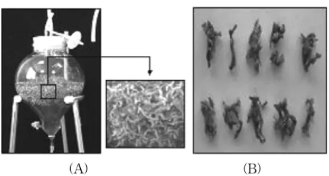
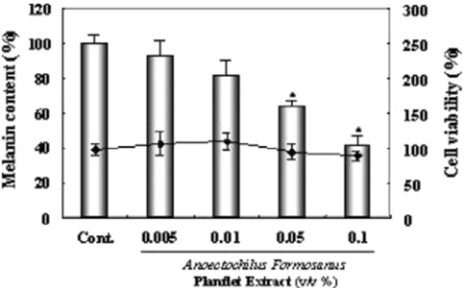
요 약: 일반적으로 보석란으로 알려진 금선련은 대만에서 폐나 간의 질병 및 발열이나 두통 치료를 위한 전통식물약제 로 사용되어 왔다. 본 연구에서는 생물반응장치를 이용하여 조직배양된 금선련 식물체에 대하여 화장품 성분으로써 응 용 가치를 평가하였다. 이미 몇몇 보고 된 논문에서 금선련은 항암활성, 면역 활성, 간 보호 활성 및 지질대사의 약리학 적 활성 등에 대한 연구가 되고 있지만 화장품 성분으로 효능들에 대한 연구는 알려져 있지 않다. 따라서 본 연구에서는 생물반응장치를 이용하여 조직배양된 금선련 추출물에 대하여 미백 및 항비만 관련한 효능 효과를 평가하였다. 실험 결과 조직배양된 금선련 추출물은 tyrosinase 활성 및 멜라닌 합성 억제 효과뿐만 아니라 지방 전구 세포의 지방세포로 의 분화를 억제시킴으로써 세포 내 지질 축적을 억제하였다. 이러한 결과들은 피부보호를 위한 화장품 성분으로서 응용 가능성을 제공 할 수 있을 것으로 사료된다.

Abstract:

Anoectochilus formosanus

, commonly known as “Jewel Orchids”, which has been used in traditional folk medicines for feber, pain, and diseases of the lung and liver in Taiwan. We artificially cultured

Anoectochilus for- mosanus

plantlet by using the bioreactor culture system for this study from

Anoectochilus formosanus

. Previously, several studies have been reported on pharmacological activities of lipid-metabolism, hepatoprotective activity, an- ti-tumor activity and immuno-stimulating effects but other efficacy were not well known as a cosmetic ingredient for skin care. In this study, we investigated the effect of melanogenesis in B16 mouse melanoma cells and lipid droplet accumulation in 3T3-L1 preadipocytes about

Anoectochilus formosanus

plantlet extract. We report that

Anoectochilus formosanus

plantlet extract inhibits the cytoplasmic lipid droplet accumulation through adipogenic differentiation of preadipocytes as well as inhibition of tyorsinase activity and melanogenesis. As a result, our findings indicate that

Anoectochilus formosanus

plantlet extract may be the potential natural ingredient for whitening and slimming cos- metic products.

Keywords: Anoectochilus formosanus, melanogenesis, adipogenesis, plantlet, tissue-culture

1. Introduction

1)

The genus Anoectochilus (Orchidaceae) grows in deep

† 주 저자 (e-mail: [email protected])

shade on moist forest floors with a rich layer of humus and consists of approximately 40 species[1]. Several species have been used as traditional folk medicines.

Among the genus Anoectochilus, Anoectochilus for-

(2)

mosanus Hayata is a native perennial and terrestrial or- chid plant, which is grown in Taiwan (China) and Okinawa (Japan)[2]. In general, Anoectochilus for- mosanus Hayata is well known as “Jewel Orchids” be- cause its leaves have network of colorful venation.

Anoectochilus formosanus is mainly used to treat hy- pertension, diabetes, heart and bright's disease and oth- er diseases as well as in traditional folk medicine against lung and liver disease in China, Taiwan and many other countries[3,4]. Anoectochilus formosanus is conventionally propagated by seed. However, the ger- mination rate is very low. Anoectochilus formosanus is also an expensive Chinese folk medicinal plant. It has great value in herbal medicines because of its pharma- cological activities of lipid-metabolism, hepatoprotective activity, anti-tumor activity and immuno-stimulating effects, anti-inflammation and antioxidant activities[5,6].

Melanin is a biological pigment found in skin, which mainly determines human skin color by the content of the pigment melanin. In addition, melanin plays an im- portant role in protecting human skin from the harmful effects of UV radiation from the sun. Melanin is pro- duced in unique organelles melanocytes in the epi- dermis of skin[7,8]. Melanogenesis is initiated with the conversion of L-tyrosine through L-dopa to dopaquinone by tyrosinase[9,10]. Although melanin plays an im- portant role as a photoprotective function of sun-in- duced skin injury, its abnormal accumulation results in serious skin problems. Adipocyte differentiation is influ- enced by a large number of growth factors. Insulin, IGF-1, fibroblast growth factor, platelet-derived growth factor and epidermal growth factor promote pre- adipocyte proliferation but only insulin or IGF-1 can in- duce adipocyte differentiation. Preadipocyte differ- entiation is characterized by changes in cell morphol- ogy, hormone sensitivity and gene expression in anti- obesity study. The cells accumulate lipid droplets[11,12].

In the present study, we investigated the effect of mel- anogenesis and anti-obesity whether Anoectochilus for- mosanus plantlet cultured by using the bioreactor cul- ture system has a merit as a cosmetic ingredient.

2. Materials and Methods

2.1. Reagents and Cell Culture

Antibodies against tyrosinase and ß-actin were pur- chased from Santa Cruz Biotechnology (Santa Cruz, CA). Insulin, dexamethasone, isobutylmethylxanthine (IBMX) and Oil Red O were purchased from sigma-al- drich (St. Louis, MO, USA). Other commercially avail- able reagents and solvents were used as received.

3T3-L1 preadipocytes procured from American Type Culture Collection and B16F10 mouse melanoma cell line procured Korean Cell Line Bank were used in this study. Preadipocyte line 3T3-L1 cells were maintained in Dulbecco’s modified Eagle’s medium (DMEM, Wel- GENE, Korea) supplemented with 10 % heat-inactivated calf serum (CS, Gibco BRL, USA), 100 U/mL pen- icillin (Gibco BRL, USA) and 100 µg/mL streptomycin (Gibco BRL, USA) at 37 ℃ in a humidified atmos- phere containing 5 % CO

2

. B16 mouse melanoma cells was maintained in Dulbecco’s modified Eagle’s medium (DMEM, WelGENE, Korea) supplemented with 10 % heat-inactivated fetal bovine serum (FBS, Gibco BRL, USA), 100 U/mL penicillin (Gibco BRL, USA) and 100 µg/mL streptomycin (Gibco BRL, USA) at 37 ℃ in a humidified atmosphere containing 5 % CO

2

.

2.2. Preparation of Anoectochilus Formosanus Plantlet Extract

Anoectochilus formosanus plantlet was obtained from Research Center for The Development of Advanced Horticultural Technology, Chungbuk National University.

Extraction : After dried, Anoectochilus formosanus plantlet was immersed in 70 % ethanol solution and 1,3- butyleneglycol (1,3-BG) for sufficient time and mixed enough for 48 h in 45 ℃ using agitator. Then, the sol- ution was filtered through a filter paper, followed by the removal of ethanol from the filtrate at 50 ~ 60 ℃ to obtain a Anoectochilus formosanus plantlet extract.

2.3. Measurement of Melanin Content in B16 Mouse Melanoma Cells

Measurement of melanin content was performed

using a modification of the method reported by Funasaka

(3)

[13]. B16 mouse melanoma cells were cultured in DMEM supplemented with 10 % FBS in humidified incubator at 37 ℃ under 5 % CO

2

in 6 well plate at density of 2 × 10

4

cells/well. After cells were attached, medium was replaced with DMEM containing 10 % FBS, 0.2 uM α-MSH, 2 mM theophylline and samples addition. After 4 days, trypsin was added and sus- pended cells were collected by centrifugation. Then cell pellets were dried and dissolved in 1 N NaOH. Melanin synthesis inhibition rates were measured 490 nm using ELISA reader.

2.4. Assay of Tyrosinase Activity in a Cell Culture-free System

A mushroom tyrosinase was dissolved in a phosphate buffer (pH 6.5) to prepare a tyrosinase enzyme solution.

L-tyrosine was dissolved in a phosphate buffer (pH 6.5) to the concentration of 1.5 mM to prepare a substrate solution. Tyrosinase enzyme solution was added to the test solution. After incubation at 37 ℃ for 10 min, sub- strate solution was added there to. Absorbance was measured using an ELISA reader at 490 nm.

2.5. Pre-adipocyte (3T3-L1) Differentiation

To induce differentiation, pre-adipocytes were grown in 24-well plates until 2 d post-confluence. Confluent cells were differentiated by incubation with MID hormone mixture (10 µg/mL insulin, 0.5 µM dexamethasone, and 1 mM IBMX) in 10 % fetal bovine serum (FBS) / DMEM for 36 h and maintained in post-differentiation medium containing 10 µg/mL insulin for more 6 d.

2.6. Oil Red O Staining Assay

The degree of anti-adipogenic effects, with and without Anoectochilus formosanus plantlet, was eval- uated by observation of the lipid accumulation with Oil Red O staining. At 6 d after the initiation, cells were washed twice with PBS and then fixed for 20 min with 3.7 % formaldehyde. Cells were washed twice in PBS and the lipidic content of the cells is evaluated by the red oil coloration method. Briefly, cells were stained for 10 min using 0.5 % oil red in isopropanol/water at room temperature. Then the stain was solubilized in pure iso-

propanol, and the absorbance was measured at 540 nm on a spectrophotometer.

2.7. Cell cytotoxicity by MTT Assay

Cells (1 × 10

5

cells/well) were seeded in 10 % BCS/DMEM medium and incubated in 5 % CO

2

in- cubator at 37 ℃ after treatment with Anoectochilus formosanus plantlet extract for indicate times. Mea- surement of mitochondrial activity to form purple for- mazan by MTT was used to assess the cytotoxicity of cell following extract treatment : MTT (0.5 mg/mL), one tenth of the original culture volume, was added to each culture and incubated for 3 h at 37 ℃ in 5 % CO

2

. The purple formazan formed by viable cells was dis- solved by the addition of DMSO and absorbance at the dual ranges of 540 nm and 630 nm was measured by using spectrophotometer.

2.8. Western Blot Analysis

Cells were treated with various dose and lysed in lysis buffer as described previously[14]. After differentiation, cells were lysed in lysis buffer. The lysates were clari- fied by centrifugation at 12,000 × g for 15 min at 4 ℃ and protein content was measured by 12.5 % SDS- PAGE and blotted to nitrocellulose membrane (0.2 mm, Amersham, Arlington Heights, IL). The membrane was blocked with 5 % non-fat skim milk in TBS-T and incubated with the primary and secondary antibodies.

Immunoblots were visualized by enhanced chemilu- minescence (Amersham, UK), according to the manu- facturer’s protocol.

2.9. Statistical Analysis

Data are presentated as mean ± SD. Comparisons be- tween groups were used to the paired Student’s t-test.

Asterisk (*, p < 0.05) was considered to bestatistically significant.

3. Results

3.1. Anoectochilus Formosanus Plantlet Cultured by the Bioreactor Culture System

Anoectochilus formosanus is precious in the Taiwanese

(4)

(A) (B)

Figure 1. The

Anoectochilus formosanus

plantlet cultured by the bioreactor culture system. Figure 1A is tissue-cultured

Anoectochilus formosanus

plantlet culturing by the bioreactor culture system using liquid medium and Figure 1B is a

picture of a dried

Anoectochilus formosanus

plantlet. Figure 2. The Effect of

Anoectochilus formosanus

plantlet on Tyrosinase Activity in Cell-culture Free System. Tyro- sinase activity were expressed as a percentage of control.

The results were represented as mean of standard deviation (S.D.) of three independent experiments. *,

p

< 0.05 versus control.

* *

Figure 3. The effect of

Anoectochilus formosanus

plantlet on the Protein Expression of Tyrosinase in B16 Melanoma Cells. B16 melanoma cells were treated with

Anoectochilus formosanus

plantlet at the concentrations indicated in the figure and incubated for 72 h. Tyrosinase protein expression was determined by a western blotting analysis using a specific antibody, as described in the manteials and methods section. Bands were subjected to densitometric scanning using the Scion image NIH image software. The results were represented as mean of standard deviation (S.D.) of three independent experiments. *,

p

< 0.05 versus control.

market but seriously is reduced its populations. Tissue culture protocols have been developed for preservation of this valuable plant. Figure 1(A) is tissue-cultured Anoectochilus formosanus plantlet cultured by the bio- reactor culture system using liquid medium. Figure 1(B) is the picture of a dried Anoectochilus formosanus plant- let cultured by the bioreactor culture system. We ex- tract the dried Anoectochilus formosanus plantlet and evaluate the efficacy as cosmetic ingredients.

3.2. Inhibitory Effect of Anoectochilus Formosanus Plantlet on Mushroom Tyrosinase Activity in a Cell Culture Free System

To investigate the whitening effect of Anoectochilus formosanus plantlet extract, we first analyzed inhibition rate of mushroon tyrosinase activity in a cell-culture free system. As shown in Figure 2, Anoectochilus for- mosanus plantlet extract inhibited the tyrosinase activ- ity in a concentration-dependent manner. The tyrosinase activity was reduced to 39 % at 0.1 % and to 28 % at 0.05 %, as compared with the non-treated control.

3.3. Inhibitory Effect of Anoectochilus Formosanus Plantlet on Melanogenesis and Tyrosinase Protein Expression in a Cell Based System

We investigated whether Anoectochilus formosanus plantlet extract inhibits melanin synthesis and ty- rosinase expression in B16 mouse melanoma cells as well as mushroom tyrosinase activity in cell-culture free system. Also, we performed western blot analysis

against the cell lysate obtained from Anoectochilus

formosanus plantlet extract-treated B16 mouse mela-

noma cells. As shown in Figure 3, Anoectochilus for-

mosanus plantlet extract decreased the expression of

(5)

Figure 4. The Effect of

Anoectochilus formosanus

plantlet extract on the Melanin Content and cell viability of Mouse B16 Melanoma Cells. B16 melanoma cells were treated with

Anoectochilus formosanus

plantlet at the concentrations indicated in the Figure 2 and then incubated for 72 h. The melanin content and cell viability were determined as described in the Manteials and Methods section. The results were represented as mean of standard deviation (S.D.) of three independent experiments. *,

p

< 0.05 versus control.

Figure 5. Inhibitory effect of

Anoectochilus formosanus

plantlet on the lipid accumulation and cell viability of 3T3-L1 cells. We compared a lipid contents after cotreatment of

Anoectochilus formosanus

plantlet extract with MID hormone mixture. Lipid accumulation after differentiation for 6 d was measured by Oil Red O staining as shown in Figure 5.

The Lipid accumulation and cell viability were determined as described in the Manteials and Methods section. All values are represented as means ± standard deviation (S.D.) of three independent experiments. *,

p

< 0.05 versus control.

tyrosinase protein in a concentration-dependent manner.

In addition, we examined melanin content of B16 mouse melanoma cells in order to confirm melanogenesis in- hibiting-effect of Anoectochilus formosanus plantlet extract. As shown in Figure 4, Anoectochilus formosanus plantlet extract inhibited the melanogenesis in a con- centration-dependent manner. The melanin content was reduced to 59 % at 0.1 % and to 36 % ant 0.05 %, as compared with the non-treated control cells at a con- centration without any cytotoxicity.

3.4. Inhibitory Effect of Anoectochilus Formosanus Plantlet on the Lipid Accumulation of 3T3-L1 Cells To investigate whether Anoectochilus formosanus plantlet extract has anti-obesity effect, we examined a cytoplasmic lipid droplet accumulation by the differ- entiation of preadipocytes into matures adipocytes in presence of MID hormone mixture. After incubation for 6 d with both MID and Anoectochilus formosanus plantlet extract, cells reduced a lipid droplet accumulation.

We investigated whether Anoectochilus formosanus plantlet extract inhibits a cytoplasmic lipid droplet by Oil red O staining assay as described in materials and methods. In result, Anoectochilus formosanus plantlet extract, tested at 0.01 %, 0.05 % and 0.1 % inhibited

by 11 %, 27 % and 49 % a lipid content, as compared with the only MID hormone mixture control after in- cubation with both MID hormone mixture and extract at indicated concentrations without any cytotoxicity as shown in Figure 5.

4. Discussion

Generally, tissue-cultured plant has a benefit to solve

the drawbacks such as a rareness, long time and high

cost for growing herb. Although the Anoectochilus

formosanus has been well known as herb for remedy a

hypertension, diabetes, heart, lung and liver disease as

a traditional medicine, we investigated whether Anoe-

ctochilus formosanus plantlet extract have efficacy as a

cosmetic ingredient for skin care in this study. In sum-

mary, using B16 mouse melanoma cells and 3T3-L1

preadipocyte, we found that Anoectochilus formosanus

plantlet extract had a inhibitory effect of melanogenesis

by reducing the protein amounts of tyrosinase and sup-

pressive effect of lipid droplet accumulation. As a re-

sults, we suggest that Anoectochilus formosanus plant-

let could be cosmetic ingredient for skin care such as

whitening and inhibition of lipogenesis.

(6)

Acknowledgments

This study was supported by a grant of the Korea Healthcare technology R&D Project, Ministry for Health, Welfare & Family Affairs, Republic of Korea.

(과제고유번호 : A092055)

References

1. I. Belitsky and V. N. A. Bersenev, Jewel Orchids, Mag. amer. Orchid Soc., 1999, 33 (1999).

2. B. Sugiharto, N. Ermawati, H. Mori, K. Aoki, K.

Yonekura-Sakakibara, T. Yamaya, T. Sugiyama, and H. Sakakibara, Identification and characterization of a gene encoding drought-inducible protein localizing in the bundle sheath cell of sugarcane, Plant Cell Physiol., 43, 350 (2002).

3. W. L. Liang, R. C. Chen, Y. J. Chiang, C. H. Su, L. L. Yang, and K. L. Yen, Study of Anoectochilus species. I. Study on the physiological activities of Jin-Sian-Lian, Formosan Sci., 43, 47 (1990).

4. C. C. Lin, P. C. Huang, and J. M. Lin, Antioxidant and hepatoprotective effects of Anoectochilus for- mosanus and Gynostemma pentaphyllum, Amer J Clin Med., 28, 87 (2000).

5. S. Y. Wang, Y. H. Kuo, H. N. Chang, P. L. Kang, H. S. Tsay, K. F. Lin, N. S. Yang, and L. F. Shyur, Profiling and characterization of antioxidant activ- ities in Anoectochilus formosanus Hayata, J Agric Food Chem., 50, 1859 (2002).

6. C. C. Tseng, H. F. Shang, L. F. Wang, B. S. u, C.

C. Hsu, H. Y. Kao, and K. T. Cheng, Antitumor and immunostimulating effects of Anoectochilus formosanus Hayata, Phytomedicine, 13, 366 (2006).

7. K. U. Schallreuter, S. Kothari, B. Chavan, and J. D.

Spencer, Regulation of melanogenesiscontroversies and new concepts, Exp. Dermatol., 17, 395 (2008).

8. S. Ito, The IFPCS presidential lecture : a chemist's view of melanogenesis, pigment Cell Res., 16, 230 (2003).

9. C. J. Cooksey, P. J. Garratt, E. J. Land, S. Pavel, C. A. Ramsden, P. A. Riley, and N. P. Smit, J. Biol.

Chem., 272, 26226 (1997).

10. F. Solano, S. Briganti, M. Picardo, and G. Ghanem, Pigment Cell Res., 19, 550 (2006).

11. B. H. Choi, I. S. Ahn, Y. H. Kim, J. W. Park, S. Y.

Lee, C. K. Hyun, and M. S. Do, Berberine reduces the expression of adipogenic enzymes and inflam- matory molecules of 3T3-L1 adipocyte, Exp Mol Med., 38(6), 599 (2006).

12. J. W. Kim, H. Monila, A. Pandey, and M. D. Lane, Upstream stimulatory factors regulate the C/EBP alpha gene during differentiation of 3T3-L1 pre- adipocytes, Biochem Biophys Res Commun., 354(2), 517 (2007).

13. Y. Funasaka, A. K. Chakraborty, M. Komoto, A.

Ohashi, and M. Ichihashi, The depigmenting effect of alpha-tocopheryl ferulate on human melanoma cells, Br. J. Dermatol., 141, 20 (1999).

14. C. M. Park, M. J. Park, H. J. Kwak, H. C. Lee, M.

S. Kim, S. H. Lee, I. C. Park, C. H. Rhee, and S.

I. Hong, Ionizing radiation enhances matrix metal- loproteinase-2 secretion and invasion of glioma cells through Src/epidermal growth factor receptor-medi- ated p38/Akt and phosphatidylinositol 3-kinase/

Akt signaling pathways, Cancer Res., 66(17), 8511

(2006).

수치

Figure 3. The effect of  Anoectochilus formosanus  plantlet  on the Protein Expression of Tyrosinase in B16 Melanoma  Cells
Figure  5.  Inhibitory  effect  of  Anoectochilus  formosanus  plantlet on the lipid accumulation and cell viability of 3T3-L1  cells

참조

관련 문서