야관문이 생쥐의 면역세포 활성에 미치는 영향
은재순*
우석대학교 약학대학
Effect of Lespedeza Cuneata G. Don on the Activity of Murine Immune Cells
Jae Soon Eun*
Department of Pharmacy, Woosuk University
The purpose of this research was to investigate the effects of 50% ethylalcohol extracts of Lespedeza cuneata G. Don (LE) on the activity of murine immune cells. LE was administered orally once a day for 7 days at the dose of 250 mg/kg. LE increased the cell viability of thymocytes, splenocytes and peritoneal macrophages in vitro and in vivosystem, but decreased the cell viability of thymocytes and splenocytes in the presence of concanavalin Ain vivo system. Also, the administration of LE was increased the production of γ-interferone, but did not affect the production of interleukin-2 and interleukin-4. Furthermore, LE decreased the phagocytic activity of peritoneal macrophagesin vitro and in vivo system, but enhanced the production of nitric oxide. These results suggest that LE has a immuno-regulative action via stimulation of immune cells proliferation.
Key words : Lespedeza cuneata G. Don, thymocytes, splenocytes, macrophages
* 교신저자 : 은재순, 완주군 삼례읍 후정리 490, 우석대학교 약학대학
․E-mail : [email protected], ․Tel : 063-290-1569
․접수 : 2011/08/26 ․수정 : 2011/09/21 ․채택 : 2011/10/07
서 론
야관문(Lespedeza cuneata G. Don, 夜關門)은 콩과 (Leguminosae)에 속하는 여러해살이 풀로서, 우리나라 이름으로 는 비수리라고 하며, 우리나라 전국에 분포되어 있다1). 주요성분 으로는 pinitol, tannin, β-sitosterol 등이 함유되어 있으며, 특히 flavonoid로서 quercetin, kaempferol, vitexin, orientin 등을 함유 하고 있다2-5). 한방에서는 肝, 腎을 補하고 肺陰을 補益하며 散瘀, 消腫 효능이 있고, 遺精, 遺尿, 白濁, 白帶, 喘哮, 胃痛, 勞傷, 小兒 疳積, 下痢, 타박상, 시력감퇴, 결막염, 급성유선염 등을 치료하는 데 사용되어 왔다6-10). 면역이란 생체가 자기 성분 이외의 물질 등이 생체의 항상성을 깨뜨리거나 자기를 위협하는 것을 배제하 기 위해 일어나는 일련의 생체방어반응을 의미하며, T- 및 B-lymphocyte가 관련된 특이적면역과 대식세포가 관련된 비특 이적면역으로 분류할 수 있다11).
임상에서 야관문은 35도 이상 되는 술로 3개월쯤 우려내어 사용하여야만 효능이 강력하다고 알려져 있다. 따라서 본 실험에
서는 다양한 용도로 사용되고 있는 야관문이 면역능에 미치는 영향을 알아보고자, 야관문을 50% 에탄올로 추출하여, 생쥐의 흉선세포, 비장세포 및 대식세포의 활성에 미치는 영향을 측정한 결과 약간의 지견을 얻었기에 이에 보고하고자 한다.
재료 및 방법
1. 실험동물
본 실험에 사용한 생쥐는 ICR계 수컷 20 ± 2 g을 대한실험 동물(주)에서 구입하여, 온도 20 ± 3℃, 습도 50 ± 5%, dark/light 12시간의 조건하에서 1 주일 이상 실험실에 적응시킨 후 사용하 였으며, 고형사료와 물을 자유스럽게 섭취하도록 하였다.
2. 시약
실험에 사용한 시약은 fetal bovine serum (FBS), RPMI medium 1640은 Gibco Co.에서, concanavalin A (Con A), lipopolysaccharide (LPS), γ-interferon (γ-IFN), lucigenin, zymosan, MTT는 Sigma Co.에서, mouse γ-IFN immunoassay kit, mouse IL-2 immunoassay kit, mouse IL-4 immunoassay kit 는 R&D Co.에서 구입하여 사용하였으며, 기타 시약은 cell
culture용 및 1급 시약을 사용하였다. 사용기구는 multi-well plate (96-well, 24-well)는 Costar Co.에서, white multi-well plate (96-well)는 Nunc Co.에서 구입하여 사용하였으며, microplate-reader (Molecular Devices, SoftMax Pro5), CO2
incubator (Vision scientific Co.), Infinite F200 (TECAN Co.) 등 을 사용하였다.
3. 검액의 조제
야관문은 전주 야산에서 채집하여 음건한 후, 100 g을 50%
에탄올 2 L로 48시간씩 2회 추출한 후, 여과하여 여액을 감압농 축한 다음, freeze dryer로 동결건조하여, 건조분말 12.2 g을 얻었 다 (이하 LE라 함, 수득률 12.2%). 동물실험 시에는 생리식염수 에 용해시켜 사용하였으며, 세포실험시에는 PBS에 용해시켜 membrane filter (0.45 μM)로 여과멸균하여 사용하였다.
4. 흉선 및 비장세포의 분리
생쥐의 흉선 및 비장세포 분리는 Wysocki12) 및 Mizel13) 등 의 방법을 이용하였다. 생쥐 5마리를 1군으로 하여, 대조군에는 생리식염수만을, 실험군에는 LE 250 mg/kg을 1일 1회씩 7일간 경구투여한 다음 생쥐를 CO2gas로 질식시켰다. 적출한 흉선 및 비장을 DPBS를 넣은 petri dish에서 잘게 분쇄하고 멸균된 stainless mesh로 여과하여 세포부유액을 얻은 후, 10% FBS와 penicillin-streptomycin (100 units/ml, 100 ㎍/ml)을 첨가한 RPMI 1640 배지로 현탁하여, 흉선 및 비장세포 부유액으로 하 였다.
5. 복강대식세포의 분리
대식세포의 분리는 LE 250 mg/kg을 1일 1회씩 7일간 경구 투여 하였고, 약물 투여 4일째 생쥐 복강에 3% thioglycollate 2 ml를 주입하였다. 생쥐를 CO2gas로 질식시켜, 복강에 DPBS 10 ml를 넣어 복강세포를 수집하였다. 수집한 세포를 4℃에서 1,300 rpm으로 10 분간 원심분리하고, 10% FBS와 penicillin- streptomycin을 첨가한 RPMI 배지로 현탁하여, 직경 120 mm petri dish에 분주하여 CO2 incubator에서 2시간 동안 배양하였 다. 부착하지 않은 세포를 제거한 다음, 부착한 세포를 복강대식 세포로 사용하였다.
6. 흉선세포, 비장세포 및 복강대식세포의 세포생존율 측정 세포생존율 측정은 Mosmann14)이 개발하여 Kotnik 등15)이 변형시킨 MTT 방법으로 측정하였다. 흉선 및 비장세포를 96-well plate의 각 well에 100 ㎕ (1.2 × 10⁷cells/ml)씩 분주하 고, T-lymphocytes를 관찰할 때는 concanavalin A (Con A) 5 ㎍ /ml를 B-lymphocytes를 관찰할 때는 lipopolysaccharide (LPS) 10 ㎍/ml를 첨가하거나 첨가하지 않은 조건으로 37℃의 CO2
incubator에서 48 시간 배양하였다. 배양 종료 4 시간 전에 5 mg/ml 농도로 DPBS에 희석된 MTT용액 20 ㎕를 각 well에 첨 가하였다. 4시간 후 0.1N-HCl에 용해시킨 10%-SDS 100 ㎕를 각 well에 첨가하여, 차광 상태에서 18 시간 더 배양한 후, 발색된
각 well의 흡광도를 microplate reader로 570 nm에서 측정하여 세포생존율을 산정하였다. 복강대식세포를 96-well plate의 각 well에 100 ㎕ (5 × 10⁵ cells/ml)씩 분주하고, 37℃의 CO2
incubator에서 48 시간 배양하였다. 배양 종료 후 MTT용액을 각 well에 첨가하고, 4시간 후 DMSO 100 ㎕를 각 well에 첨가하여, 발색된 각 well의 흡광도를 microplate reader로 570 nm에서 측 정하여 세포생존율을 산정하였다.
7. Serum 중 cytokines 측정
Cytokine 측정은 생쥐 5마리를 1군으로 하여 대조군에는 생 리식염수만을, 실험군에는 LE 250 mg/kg을 1일 1회씩 7일간 경 구투여한 다음 8일째 serum을 분리한 후, serum 50 ㎕를 취하여 mouse immunoassay kit로 cytokine양을 측정하였다16).
8. 복강 대식세포로부터 phagocytosis 측정
분리한 복강대식세포를 2 × 10⁶cells/ml가 되도록 HBS buffer 에 부유시켜, 96-well white microplate의 각 well에 세포부유액 50
㎕, lucigenin 용액 50 ㎕ 및 zymosan 용액 30 ㎕를 첨가하여, 최종 volume이 200 ㎕가 되도록 조정한 후, 37℃에서 30분간 전처리한 다음, 5분 간격으로 30분 동안 lucigenin chemiluminescence 양을 luminometer를 이용하여 측정하였다17,18).
9. 대식세포로부터 nitric oxide 생성량 측정
대식세포로 부터 생성되는 nitric oxide (NO)의 양은 Griess 법19)으로 측정하였다. 분리한 대식세포를 2 × 10⁶cells/ml로 조 제하여, 24 well plate에 100 ㎕ 씩 분주한 후, 최종 volume이 200 ㎕가 되도록 조정한 다음, 각 well에 LPS 1 ㎍/ml와 γ-IFN 25 units/ml를 첨가하여 배양하였다. 24 시간 후 배양액 100 ㎕ 와 Griess 시약 100 ㎕를 혼합하여 96 well module에 넣고, 37℃
에서 10 분간 방치한 후 570 nm에서 microplate reader로 흡광도 를 측정하여 미리 작성한 NaNO2의 검량선에 의해 NO2-의 농도 를 환산하였다.
10. 통계처리
모든 실험 결과들은 mean ± SE로 나타내었고 통계처리는 Student'st-test를 실시하여 p<0.05를 기준으로 유의성 여부를 판 정하였다.
결 과
1. 흉선세포, 비장세포 및 복강대식세포의 세포생존율에 미치는 효과
In vitro실험에서, 흉선세포에 lymphocyte mitogen을 처리 하지 않았을 때의 세포생존율을 100%로 하였을 때, LE 1.25, 5 및 20 μg/ml를 처리하였을 때 각각 108.4 ± 1.4, 123.7 ± 1.5 및 166.2 ± 1.9%로 모든 농도에서 대조군에 비해 세포생존율이 증가 하였다. Con A를 처리하였을 때 대조군의 세포생존율은 126.5 ± 1.8%로 증가하였으며, Con A와 LE 1.25, 5 및 20 μg/ml를 처리
하였을 때 136.2 ± 1.3, 155.8 ± 1.2 및 190.3 ± 1.9%로 모든 농도 에서 Con A를 처리한 대조군에 비해 세포생존율이 증가하였다 (Table 1).
Table 1. Effects ofLespedeza cuneata G. Don ethylalcohol extracts (LE) on the cell viability of thymocytes in vitro system
Samples Dose
(μg/ml)
Cell viability of thymocytes(%) Concanavalin A
- +
Control - 100.0 ± 1.2 126.5 ± 1.8**
LE
1.25 108.4 ± 1.4* 136.2 ± 1.3# 5 123.7 ± 1.5*** 155.8 ± 1.2###
20 166.2 ± 1.9*** 190.3 ± 1.9###
The various concentration of LE were treated in thymocytes and then the cells were cultured for 48 hrs. The data represents the mean ± SE of 3 experiments. *; Significantly different from control group (*; p<0.05, **; p<0.01, ***; p<0.001). #; Significantly different from Con A-treated control group (#; p<0.05,###; p<0.001).
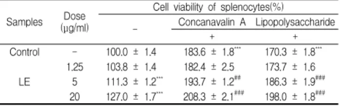
분리한 비장세포에 lymphocyte mitogen을 처리하지 않았을 때의 세포생존율을 100%로 하였을 때, LE 1.25, 5 및 20 μg/ml를 처리하였을 때 각각 103.8 ± 1.4, 111.3 ± 1.2 및 127.0 ± 1.7%로 5 μg/ml 이상의 농도에서 대조군에 비해 세포생존율이 증가하 였다. Con A를 처리하였을 때 대조군의 세포생존율은 183.6 ± 1.8%로 증가하였으며, Con A와 LE 1.25, 5 및 20 μg/ml를 처리 하였을 때 182.4 ± 2.5, 193.7 ± 1.2 및 208.3 ± 2.1%로 5 μg/ml 이상의 농도에서 Con A를 처리한 대조군에 비해 세포생존율이 증가하였다. LPS를 처리하였을 때 세포생존율은 170.3 ± 1.8%로 증가하였으며, LPS와 LE 1.25, 5 및 20 μg/ml를 처리하였을 때 각각 173.7 ± 1.6, 186.3 ± 1.9 및 198.0 ± 1.8%로 5 μg/ml 이상의 농도에서 LPS를 처리한 대조군에 비해 세포생존율이 증가하였 다(Table 2).
Table 2. Effects of LE on the cell viability of splenocytes in vitro system
Samples Dose (μg/ml)
Cell viability of splenocytes(%)
- Concanavalin A Lipopolysaccharide
+ +
Control - 100.0 ± 1.4 183.6 ± 1.8*** 170.3 ± 1.8***
LE
1.25 103.8 ± 1.4 182.4 ± 2.5 173.7 ± 1.6 5 111.3 ± 1.2*** 193.7 ± 1.2## 186.3 ± 1.9###
20 127.0 ± 1.7*** 208.3 ± 2.1### 198.0 ± 1.8###
The various concentration of LE were treated in splenocytes and then the cells were cultured for 48 hrs. The data represents the mean ± SE of 3 experiments. *; Significantly different from control group (*; p<0.05, **; p<0.01, ***; p<0.001). #; Significantly different from mitogen-treated control group (###; p<0.001).
분리한 복강대식세포의 대조군 세포생존율을 100%로 하였 을 때 LE 1.25, 5 및 20 μg/ml를 처리하였을 때 각각 101.5 ± 1.7, 105.5 ± 1.8 및 116.4 ± 1.7%로 20 μg/ml 농도에서 대조군에 비 해 세포생존율이 증가하였다(Table 3).
In vivo실험에서 대조군의 흉선세포에 lymphocyte mitogen 을 처리하지 않았을 때의 세포생존율을 100%로 하였을 때, LE를 투여하고 분리한 흉선세포의 세포생존율은 114.1 ± 2.5%로 증가 하였다. 대조군의 흉선세포에 Con A를 처리하였을 때의 세포생 존율은 123.0 ± 2.9%로 증가하였으며, LE를 투여하고 분리한 흉 선세포에 Con A를 처리하였을 때 111.2 ± 2.3%로 Con A를 처리
한 대조군에 비해 감소하였다(Table 4).
Table 3. Effects of LE on the cell viability of peritoneal macrophages in vitro system
Samples Dose (μg/ml) Cell viability of peritoneal macrophages(%)
Control - 100.0 ± 1.5
LE
1.25 101.5 ± 1.7
5 105.5 ± 1.8
20 116.4 ± 1.7***
The various concentration of LE were treated in peritoneal macrophages and then the cells were cultured for 48 hrs. The data represents the mean ± SE of 3 experiments.
*; Significantly different from control group (***; p<0.001).
Table 4. Effects of the administration of LE on the cell viability of thymocytes
Samples Dose
(mg/kg, p.o.)
Cell viability of thymocytes(%) Concanavalin A
- +
Control - 100.0 ± 2.2 123.0 ± 2.9***
LE 250 114.1 ± 2.5** 111.2 ± 2.3##
LE (250 mg/kg) was administered p.o. once a day for 7 days, and the separated thymocytes (1.2 × 10⁷cells/ml) were cultured for 48 h in RPMI1640 media mixed with an activating mitogen of concanavalin A (Con A). The data represents the mean ± SE of 5 mice. *; Significantly different from control group (**; p<0.01). #; Significantly different from Con A-treated control group (##; p<0.01).
대조군의 비장세포에 lymphocyte mitogen을 처리하지 않았 을 때의 세포생존율을 100%로 하였을 때, LE를 투여하고 분리한 비장세포의 세포생존율은 113.5 ± 2.7%로 증가하였다. 대조군의 비장세포에 Con A를 처리하였을 때의 세포생존율은 184.9 ± 2.7%로 증가하였으며, LE를 투여하고 분리한 비장세포에 Con A 를 처리하였을 때 157.5 ± 2.6%로 Con A를 처리한 대조군에 비 해 감소하였으며, 대조군의 비장세포에 LPS를 처리하였을 때의 세포생존율은 124.7 ± 2.5%로 증가하였으며, LE를 투여하고 분 리한 비장세포에 LPS를 처리하였을 때 127.3 ± 2.3%로 LPS를 처 리한 대조군에 비해 별 차이가 없었다(Table 5).
Table 5. Effects of the administration of LE on the cell viability of splenocytes
Samples
Cell viability of splenocytes(%)
Concanavalin A Lipopolysaccharide
- + +
Control 100.0 ± 2.6 184.9 ± 2.7*** 124.7 ± 2.5***
LE 113.5 ± 2.7** 157.5 ± 2.6## 127.3 ± 2.3 LE (250 mg/kg) was administered p.o. once a day for 7 days, and the separated splenocytes (1.2 × 10⁷cells/ml) were cultured for 48 h in RPMI1640 media mixed with an activating mitogen of concanavalin A (Con A) or lipopolysaccharide (LPS). The data represents the mean ± SE of 5 mice.*; Significantly different from control group (**; p<0.01, ***; p<0.01). #; Significantly different from Con A-treated control group (##; p<0.01).
Table 6. Effects of the administration of LE on the cell viability of peritoneal macrophages
Samples Cell viability of peritoneal macrophages(%)
Control 100.0 ± 1.6
LE 121.5 ± 1.5***
LE (250 mg/kg) was administered p.o. once a day for 7 days, and then 3%
thioglycollate was injectedi.p. at the 4th day. Peritoneal macrophages (2 x 10⁶cells/ml) obtained after 2 hours adherence period were cultured for 24 hours. The data represents the mean ± SE of 5 mice.*; Significantly different from control group (***; p<0.001).
복강대식세포 대조군의 세포생존율을 100%로 하였을 때 LE
를 투여하고 분리한 복강대식세포의 세포생존율은 121.5 ± 1.5%
로 대조군에 비해 세포생존율이 증가하였다(Table 6).
2. Serum 중 cytokine의 함량에 미치는 효과
대조군의 serum 중 γ-interferone, interleukin-2 및 interleukin-4의 양은 각각 45.6 ± 3.8, 62.8 ± 4.2 및 52.9 ± 5.8 pg/ml 이었으며, LE를 투여하고 분리한 serum 중 중 γ -interferone, interleukin-2 및 interleukin-4의 양은 72.5 ± 4.8, 69.6 ± 4.5 및 54.1 ± 3.2 pg/ml로 대조군에 비해 γ-interferone의 양이 증가하였다(Table 7).
Table 7. Effects of the administration of LE on the production of cytokines in serum
Samples Production of cytokines(pg/ml)
γ-Interferone Interleukin-2 Interleukin-4
Control 45.6 ± 3.8 62.8 ± 4.2 52.9 ± 5.8
LE 72.5 ± 4.8* 69.6 ± 4.5 54.1 ± 3.2
LE (250 mg/kg) was administeredp.o. once a day for 7 days, and the production of cytokines were determined in serum with ELISA kit. The data represents the mean ± SE of 5 mice.*; Significantly different from control group (*; p<0.05).
3. 복강대식세포의 탐식능에 미치는 효과
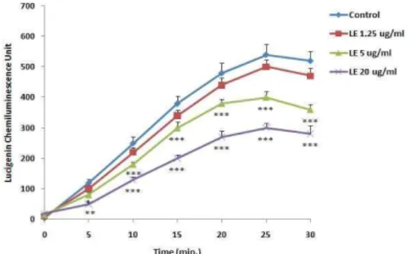
In vitro실험에서 LE 1.25, 5 및 20 μg/ml를 처리하였을 때, 5 μg/ml 및 10 μg/ml 농도에서 복강대식세포로부터 생성되는 lucigenin chemiluminescence양이 대조군에 비해 감소하였으며 (Fig. 1),In vivo실험에서도 LE를 투여하고 분리한 복강대식세포 에서 생성되는 lucigenin chemiluminescence양이 대조군에 비해 감소하였다(Fig. 2).
Fig. 1. Effects of LE on the level of lucigenin chemiluminescence in murine peritoneal macrophagesin vitro system.Peritoneal macrophages (2 x 10⁶cells/ml) obtained after 2 hr adherence period were cultured for 24 hrs.
The collected peritoneal macrophages were cultured in HBS buffer mixed with lucigenin and opsonized zymosan treated with several concentration of LE for 30 min. The data represents the mean ± SE of 3 experiments. *; Significantly different from control group (*; p<0.05, **; p<0.01,***; p<0.001).
4. 복강대식세포로 부터 nitric oxide의 생성에 미치는 효과 대조군의 대식세포에 LPS와 γ-IFN을 처리하지 않고 24 시 간 배양하였을 때 nitric oxide(NO) 생성양은 1.8 ± 0.2 μM 이었 으며, LPS와 γ-IFN을 처리하였을 때 대조군의 NO 생성양은 27.1
± 1.4 μM로 증가하였다. LE 1.25, 5 및 20 μg/ml를 처리하고 LPS와 γ-IFN을 처리하였을 때 각각 25.2 ± 1.3, 28.7 ± 1.5 및 37.5 ± 1.8 μM로 20 μg/ml 농도에서 LPS와 γ-IFN을 처리한 대
조군에 비해 증가하였다(Table 8). LE를 투여하고 분리한 대식세 포에서 NO 생성양은 2.5 ± 0.3 μM로 대조군과 별 차이가 없었으 나, LPS와 γ-IFN을 처리하였을 때 대조군의 NO 생성양은 27.9 ± 1.1 μM로 증가하였으며, LE를 투여하고 분리한 대식세포에서 NO 생성양은 35.5 ± 1.8 μM로 LPS와 γ-IFN를 처리한 대조군에 비해 증가하였다(Table 9).
Fig. 2. Effects of the administration of LE on the level of lucigenin chemiluminescence in murine peritoneal macrophages.LE (250 mg/kg) was administeredp.o. once a day for 7 days, and then 3% thioglycollate was injectedi.p. at the 4th day. Peritoneal macrophages (2 x 10⁶cells/ml) obtained after 2 hr adherence period were cultured for 24 hrs. The collected peritoneal macrophages were cultured in HBS buffer mixed with lucigenin and opsonized zymosan for 30 min. The data represents the mean ± SE of 5 mice. *; Significantly different from control group (**; p<0.01,***; p<0.001).
Table 8. Effects of LE on the production of nitric oxide from murine peritoneal macrophages in vitro system
Samples Dose(μg/ml) LPS and γ-IFN Nitric oxide production (μM)
Control - - 1.8 ± 0.2
- + 27.1 ± 1.4***
LE
1.25 + 25.2 ± 1.3
5 + 28.7 ± 1.5
20 + 37.5 ± 1.8##
The various concentration of LE were treated into peritoneal macrophages (2 x 10⁶ cells/ml) for 24 hrs. in the presence or in the absence of LPS and γ-IFN. The production of nitric oxide was determined with a Griess reagent. The data represents the mean ± SE of 3 experiments. *; Significantly different from control group (***; p<0.001).#; Significantly different from LPS and γ-IFN treated group (##; p<0.01).
Table. 9. Effects of the administration of LE on the production of nitric oxide from murine peritoneal macrophages
Samples Dose(mg/kg) LPS and γ-IFN Nitric oxide production (μM)
Control - - 2.1 ± 0.3
+ 27.9 ± 1.1***
LE 250 - 2.5 ± 0.3
+ 35.5 ± 1.8#
LE (250 mg/kg) was administered p.o. once a day for 7 days, and then 3%
thioglycollate was injectedi.p. at the 4th day. Peritoneal macrophages (2 x 10⁶cells/ml) obtained after 2 hr adherence period were cultured for 24 hrs. in the presence or in the absence of LPS and γ-IFN. The production of nitric oxide was determined with a Griess reagent. The data represents the mean ± SE of 5 mice.*; Significantly different from control group (***; p<0.001). #; Significantly different from LPS and γ-IFN treated control group (#; p<0.05).
고 찰
In vitro계에서 흉선세포에 LE를 처리하고 T lymphocytes mitogen인 concanavalin A (Con A)를 처리하지 않았을 때나 처 리하였을 때 모두 농도의존적으로 대조군에 비해 세포생존율이 증가하였다. 비장세포에 LE를 가하고 Con A와 B lymphocytes
mitogen인 lipopolysaccharide (LPS)를 처리하지 않았을 때나 처 리하였을 때 모두 농도의존적으로 대조군에 비해 세포생존율이 증가하였다. 또한, 복강대식세포에 LE를 처리하였을 때 고농도 에서 세포생존율이 증가하였다. 이는 LE가 in vitro계에서 면역세 포의 증식을 촉진하고 있음을 의미하는 것이다.
LE를 생쥐에 투여하고 분리한 흉선세포는 대조군에 비해 세 포생존율이 증가하였으나, Con A를 동시에 처리하였을 때는 Con A를 처리한 대조군에 비해 세포생존율이 감소하였으며, LE 를 생쥐에 투여하고 분리한 비장세포는 대조군에 비해 세포생존 율이 증가하였으나, Con A를 동시에 처리하였을 때는 Con A를 처리한 대조군에 비해 세포생존율이 감소하였고, LPS를 동시에 처리하였을 때는 LPS를 처리한 대조군에 비해 별 차이가 없었 다. 또한, LE를 생쥐에 투여하고 분리한 복강대식세포는 대조군 에 비해 세포생존율이 증가하였다. 이상의 실험결과는 LE가 in vitro및in vivo계에서 모두 흉선, 비장 및 복강대식세포의 생존 율을 증가시켜 면역능을 증가시킬 수 있음을 시사하는 것이다.
T lymphocyte는 분화과정을 거쳐 helper T lymphocyte 및 cytotoxic T lymphocyte로 분화되며, 분화된 helper T (Th) 세포 중 Th1 세포에서는 γ-IFN 및 IL-2가, Th2 세포에서는 IL-4, IL-6 및 IL-10 등의 cytokine이 분비되어, 다른 T, B 임파구 및 macrophage의 증식과 분화를 촉진하는 것으로 알려져 있다20). LE를 투여하고 분리한 혈청 중 γ-IFN양은 대조군에 비해 증가하 였으나, IL-2 및 IL-4양은 별 차이가 없었다. 이는 LE가 주로 Th1 세포를 활성화 할 수 있음을 의미하는 것이라 할 수 있다.
Lymphocytes에서 분비되는 cytokines들은 대식세포를 조절 하는 것으로 알려져 있기 때문에21), 복강대식세포에서 분비되는 phagocytic activity 및 nitric oxide양을 측정하였다. 외부로부터 이물질이 침입하게 되면 생체는 자기방어를 위해 대식세포가 활 성화되어 탐식능이 증가한다. LE를 in vitro계에서 처리하였을 때 농도의존적으로 lucigenin chemiluminescence양이 감소하였다.
대식세포는 particle을 탐식하는 동안 oxygen radical을 생성하는 데, 이때 생성된 oxygen radical과 lucigenin이 반응하여 lucigenin chemiluminescence를 발광하게 된다22). LE는 in vitro 계에서 처리하였을 때 lucigenin chemiluminescence양이 감소하 였으며, LE를 투여하고 복강대식세포를 분리하여 lucigenin chemiluminescence양을 측정하였을 때도 감소하였다. 이 결과는 LE가in vitro및in vivo계에서 모두 복강대식세포의 탐식능을 감 소시킴을 의미하는 것이다.
Nitric oxide (NO)는 활성화된 대식세포의 pseudopodia 형 성을 억제하여 탐식능을 감소시킨다고 알려져 있다23). 복강대식 세포에서 분비되는 NO양을 측정한 결과 NO양은 LPS와 γ-IFN 을 처리하였을 때 현저히 증가하였으며, LE를 in vitro계에서 처 리하였을 때 고농도에서 NO양이 증가하였으며, LE를 경구투여 하고 분리한 복강대식세포에서도 NO양이 대조군에 비해 증가하 였다. 이는 LE가 in vitro및 in vivo계에서 모두 NO 생성을 증가 시킴을 의미하는 것이다. LE가 NO 생성을 촉진하여 in vitro계 및 in vivo계 모두 탐식능을 감소시켰는지의 여부는 추후 연구되 어야 할 과제이다.
결 론
야관문 에탄올 추출물 (LE)은 생쥐의 흉선세포, 비장세포 및 복강대식세포의 세포생존율을 in vitro및 in vivo계에서 모두 증 가시켰으며, LE는 혈청 중 γ-IFN 양은 증가시켰으나, IL-2 및 IL-4양에는 별 영향을 주지 않았다. 또한, LE는 복강대식세포의 탐식능을 in vitro 및 in vivo계에서 모두 감소시켰으나, nitric oxide 생성은 증가시켰다. 이상의 실험결과 야관문은 면역세포의 증식을 촉진시켜 면역능을 조절할 수 있는 약재라 사료된다.
감사의 글
이 논문은 2011년도 우석대학교 교내학술연구비 및 교육과 학기술부로부터 지원받아 수행된 연구임(지역거점연구단육성사 업/헬스케어기술개발사업단).
참고문헌
1. Shin, M.K., Kim, C.M., An, D.G. and Lee, J.S. Dictionary of oriental medicine, Jungdam Press, Seoul, Korea, pp 2770-2773, 1997.
2. Atsushi, N. and Kazuko, H. C-Glycoylfalavones in Lespedeza cuneata, Chem. Pharm. Bull., 28: 964-965, 1980.
3. Kwon, D.J., Kim, J.K., Ham, Y.H. and Bae, Y.S., Flavone glycosides from the aterial parts of Lespedeza cuneata G.
Don, J. Kor. Soc. Appl. Biol. Chem., 50(4):344-347, 2007.
4. Shin, M., Munckazu, L., Emiko, I., Hiromasa, T. and Kengo, K. Studies on the constituents of the useful plants. VIII.
The constituents of Lespedeza cuneata G. Don, Yakugaku Zasshi, 98: 1542-1544, 1978.
5. Deng, F., Chang, J., Zhang, J.S., New flavonoids and other constituents from Lespedeza cuneata. J. Asian. Nat. Prod.
Res., 9(6-8):655-658, 2007.
6. 강소신의학원. 중약대사전, 상해, 상해인민출판사, pp 3006- 3006, 1977.
7. Lee, J.K., Kang, D.G. and Lee, H.S. Vascular relaxation induced by aqueous extract of Lespedeza cuneata via the NO-cGMP pathway. J. Nat. Med., May 24, in press. 2011.
8. Kim, S.M., Kang, K., Jho, E.H., Jung, Y.J., Nho, C.W., Um, B.H. and Pan, C.H. Hepatoprotective Effect of Flavonoid Glycosides from Lespedeza cuneata against Oxidative Stress Induced by tert-Butyl Hyperoxide. Phytother. Res., Jan. 12. in press, 2011.
9. Lee, H.J., Lim, G.N., Park, M.A. and Park, S.N., Antibacterial and antioxidative activity of Lespedeza cuneata G. Don extracts. Kor. J. Microbiol. Biotechnol., 39(1):63-69, 2011.
10. Chung, Y.H. and Lim, C.H. Bioactivity of an extract of
Lespedeza cuneata G. Don to rabbit corpus cavernosum smooth muscle tone. J. Agri. Sci., 32(1):63-70, 2005.
11. Abbas, A.K., Lichtman, A.H. and Pober, J.S. Cellular and Molecular Immunology. 2ed. Saunders, p 5, 1994.
12. Wysocki, L.J. and Sato, V.L. Planning for lymphocytes: A method for cell selection. Proc. Nat1. Acad. Sci. USA. 75:
2844, 1978.
13. Mizel, S.B., Openheim, J.J. and Rosensteich, D.L.
Characterization of lymphocyte-activating factor(LAF) produced by the macrophage cell line P388D1. J. Immunol.
120: 1497, 1979.
14. Mosmann, T. Rapid colorimetric assay for cellular growth and survival: application to proliferation and cytotoxic assays. J. Immunol. methods. 65: 55, 1983.
15. Kotnic, V. and Fleischmann, W.R.Jr. A simple and rapid method to determine hematopoietic growth factor activity.
J. Immunol. methods. 129: 23, 1990.
16. Eun, J.S., Lee, D.H., Jeon, Y.K., Kwon, Y.A. and Kwon, J.
Effect of Kamichungbieum on immune reaction. Kor. J.
Oriental Physiology & Pathology, 18(5):1391-1396, 2004.
17. Boudard, F., Vallot, N., Cabaner, C. and Bastide, M.
Chemiluminenscence and nitrite determinations by the
MALU macrophage cell line. J. Immunol. Methods, 174:
259, 1994.
18. Blair, A.L., Cree, I.A., Beck, J.S. and Hating, M.J.G.
Measurement of phagocyte chemiluminenscence in a microtiter plate format. J. Immunol. Methods, 112: 163, 1988.
19. Rockett, K.A., Awburn, M.M., Cowden, W.B. and Clark, I.A. Killing of Plasmodium faciparum in vitro by nitric oxide derivatives. Infec. Immunity, 59(9):3280, 1991.
20. Miceli, M.C. and Parnes, J.R. The role of CD4 and CD8 in T cell activation and differentiation. Advances in Immunology, 53: 59, 1993.
21. Charles, A. J., Paul, T., Mark, W. The immune system in health and disease. 4ed, Garland Pub., p 463, 2000.
22. Channon, J. Y., Leslie, C. C. and Johnston, Jr. R. B.
Zymosan-stimulated production of phosphatidic acid by macrophages: relationship to release of superoxide anion and inhibition by agents that increase intracellular cyclic AMP. J. Leucocyte Biol. 41: 450-455, 1987.
23. Jun, C.D., Park, S.K., Kim, J.M., Kim, J.D. and Kim, S.H.
Nitric oxide inhibits macrophage pseudopodia formation in the activated macrophages. Kor. J. Immunol. 18: 635-644, 1996.