∙ Received: June 29, 2011. Accepted: July 5, 2011.
∙ Corresponding author: Chae Ho Shin
Department of Nuclear Medicine, Inha University Hospital, 7-206 3rd ST, Jung-gu, Incheon, Korea
Tel: +82-32-890-3170, Fax: +82-32-890-3164 E-mail: dolpals@hanmail.net
Original Article 99m
Tc을 이용한 심근 관류 SPECT에서 Multiple Confocal SPECT System의 유용성인하대학교병원 핵의학과
신채호⋅표성재⋅김봉수⋅조용귀⋅조진우⋅김창호
Usability of Multiple Confocal SPECT SYSTEM in the Myocardial Perfusion SPECT Using
99mTc
Chae ho Shin, Sung Jai Pyo, Bong Su Kim, Yong Gyi Cho, Jin Woo Jo and Chang Ho Kim Department of Nuclear Medicine, Inha University Hospital, Incheon, Korea
Purpose: The recently adopted multiple confocal SPECT SYSTEM (hereinafter called IQ SPECTTM) has a high difference from the conventional myocardial perfusion SPECT in the collimator form, image capture method, and image reconstruction method. This study was conducted to compare this novice equipment with the conventional one to design a protocol meeting the IQ SPECT, and also determine the characteristics and usefulness of IQ SPECT. Materials and Methods: 1. For the objects of LEHR (Low energy high resolution) collimator and Multiple confocal collimator, 99mTc 37MBq was put in the acrylic dish then each sensitivity (cpm/µCi) was measured at the distance of 5 cm, 10 cm, 20 cm, 30 cm, and 40 cm respectively. 2. Based on the sensitivity measure results, IQ SPECT Protocol was designed according to the conventional general myocardial SPECT, then respectively 278 kBq/mL, 7.4 kBq/mL, and 48 kBq/mL of 99mTc were injected into the myocardial and soft tissues and liver site by using the anthropomorphic torso phantom then the myocardial perfusion SPECT was run. 3. For the comparison of FWHMs (Full Width at Half Maximum) resulted from the image reconstruction of LEHR collimator, the FWHMs (mm) were measured with only algorithms changed, in the case of the FBP (Filtered Back projection) method- a reconstruction method of conventional myocardial perfusion SPECT, and the 3D OSEM (Ordered subsets expectation maximization) method of IQ SPECT, by using 99mTc Line source. Results: 1. The values of IQ SPECT collimator sensitivity (cpm/µCi) were 302, 382, 655, 816, 1178, and those of LEHR collimator were measured as 204, 204, 202, 201, 198, both at the distance of 5 cm, 10 cm, 20 cm, 30 cm, and 40 cm respectively. It was found the difference of sensitivity increases up to 4 times at the distance of 30 cm in the cases of IQ SPECT and LEHR. 2. The myocardial perfusion SPECT Protocol was designed according to the geometric characteristics of IQ SPECT based on the sensitivity results, then the phantom test for the aforesaid protocol was conducted. As a result, it was found the examination time can be reduced 1/4 compared to the past. 3. In the comparison of FWHMs according to the reconstructed algorithm in the FBP method and 3D OSEM method followed after the SEPCT test using a LEHR collimator, the result was obtained that FWHM rose around twice in the 3D OSEM method. Conclusion : The IQ SPECT uses the Multiple confocal collimator for the myocardial perfusion SPECT to enhance the sensitivity and also reduces examination time and contributes to improvement of visual screen quality through the myocardial-specific geometric image capture method and image reconstruction method. Due to such benefits, it is expected patients will receive more comfortable and more accurate examinations and it is considered a further study is required using additional clinical materials. (Korean J Nucl Med Technol 2011;15(2):65-71)
Key Words : Myocardial perfusion SPECT, IQ SPECTTM, Multiple confocal collimator, LEHR collimator, FWHM
A B C
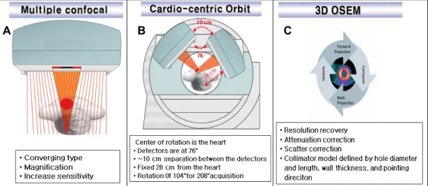
Fig. 1. The images that represents the characteristics of collimator (A), image acquigition method (B), and image reconstruction method (C) of IQ SPECT.
Fig. 2. Symbia T6 SPECT/CT (Siemens Medical Solution, USA)
서 론
근래의 심근관류 SPECT는 검사 장비와 소프트웨어의 발 달로 인해 진단의 정확도는 보다 향상 되었을 뿐만 아니라 검사 시간을 단축함으로써 환자의 편의성을 높이고 움직임 에 대한 Artifact를 줄이는 방향으로 발전하고 있다.1-2) 최근 본원에 도입된 multiple confocal SPECT system (이하 IQ SPECTTM)은 Symbia T6 SPECT/CT (Siemens Medical Solution, USA)의 심근관류 SPECT system으로 IQ SPECT 는 기존의 심근 관류 SPECT와 collimator 형태, 영상획득방 법 및 영상 재구성 방법에 큰 차이가 있다(Fig. 1). 기존의 SPECT가 90도의 회전형 구조라고 한다면, IQ SPECT에서 는 두 개의 검출기의 각도가 76도이고 서로 10 cm가량 떨어 져 있으면서 심장과 일정한 거리에서 영상을 얻는 것이 특징 이다. 또한 회전각도도 90도나 180도가 아니고 104도와 208 도로 심장의 구조 및 검출기 위치에 최적화 시킨 형태를 보 여주고 있다.1,3)
본 연구에서는 기존의 심근 관류 SPECT와 비교하여 IQ-SPECT에 맞는 protocol을 design 하고 IQ SPECT의 특 성과 유용성에 대하여 평가하고자 한다.
실험재료 및 방법
1. 실험 재료
장비는 Symbia T6 SPECT/CT를 사용하였으며(Fig. 2), collimator는 LEHR (Low energy high resolution) collimator 와 multiple confocal collimator를 사용하여 실험 하였다.
2. Collimator sensitivity 측정
기존의 심근관류 SPECT의 LEHR collimator와 IQ SPECT의 multiple confocal collimator의 감도 비교를 위하여 acrylic dish에 99mTc 37 MBq을 넣고 5 cm, 10 cm, 20 cm, 30
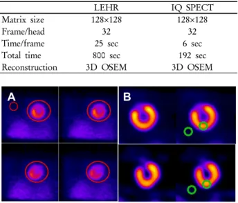
Table 1. Acquisition protocol of conventional LEHR myocardial perfusion SPECT and IQ SPECT
LEHR IQ SPECT
Matrix size 128×128 128×128
Frame/head 32 32
Time/frame 25 sec 6 sec
Total time 800 sec 192 sec
Reconstruction 3D OSEM 3D OSEM
A B
Fig. 5. A method to set ROIs of total counts measuring method (A) of projection image and reconstruction image (B) then assess (%) contrast.
Fig. 3. Shows how to measure sensitivity for measuring the sensitivity of collimator.
Fig. 4. Anthropomorphic torso phantom.
cm, 40 cm 거리에서 각각 sensitivity (cpm/μCi)를 측정 하였 다(Fig. 3).
3. Phantom 실험 1) SPECT 영상획득
Sensitivity 측정 결과를 바탕으로 기존의 일반적인 심근 관류 SPECT를 기준으로 IQ SPECT의 acquisition protocol 을 design 하였다. 그리고 Anthropomorphic torso phantom (Fig 4.)을 사용하여 심근과 연부조직, 간 영역에 99mTc을 각 각 278 kBq/mL, 7.4 kBq/mL, 48 kBq/mL 투여 후 기존의 LEHR collimator를 사용한 심근 관류 SPECT와 total acquisition time을 약 1/4로 단축한 IQ SPECT를 시행하여 비교하였다(Table 1).
2) SPECT 영상분석
기존의 LEHR 심근관류 SPECT와 IQ SPECT의 projection image에 대하여 획득된 total counts를 측정하고, reconstruction image에서 ROI (Region of Interest)를 설정하 여 (%) contrast를 측정하였다(Fig. 5).
통계분석은 Medcalc 프로그램 version 11.5 (Medcalc software, Belgium)를 이용하여 상관분석을 실시하였으며, p 값 유의성은 0.05 미만으로 설정하였다.
4. SPECT 공간 분해능 비교
LEHR collimator의 영상 재구성에 따른 FWHM (Full Width at Half Maximum) 비교를 위해 2개의 capillary tube 에 99mTc 18.5 MBq/mL를 투여한 line source를 100 mm 수평 간격으로 위치시켜 기존의 심근 관류 SPECT의 재구성법인 FBP (Filtered Back projection)과 IQ SPECT의 3D OSEM
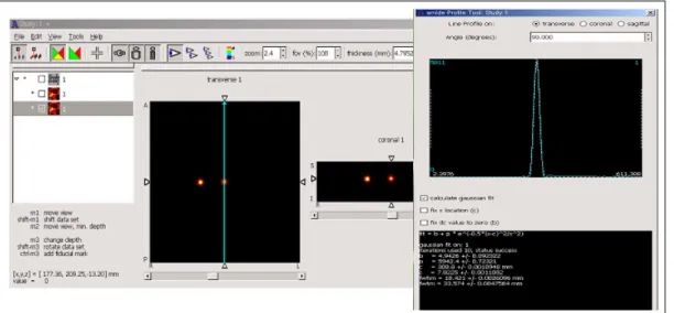
Fig. 6. Measures the FWHM of profile curve by using an Amide. exe program.
Table 2. A sensitivity comparison result of the LEHR collimator and Multiple confocal collimator.
5 cm 10 cm 20 cm 30 cm 40 cm
LEHR 204 204 202 201 198
Multiple confocal 302 382 655 816 1178
Ratio 1.48 1.87 3.24 4.06 5.95
Fig. 7. A sensitivity comparison graph of LEHR collimator and Multiple confocal collimator, which shows around 4 times of difference of sensitivity at the distance of 30 cm.
(Ordered subsets expectation maximization)법에 대하여 비교 하였다. 영상획득은 128x128 matrix size로 frame 당 20 sec, head 당 64 frame을 획득하였으며, 영상재구성은 FBP법은 butterworth filter를 사용하여 order 5, cut off 0.38로 설정하 였고, 3D OSEM법은 Gaussian filter(FWHM 8.4 mm)를 사 용하여, subset 1, iteration 30을 설정하였다. FWHM 측정은 amide. exe 프로그램 (Geeknet, Inc., USA)을 이용하여 profile curve의 FWHM을 측정하였다(Fig. 6).
결 과
1. Collimator sensitivity 측정 결과
기존의 LEHR collimator sensitivity (cpm/μCi)는 5 cm, 10 cm, 20 cm, 30 cm, 40 cm에서 각각 204, 204, 202, 201, 198 이었으며, IQ SPECT의 collimator는 302, 382, 655, 816, 1178 로 측정되었다. IQ SPECT는 환자 체표면에서 약 30 cm 거 리에서 회전하게 되므로 IQ SPECT의 multiple confocal collimator는 기존의 LEHR collimator와 비교하여 sensitivity 차이는 약 4배 정도 많아짐을 알 수 있다(Fig. 7, Table 2).
2. Phantom 실험 결과
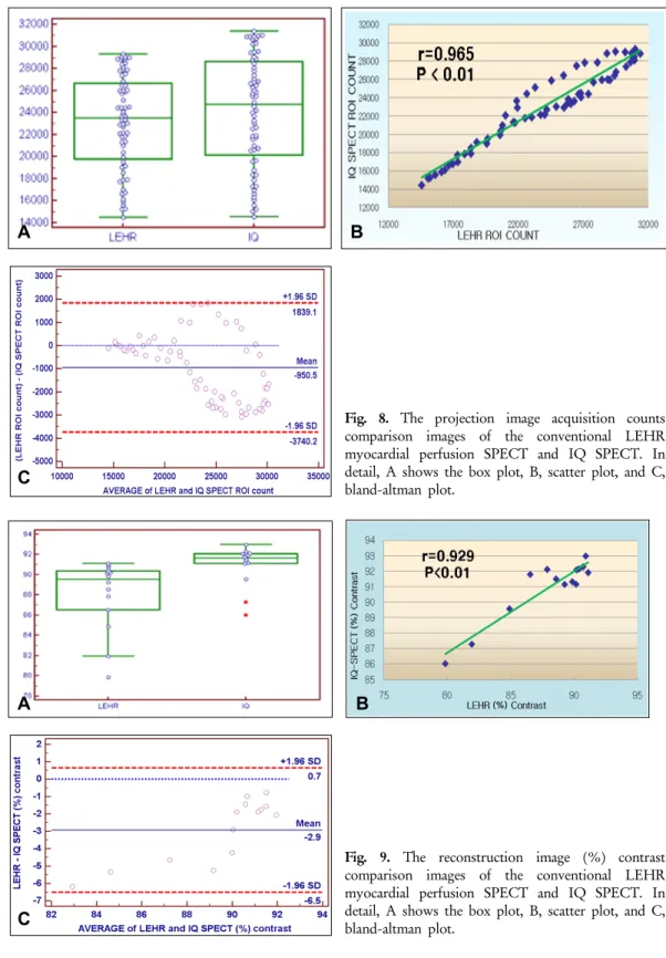
1) Sensitivity 측정 결과를 바탕으로 기존의 LEHR 심근관 류 SPECT와 비교하여 영상획득 시간을 약 1/4로 단축하여 IQ SPECT를 실시한 결과 projection image의 획득된 total counts는 LEHR에서 평균과 표준편차가 23118 ± 4325이고, IQ SPECT에서는 24069 ± 5054로 측정되었으며, 두 측정결 과의 상관계수 r은 0.965로 높은 상관관계를 보였으며, 상관 분석의 p-value는 0.01 미만으로 통계적으로 유의성이 있었 다(Fig. 8).
A B
C
Fig. 8. The projection image acquisition counts comparison images of the conventional LEHR myocardial perfusion SPECT and IQ SPECT. In detail, A shows the box plot, B, scatter plot, and C, bland-altman plot.
A B
C
Fig. 9. The reconstruction image (%) contrast comparison images of the conventional LEHR myocardial perfusion SPECT and IQ SPECT. In detail, A shows the box plot, B, scatter plot, and C, bland-altman plot.
2) Reconstruction image의 (%) contrast 측정 결과는 기존 의 LEHR 심근관류는 평균과 표준편차가 88.01 ± 3.52이고 IQ SPECT에서는 90.94 ± 1.99로 측정되었으며, 두 측정 결 과의 상관계수 r은 0.929로 높은 상관관계를 보였으며, 상관
분석의 p-value는 0.01 미만으로 통계적으로 유의성이 있었 다(Fig. 9).
Table 3. The FWHM (mm) results of the FBP method and 3D OSEM method, which were produced after the SPECT test.
Radial Tangential
FBP 17.48 18.42
3D-OSEM 8.57 9.12
Ratio 2.04 2.02
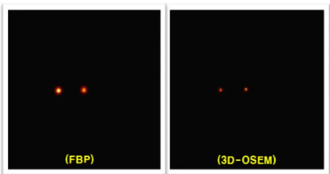
Fig. 10. As a result of visible comparison of reconstruction images of the conventional FBP method and 3D OSEM method of IQ SPECT, it can be checked the image is more clear in the 3D OSEM method.
결 론
IQ SPECT는 심근 관류 SPECT에서 Multiple confocal collimator의 사용으로 기존의 LEHR collimator를 사용한 심 근 관류 SPECT보다 약 4배 향상된 감도를 가진다. 그리고 심장에 특화된 기하학적인 영상 획득 방식과 영상 재구성 방 법을 통하여 검사 시간을 약 1/4로 단축할 수 있었으며, 약 2배의 공간 분해능 향상을 보임으로써 전체 검사 시간의 단 축과 영상의 화질 개선에 도움을 준다. 이로 인해 환자의 편 의성 향상과 진단의 정확도 개선을 기대 할 수 있을 것이다.
하지만 본 연구는 실험을 통한 연구 자료로써 추가적인 임상 자료의 지속적인 연구를 통한 임상 적용이 필요할 것으로 생 각된다.
요 약
근래의 심근 관류 SPECT를 위한 검사 장비는 진단의 정 확도가 높아졌을 뿐만 아니라 검사 시간을 단축함으로써 환 자의 편의성을 높이고 움직임에 대한 artifact를 줄이는 방향 으로 발전되고 있다. 본 연구에서는 기존의 심근 관류 SPECT와 비교하여 IQ SPECT에 맞는 protocol을 design 하 고 IQ SPECT의 특성과 유용성에 대하여 알아 보고자 하였 다. Simens사의 Symbia T6 SPECT/CT를 이용하여 LEHR collimator와 Multiple confocal collimator에 대하여 acrylic dish에 99mTc 37MBq을 넣고 5 cm, 10 cm, 20 cm, 30 cm, 40 cm 거리에서 각각 sensitivity (cpm/μCi)를 측정 하였다. 그리 고 Sensitivity 측정 결과를 바탕으로 기존의 일반적인 심근관 류 SPECT를 기준으로 IQ SPECT protocol을 design 후 Anthropomorphic torso phantom을 사용하여 심근 관류 SPECT를 시행하여 비교하고, LEHR collimator의 영상 재구 성에 따른 FWHM 비교를 위해 99mTc Line source 를 이용하 여 기존의 심근 관류 SPECT의 재구성법인 FBP법과 IQ SPECT의 3D OSEM법에 대하여 알고리즘만 변화시켜 FWHM을 측정 비교하였다. Collimator senstivity 측정 결과 IQ SPECT의 multiple confocal collimator의 sensitivity가
LEHR collimator와 비교하여 30 cm 거리에서 sensitivity가 약 4배 정도 더 많아짐을 알 수 있었다. Sensitivity 결과를 바 탕으로 IQ SPECT의 기하학적 특성에 맞게 심근 관류 SPECT protocol을 design 하여 phantom 실험을 시행한 결과 기존에 비해 검사시간을 1/4로 단축할 수 있었으며, LEHR collimator를 사용하여 SPECT 검사 후 FBP법과 3D OSEM 법의 재구성 알고리즘에 따른 FWHM 비교에서는 3D OSEM법에서 FWHM이 2배 정도 향상된 결과를 얻었다. 본 연구를 통해 IQ SPECT는 심근관류 SPECT에서 Multiple confocal collimator를 사용하여 감도를 향상시키고 심장에 특화된 기하학적인 영상 획득 방식과 영상 재구성 방법을 통 하여 검사 시간을 단축하고 영상의 화질 개선에 도움을 준다. 이로 인해 환자는 전보다 더욱 편안하고 정확한 검사를 수행 할 수 있을 것이며, 추가적으로 더 많은 임상 자료를 통한 연 구가 필요할 것이다.
REPERENCES
1. Byeong il Lee. State of the art of imaging Equipment and Tools for Nuclear Cardiology. Nucl Med Mol Imaging 2009;43:
165-173
2. Ernest V Garcia, Tracy L Raber, and Fabio P Esteves. Cardiac Dedicated Ultrafast SPECT Cameras: New Designs and Clinical Implications. J Nucl Med 2011;52:210-217
3. Slomka. Advances in technical aspects of myocardial perfusion SPECT imaging. J Necl Cardiology 2009; 16:255-76
4. H Everaert, C Vanhove, J J Hamill, P R Franken. Cardiofocal collimators for gated single-photon emission tomographic myocardial perfusion imaging. Euro J Necl Med 1998;25:3-7 5. Michael K,Kalpana M Kanal, Mark W Gebgard and Philip J
Rossman. Comparison of Four Motion Correction Techniques in SPECT Imaging of the Heart: A cardiac Phantom Study. J Necl Med 1998;39:2027-34
6. Mark W, Groch, Yoshio Takamiya, Paul J, Groch and William D Erwin. Quantitative Gated Myocardial SPECT:Effect of Collimation on Left-Ventricular Ejection Fraction. J Nucl Med Technol 2000;28:36-40
7. Lai K Leong, Randall L Kruger, and Michael K O'Connor. A Comparison of the Uniformity Requirements for SPECT Image Reconstruction Using FBP and OSEM Techniques. J Nucl Med Technol 2001;29:79-83