• 검색 결과가 없습니다.

Forced Eruption of Severe Angulated and Impacted Permanent Teeth after Marsupialization of Dentigerous Cyst: Case Report

N/A
N/A
Protected

Academic year: 2021

Share "Forced Eruption of Severe Angulated and Impacted Permanent Teeth after Marsupialization of Dentigerous Cyst: Case Report"

Copied!
6
0
0

로드 중.... (전체 텍스트 보기)

전체 글

(1)

Case Report

원고 접수일 2010년 10월 14일, 게재 확정일 2011년 1월 2일 책임저자 남정훈

(150-950) 서울시 영등포구 대림1동 948-1, 한림대학교 의과대학 강남성심병원 치과/구강외과학교실

Tel: 02-829-5233, Fax: 02-846-9789, E-mail: [email protected]

RECEIVED October 14, 2010, ACCEPTED January 2, 2011 Correspondence to Jeong-Hun Nam

Department of Oral and Maxillofacial Surgery, Kangnam Sacred Heart Hospital, College of Medicine, Hallym University

948-1, Daerim 1-dong, Yeongdeungpo-gu, Seoul 150-950, Korea Tel: 82-2-829-5233, Fax: 82-2-846-9789, E-mail: [email protected]

CC This is an open access article distributed under the terms of the Creative Commons Attribution Non-Commercial License (http://creativecommons.org/licenses/

by-nc/3.0) which permits unrestricted non-commercial use, distribution, and reproduction in any medium, provided the original work is properly cited.

함치성 낭종의 조대술 후 미맹출 변위 영구치의 교정적 정출: 증례보고

남정훈1,3ㆍ노경록1ㆍ유우근1ㆍ이병민1ㆍ전지현1ㆍ박수현1ㆍ안장훈1ㆍ김정희2

1한림대학교 의과대학 강남성심병원 구강악안면외과학교실, 2한림대학교 임상치의학대학원 치과병원,

3스타28치과그룹 일산센터 치과임플란트과

Abstract

Forced Eruption of Severe Angulated and Impacted Permanent Teeth after Marsupialization of Dentigerous Cyst: Case Report

Jeong-Hun Nam 1,3 , Kyung-Lok Noh 1 , Woo-Geun Yoo 1 , Byeong-Min Lee 1 , Ji-Hyeon Jeon 1 , Su-Hyun Park 1 , Jang-Hoon Ahn 1 , Jung-Hee Kim 2

1

Department of Oral and Maxillofacial Surgery, Kangnam Sacred Heart Hospital, College of Medicine,

2

Dental Hospital, Graduate School of Clinical Dentistry, Hallym University,

3

Department of Dental Implantology, Ilsan Center, STAR 28 Dental Group

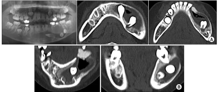
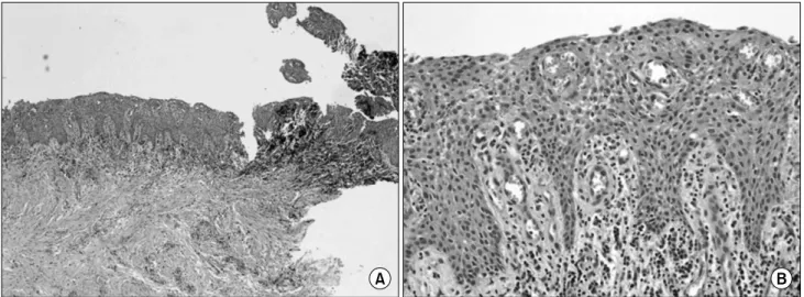
The goal of this treatment is the surgical-orthodontic eruption of impacted permanent teeth associated with a large dentigerous cyst in a preadolescent patient. Although enucleation of the entire cyst and the extraction of impacted teeth are common treatments, missing permanent teeth cause several problems in young patients. In this report, an 11-year-old female visited with the chief complaint of a large radiolucent lesion from the mandibular anterior area to the left mandibular posterior area. The permanent left canine and premolars were displaced toward the mandibular inferior border area. The extraction of infected deciduous teeth and marsupialization were performed. After 4 months, orthodontic buttons for forced eruption were applied to the impacted permanent teeth. The teeth emerged into the oral cavity 3 months after the orthodontic treatment.

Although the root form was abnormal, there were no other pathogenic signs. The alveolar bone had a normal trabecular pattern and the teeth appeared to be well maintained at postoperative 24 months.

Key words: Forced eruption, Marsupialization, Dentigerous cyst

(2)

Fig. 1. (A) Pre-operative panoramic view. Left mandibular canine, first premolar, second premolar were displaced toward mandibular

inferior border due to dentigerous cyst. Note the cystic lesion was extended right side cross the midline. (B) Pre-operative coronal and axial CT scan. These images were showing cystic lesion with inner wall thickening, bucco-lingual bone expansion, and displacement of permanent teeth and inferior alveolar nerve and vessel complex.

inclination and root formation[2-4].

 This report describes a case of preservation of permanent teeth displaced by dentigerous cyst which occurred from mandibular anterior area to left posterior area of an 11-year-old female with surgical-orthodontic treatment.

Case Report

 An 11-year-old female visited with chief complaint of swelling of left facial and lower vestibular area. The clinical examination showed a late stage of mixed dentition and malposition of permanent canine and premolars. There were no abnormal findings on general examination and

degree (Fig. 1A). The CT scan showed that permanent canine and first premolar displaced lingual, second pre- molar displaced distally and cortical bone was not des- tructed, but expanded bucco-lingually. The inferior alveo- lar nerve was involved in lower margin of cyst (Fig. 1B).

 The deciduous premolars were extracted and marsupiali- zation was done under local anesthesia, and oral mucosa on the extraction socket was communicated with the cyst.

An incisional biopsy was taken from the cyst wall for histo-

logical examination. A silastic drain was inserted into the

cystic cavity for depression of cavity during postoperative

two weeks, and an obturator which was made of sili-

cone-based impression materials (Putty

) was applied in

(3)

Fig. 2. This inflamed dentigerous cyst showed a thicker epithelial lining with hyperplastic rete ridges. Note a diffuse chronic inflammatory

infiltrate. (A) ×40 and (B) ×200 magnification.

Fig. 3. Panoramic view at 3 months after marsupialization. There

was a definite change on canine angulation. Spontaneous erup- tion was occurred.

Fig. 4. Four months after marsupia-

lization. Lingual arch and hooks were located just above impacted teeth. Orthodontic traction was ap- plied using elastic rubbers between arch and orthodontic button on im- pacted teeth.

Fig. 5. Three months after forced

eruption. The teeth emerged into the oral cavity. A residual radio- lucency was seen at mesial side of canine (arrow mark).

marsupialization opening site during postoperative three months. The histopathologic diagnosis was inflamed den- tigerous cyst (Fig. 2).

 There were several changes related with cyst cavity and impacted permanent teeth in 3 months after marsupializa- tion and size-reductive changing of obturator. The tooth axis angulations were decreased and spontaneous erup- tions were noted by shrinkage of the cyst and new bone formation (Fig. 3).

 Then, space maintainer with obturating flange was applied

(4)

Fig. 6. Eighteen months after forced

eruption. A small-sized residual ra- diolucency was remained at mesial side of canine.

Fig. 7. Two years after forced eruption. There was a mild crowding at left mandibular posterior area, but the permanent teeth related

dentigerous cyst erupted up to occlusal plane level and functioned as chewing.

(5)

Fig. 8. Two year after forced erup-

tion. The residual radiolucency be- came extinct, bone trabecular pat- terns showed normal form, and roots matured soundly (arrow mark).

radiolucency became extinct and the teeth erupted up to occlusal plane level at postoperative 2 years (Fig. 6).

During postoperative follow-up period, bony expansion with cystic lesion disappeared and normal bony structure and new bone formation were occurred at operation site and there was no recurrence at 24 months after surgery (Fig. 7 and 8).

Discussion

 It was a common treatment option that removing entire cyst and extracting involved teeth. However, it needed a considerable judgment at large cyst or in young patient.

In large cysts, serious bone loss would be induced by entire cyst enucleation. If the patient was young, this treatment was not best choice because the permanent teeth would be missed. Marsupialization, which was alternative treat- ment option in the above cases, decreased intracystic pres- sure and promoted shrinkage of the cyst as well as bone fill[5]. It was possible to preserve permanent teeth and important anatomic structures such as inferior alveolar nerve and adjacent paranasal sinuses by marsupialization.

It was suggested that marsupialization promoted the natural eruption of a cyst associated tooth and the clinical study revealed the probability of natural eruption was 72.4%[5].

 It was known that the average time to tooth eruption after marsupialization is about 100 days[6]. It had a ten- dency that over time, the natural eruption speed decreased after marsupialization and a period of 3 months after marsu- pialization was regarded as a critical time for deciding whether to extract or treat orthodontically[5-7]. In this case, for the first 3 months, the cystic cavity became shrinkage, showed changes of the angulation of impacted teeth, and new bone formation around cystic wall.

 The eruption of cyst associated teeth was significantly related with patient age, root maturity, teeth depth and

teeth angulation, but not in cyst size, available space[2].

The eruption potential was closely related to root for- mation[8]. Normally, a permanent tooth breaks through the alveolar bone and erupts when approximately two thirds of root formation has taken place, and then the tooth emerges into the oral cavity at approximately three fourths to full root formation with open apex[8]. Like the preced- ing, the cyst associated teeth which has incomplete root formation with an open apex has considerable potential to erupt than that of complete formed root. In this case, the root maturity of canine was half to three quarters and that of premolars was less than half. Thus, it was assumed that they had a potential to erupt spontaneously when the cyst size decreased after marsupialization.

 Orthodontic traction of the cyst associated impacted teeth have often been performed after marsupialization in pa- tients with a large cyst, matured roots, ectopic erupted teeth[2-4]. The tooth axis and depth can be changed by bone formation accompanied by pressure relief after mar- supialization[6]. It was reported that the averages of angula- tion were 21.8±20.4

o

in eruption group, 67.7±49.8

o

in non-eruption group[5]. In our case, the angulations of im- pacted teeth were about 90 degrees. So, orthodontic trac- tion was indicated 3 months after marsupialization.

 In young patients, conservative treatment of dentigerous

cyst was mainly performed with marsupialization and

post-operative spontaneous eruption of impacted perma-

nent teeth[9-11]. However, orthodontic treatment or trans-

plantation of impacted permanent teeth were performed

in cases of large dentigerous cyst or odontogenic kerato-

cyst[12-14]. In retrospective clinical study of more three

crown-sized dentigerous cysts during ten years, reduction

rate of cystic cavity in case of marsupialization (83.65%)

was more than that of odontogenic keratocyst (46.04%)

or radicular cyst (53.82%). Moreover, spontaneous erup-

(6)

1. Takagi S, Koyama S. Guided eruption of an impacted pre- molar associated with a dentigerous cyst in the maxillary si- nus of a 6-year-old child. J Oral Maxillofac Surg 1998;56:

237-9.

2. Neville BW, Damm DD, Allen CM, et al. Oral & maxillo- facial pathology. 2nd ed. Philadelphia: WB Saundersl; 2002.

p. 593.

3. Clauser C, Zuccati G, Barone R, Villano A. Simplified surgi- cal-orthodontic treatment of dentigerous cyst. J Clin Orthod 1994;28:103-6.

4. Sain D, Hollis WA, Togrye AR. Correction of superiorly dis- placed impacted canine due to a large dentigerous cyst. Am J Orthod Dentofacial Orthop 1992;102:270-6.

5. Hyomoto M, Kawakami M, Inoue M, Kirita T. Clinical con- ditions for eruption of maxillary canines and mandibular premolars associated with dentigerous cyst. Am J Orthod Dentofacial Orthop 2003;124:515-20.

6. Miyawaki S, Hyomoto M, Tsubouchi J, Kirita T, Sugimura

tigerous cyst by decompression: 5 cases report. J Korean Oral Maxillofac Surg 2009;35:271-5.

12. Kim WS, An KM, Sohn DS. Surgical and orthodontic treat- ment of impacted teeth associated with dentigerous cysts:

case report. J Korean Maxillofac Plast Reconstr Surg 2009;

31:173.

13. Gang TI, Park YJ, Nam JH, et al. Case report of treatment of multiple odontogenic keratocysts with basal cell nevus syndrome using preoperative marsupialization and ortho- dontic extrusion. J Korean Maxillofac Plast Reconstr Surg 2009;31:429-34.

14. Choi JW, Choi JM, Cha IH, et al. Eruption and auto- transplantation of a permanent teeth related to dentigerous cyst in mixed dentition. J Korean Oral Maxillofac Surg 2009;

35:462.

15. Kim YH, Lee EW. Comparison of clinico-histopathologic

findings before and after decompression of odontogenic cyst

in the jaw. J Korean Oral Maxillofac Surg 2005;31:150-60.

수치

Fig. 1. (A) Pre-operative panoramic view. Left mandibular canine, first premolar, second premolar were displaced toward mandibular inferior border due to dentigerous cyst
Fig. 2. This inflamed dentigerous cyst showed a thicker epithelial lining with hyperplastic rete ridges
Fig. 6. Eighteen months after forced eruption. A small-sized residual  ra-diolucency was remained at mesial side of canine.
Fig. 8. Two year after forced erup- erup-tion. The residual radiolucency  be-came extinct, bone trabecular  pat-terns showed normal form, and  roots matured soundly (arrow mark).

참조

관련 문서

Keywords: Loss of posterior teeth; Vertical dimension increase; Anterior tooth wear.. *Corresponding

Crown fractures are a common type of dental injuries and very frequently occurred on maxillary anterior teeth, area of requiring a rapid aesthetic recovery. Crown

Pontic area of temorary crown is adjusted to modified ridge lap type of anterior teeth and hygienic type of posterior teeth for oral hygiene control.... 최종 보철물은

Pontic area of temorary crown is adjusted to modified ridge lap type of anterior teeth and hygienic type of posterior teeth for oral hygiene control.... 최종

We report 3 uncommon cases of localized lipodystrophy on face which distinguished 1 year after cyst enucleation of mandibular posterior area.. Key words: Lipodystrophy,

Use of a Miniplate for Skeletal Anchorage in the Forced Eruption of a Severely Impacted Mandibular Second Molar: Case Report.. Jae-Sung Lim, Hyun-Joong

This case report describes a treatment for a 4-year-2-month-old girl whose primary and permanent dentition were affected by Dentinogenesis Imperfecta. Shell teeth were observed in