Tuberc Respir Dis 2013;74:169-176
CopyrightⒸ2013. The Korean Academy of Tuberculosis and Respiratory Diseases. All rights reserved.
Endobronchial Metastases from Extrathoracic Malignancies: Recent 10 Years' Experience in a Single University Hospital
Jung-Hyun Kim, M.D., Daniel Min, M.D., Sang-Hee Song, M.D., Ji-Hyun Lee, M.D., Hye-Cheol Jeong, M.D., Eun-Kyung Kim, M.D.
Department of Internal Medicine, Bundang CHA Medical Center, CHA University College of Medicine, Seongnam, Korea
Background: Although the lung is a common site of metastasis, endobronchial metastases (EBM) from extrathoracic malignancies are rare. Previous studies were retrospective reviews of the cases from each single institute, and the last one was performed between 1992 and 2002. We evaluated the characteristics of patients with EBM who had been diagnosed in recent 10 years in our hospital.
Methods: We retrospectively reviewed 1,275 patients who had undergone diagnostic bronchoscopic procedures between 2001 and 2011. An EBM was defined as bronchoscopically notable lesion, which was histopathologically identical to the primary tumor.
Results: A total of 18 cases of EBM were identified. The mean age was 53 years, and 12 cases of the 18 patients were female. The most common primary malignancies were colorectal cancer and breast cancer (4 cases each), followed by cervix cancer (3 cases) and renal cell carcinoma (2 cases). Cough was the most common symptom.
The most common radiologic finding was atelectasis, which was identified in 27.7% of the cases. The median interval from the diagnosis of primary malignancy to the diagnosis of EBM was 14 months (range, 0–112 months).
The median survival time from the diagnosis of EBM was 10 months (range, 1–39 months).
Conclusion: EBM from extrathoracic malignancies were rare. Colorectal cancer and breast cancer were common as primary malignancies. Fiberoptic bronchoscopy should be performed in all patients, who are suspected of having EBM. If atypical clinical and pathological features are present, appropriate diagnostic studies should be undertaken.
Key Words: Bronchi; Neoplasm Metastasis; Neoplasms
Address for correspondence: Eun-Kyung Kim, M.D.
Department of Internal Medicine, CHA Bundang Medical Center, CHA University College of Medicine, 59 Yatap-ro, Bundang-gu, Seongnam 463-712, Korea
Phone: 82-31-780-5205, Fax: 82-31-780-6143 E-mail: [email protected]
Received: Jan. 7, 2013 Revised: Feb. 22, 2013 Accepted: Mar. 18, 2013
CCIt is identical to the Creative Commons Attribution Non-Commercial License (http://creativecommons.org/licenses/by-nc/3.0/).
Introduction
Although the lung is a common site for metastases from various extrapulmonary malignancies, endotra- cheal or endobronchial metastases (EBM) are rare. The frequency of EBM varies according to the definition (range, 2–28%)
1,2. Rosenblatt et al.
1reported that the in- cidence of EBM was as high as 50% in their postmortem
examination of pulmonary metastatic disease. Braman
and Whitcomb
2reviewed the autopsies of patients who
had died with solid tumors and reported that the in-
cidence of EBM was as low as 2%. They counted only
grossly notable endobronchial lesions. Various tumors
have been associated with EBM. Treatment and man-
agement should be planned, according to the histology
of the primary tumor, anatomic location, evidence of
other metastasis, and the performance status of the
patient. We have identified 13 studies in the literature
that reviewed the prevalence and characteristics of
EBM
2-14. Because of low incidence of the EBM, as well
as the different characteristics of each institute and the
difference in time decades, the results vary from study
to study. Since the last study
14, which was performed
between 1992 and 2002, there was no further study
published in the literature. Therefore, we evaluated the
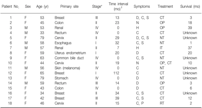
Table 1. Patients characteristics
Patient No. Sex Age (yr) Primary site Stage* Time interval
(mo)† Symptoms Treatment Survival (mo)
1 F 53 Breast III 13 D, C, S CT 3
2 F 45 Colon II 23 N OP 18
3 M 53 Renal IV 0 H OP 39
4 M 33 Rectum IV 0 C CT Unknown
5 F 79 Cervix II 29 D, C, S NT Unknown
6 M 58 Esophagus I 32 C, S NT 1
7 M 57 Renal II 7 H IT 37
8 F 59 Uterus endometrium I 20 D CT 20
9 F 63 Common bile duct IV 0 C, S NT Unknown
10 F 44 Cervix II 19 N OP, CT 10
11 M 56 Skin (melanoma) IV 0 C NT Unknown
12 F 65 Breast I 112 C CT Unknown
13 F 79 Stomach IV 0 D NT Unknown
14 M 59 Rectum III 14 D OP 3
15 F 43 Colon IV 0 D CT 6
16 F 34 Breast II 34 C, S CT Unknown
17 F 42 Breast III 39 C, S CT 12
18 F 46 Cervix II 15 C, P RT 2
*TNM stage at initial diagnosis of primary malignancy. †Time interval from initial diagnosis to endobronchial metastasis.
F: female; M: male; D: dyspnea; C: cough; S: sputum; N: no symptom; H: hemoptysis; P: pain; CT: chemotherapy; OP: operation;
NT: no treatment; IT: immunotherapy; RT: radiotherapy.
clinical, radiographic and bronchoscopic aspects of pa- tients with EBM, who had been diagnosed in recent 10 years in our hospital. We reviewed the literature and compared our cases with the previously reported series.
Materials and Methods
Between Jan 1, 2001 and Dec 31, 2011, we retro- spectively reviewed the patients who had undergone di- agnostic procedures using fiberoptic bronchoscopy at Bundang CHA Medical Center (Seongnam, Korea). In the study period, a total of 1,271 biopsy procedures were performed using fiberoptic bronchoscopy. The procedures were diagnostic in 641 cases. Among them, 438 cases of malignancies were identified. An EBM was defined as bronchoscopically notable lesion, which was histopathologically identical to the primary tumor.
Slides and reports of biopsy of the primary tumor and endobronchial biopsy material were compared to con- firm the diagnosis of EBM. We investigated the estro-
gen/progesterone receptor status if the primary malig- nancy was breast cancer, and the human papillomavirus (HPV)-DNA pattern in case of uterine cervix cancer. If the histological differentiation of the endobronchial tis- sue is still unclear, we compared the immunohisto- chemical staining, HPV-DNA pattern for cervix cancer, or estrogen/progesterone receptor status for breast cancer. Data regarding the patients' clinical character- istics, symptoms, radiographic and bronchoscopic find- ings were evaluated.
Results
A total of eighteen patients were identified as having
EBM from extrapulmonary malignancies. Among them,
twelve patients were women (66.7%). The range of age
was from 34 to 79 years with the median age of 53
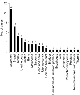
years. The primary malignancies were 4 breast cancers,
4 colorectal cancers, 3 uterine cervix cancers, 2 renal
cell carcinomas, 1 esophageal cancer, 1 cholangiocar-
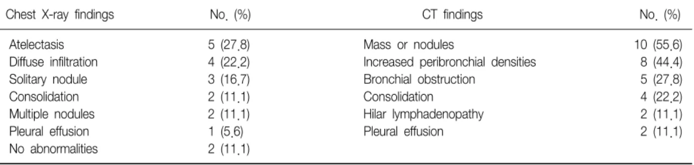
Table 2. Radiologic findings
Chest X-ray findings No. (%) CT findings No. (%)
Atelectasis 5 (27.8) Mass or nodules 10 (55.6)
Diffuse infiltration 4 (22.2) Increased peribronchial densities 8 (44.4)
Solitary nodule 3 (16.7) Bronchial obstruction 5 (27.8)
Consolidation 2 (11.1) Consolidation 4 (22.2)
Multiple nodules 2 (11.1) Hilar lymphadenopathy 2 (11.1)
Pleural effusion 1 (5.6) Pleural effusion 2 (11.1)
No abnormalities 2 (11.1)
CT: computed tomography.
Table 3. Location of lesions
Location No. of cases
Right bronchus Right main bronchus 0
Right upper lobar bronchus 1
Segmental bronchus 4
Right middle lobar bronchus 2
Segmental bronchus 1
Right lower lobar bronchus 2
Segmental bronchus 2
Right main and right lower lobe segmental 1
Total (%) 13 (72.2)
Left bronchus Left main bronchus 1
Left upper lobar bronchus 1
Left lower lobar bronchus 0
Segmental bronchus 3
Total (%) 5 (27.8)
cinoma, 1 melanoma, 1 uterine endometrial cancer, and 1 advanced gastric cancer. Table 1 shows clinical char- acteristics of the patients.
Cough was the most frequent symptom (10 patients, 55.6%), followed by dyspnea (6 patients, 33.3%), puru- lent sputum (6 patients, 33.3%), and hemoptysis (2 pa- tients, 11.1%).
The most frequent chest radiographic findings were obstructive atelectasis (5 cases) and diffuse infiltration (4 cases), followed by solitary nodule (3 cases), con- solidation (2 cases), and multiple nodules (2 cases). In two cases, chest radiograph showed no abnormalities at all. The computed tomography scan of the chest was performed on all of the patients. It revealed lung mass or nodules (10 cases), increased peribronchial densities (8 cases), bronchial obstruction (5 cases), and con-
solidations (4 cases) (Table 2).
On bronchoscopic examination, all of the cases showed single lesion, except one case. The lesions were located in the segmental bronchus (10 cases), lo- bar bronchus (6 cases), and main bronchus (1 case).
In one case, the lesions were found in multiple loca- tions (right main bronchus and right lower lobe basal segmental bronchus) (Table 3).
The median time from the diagnosis of the primary
malignancy to the diagnosis of EBM was 14.5 months
(range, 0–112 months). Six patients were found to have
EBM at the initial evaluation. The median time from the
diagnosis of EBM to death or following loss was 3
months (range, 1–39 months). When we excluded the
patients who were not followed after diagnosis, the me-
dian survival time was 10 months (range, 1–39 months)
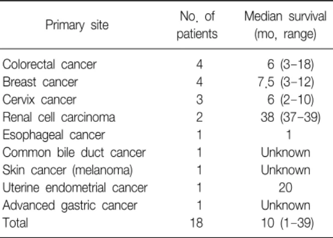
Table 4. Survival times according to primary sites
Primary site No. ofpatients
Median survival (mo, range)
Colorectal cancer 4 6 (3–18)
Breast cancer 4 7.5 (3–12)
Cervix cancer 3 6 (2–10)
Renal cell carcinoma 2 38 (37–39)
Esophageal cancer 1 1
Common bile duct cancer 1 Unknown
Skin cancer (melanoma) 1 Unknown
Uterine endometrial cancer 1 20
Advanced gastric cancer 1 Unknown
Total 18 10 (1–39)
Table 5. Summary of previous studies
Author Year Nation Study
period
No. of patients
Time interval (mo)
Most common primary sites (No.)
Median survival (mo, range) Braman and Whitcomb2 1975 US 1968–1971 5 36 (median) No dominant primary site Unavailable
Fitzgerald3 1977 US 1954–1972 17 58.5 Breast (6) 10 (2–58)
Sarcoma (3)
Baumgartner and Mark4 1980 US 1974–1979 8 60 (median) Breast (4) 13 (8–150)
Colorectal (2)
Shepherd5 1982 UK 1969–1979 25 36 (median) Colorectal (5) 11 (2–120)
Cervix (5) Breast (5)
Bourke et al.6 1989 UK Unavailable 10 48 (median) Breast (6) Unavailable
Kidney (2)
Lee et al.7 1992 Korea 1985–1992 17 40.6 (mean) Breast (4) 8.5
Cervix (3) Thyroid (2)
Heitmiller et al.8 1993 US 1971–1993 23 59.9 (mean) Breast (12) 12.5 (mean)
Kidney (4) Colorectal (3)
Salud et al.9 1996 Spain Mid 1980s 32 50.4 (median) Breast (20) 7
to 1996 Colorectal (3)
Melanoma (2)
Wang et al.10 1999 Taiwan Unavailable 40 Unavailable Head and neck (16) 12 (6–18) Katsimbri et al.11 2000 Greece 1990–2000 8 34 (9–106.5) Kidney (3) 9 (1.5–42)
Colorectal (2)
Kiryu et al.12 2001 Japan 1990–2001 16 65.3 (mean) Colorectal (6) 15.5 (mean)
Breast (3)
Kim et al.13 2002 Korea 1991–2001 27 43.8 (mean) Colorectal (7) 10 (3–49)
Uterus (4) Breast (3) Stomach (3)
Akoglu et al.14 2005 Turkey 1992–2002 15 32.8 (mean) Colorectal (3) 18 (4–84) Breast (3)
Kidney (2)
Present study 2012 Korea 2001–2011 18 14 (0–112) Colorectal (4) 10 (1–39)
Breast (4) Cervix (3)