The N-terminal Region of the Porcine Epidemic Diarrhea Virus Spike Protein is Important for the Receptor Binding
Lee, Dong-Kyu1, Se-Yeoun Cha2, and Changhee Lee1*
1Department of Microbiology, College of Natural Sciences, Kyungpook National University, Daegu 702-701, Korea
2Colledge of Veterinary Medicine, Chonbuk National University, Jeonju 561-756, Korea
Received: February 15, 2011 / Accepted: February 17, 2011
Porcine epidemic diarrhea virus (PEDV) infection causes acute enteritis with lethal watery diarrhea resulting in a high mortality rate in piglets. As with the other members of group 1 coronaviruses, PEDV also utilizes the host aminopeptidase N (APN) as the major cellular receptor for entry into target cells. The coronavirus spike (S) protein is known to interact with the cellular surface for viral attachment and the S1 domain of all charac- terized coronaviruses contains a receptor-binding domain (RBD) that mediates a specific high-affinity interac- tion with their respective cellular receptors. Although the RBDs of several coronaviruses have been mapped, the location of the PEDV RBD has to date not been defined. As a first step toward the identification of the region of the S protein of the PEDV that is critical for recognition with the cellular receptor, we generated a series of S1-truncated variants and examined their abilities to bind to the porcine APN (pAPN) receptor. Our data indicate that the N-terminus of the S1 domain is required for pAPN association. The results from the present study may assist in our understanding of the molecular interactions between the PEDV S protein and the pAPN receptor.
Key words: Porcine epidemic diarrhea virus (PEDV), spike protein, pAPN, receptor binding domain
Introduction
Coronaviruses are large enveloped, positive-sense, single- strand RNA viruses and possess a broad range of important viral pathogens that can mammalian and avian species [9].
They have a restricted tissue tropism which is determined by the interaction of the viral spike (S) glycoprotein with the receptor protein on the host cell surface [2, 5]. Corona- viruses employ a variety of cellular receptors as the binding partner of their S proteins, including carcinoembryonic antigen-related cell adhesion molecule 1 (CEACAM1), angiotensin-converting enzyme 2 (ACE2), and aminopeptidase N (APN). Remarkably, most members in group 1 corona- viruses, human coronavirus-229E (HCoV-229E), feline infectious peritonitis virus (FIPV), canine coronavirus (CCoV), transmissible gastroenteritis virus (TGEV), and
porcine epidemic diarrhea virus (PEDV), are known to commonly use the aminopeptidase N (APN) of their natural host species as a functional receptor for virus entry [3, 7, 14, 19, 20].
The S glycoprotein of coronaviruses can be functionally divided into two domains, S1 and S2; the former is responsible for binding to their host-specific receptor, while the latter appears to be involved in a direct fusion process between viral and cellular membranes [1, 5]. Therefore, the S1 domain of coronaviruses has been suggested to contain receptor-binding domains (RBDs) that could directly determine recognition of their respective receptors. Indeed, the RBDs of the S proteins of several coronaviruses have been mapped in their S1 subunit, even though the location of the RBD was found to differ depending upon the virus species [1, 6, 8, 16, 20].
PEDV, a causative agent of PED, is a devastating swine- specific enteric coronavirus causing lethal watery diarrhea and severe dehydration in piglets [18]. The S protein of PEDV is a type I membrane glycoprotein composed of
*Corresponding author
Tel: +82-53-950-7365, Fax: +82-53-955-5522 E-mail: [email protected]
1,383 amino acids (aa) and is predicted to contain a signal peptide (1-24 aa), a large extracellular region, a single transmembrane domain (1,334-1,356 aa), and a short cytoplasmic tail [12]. To date, the S1 domain of PEDV, that is critical for receptor binding, has not been characterized and the location of the PEDV RBD remains undefined.
Thus, in the present, we generated a series of S1-truncated variants and assayed their abilities to associate with the porcine APN (pAPN) receptor. Our results suggest that the N terminal region of the PEDV S1 domain is required for pAPN association.
Materials and Methods
Cells, antibodies, and plasmids
Human embryonic kidney (HEK) 293T cells were maintained in Dulbecco's modified Eagle medium (DMEM) with high glucose (Invitrogen, Carlsbad, CA, U.S.A.) containing 10% fetal bovine serum (FBS, Invitrogen) and antibiotic-antimycotic solution (100×; Invitrogen). The cells were maintained at 37oC with 5% CO2. A monoclonal antibody to six histidine residues and the goat anti-human IgG-horseradish peroxidase (HRP) were purchased from IG Therapy (Chuncheon, Korea) and Santa Cruz Biotechnology (Santa Cruz, CA, U.S.A.), respectively. Plasmids encoding the S1 fragment of severe acute respiratory syndrome coronavirus (SARS-CoV) or human ACE2 (hACE2), pCDM8 -S1-Ig and pCDM8-hACE2-Ig, were kindly provided by Hyeryun Choe [20] (Department of Pediatrics, Harvard Medical School, Boston, MA, U.S.A.).
Construction of expression plasmids
The deduced domain structure of the PEDV S protein was defined using a computer program from the web site of SOSUI (http://bp.nuap.nagoya-u.ac.jp/sosui/sosui_subunit.
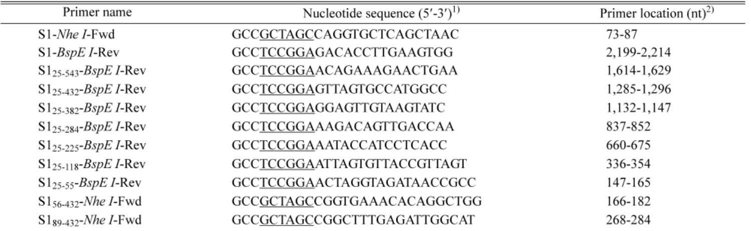
html). Plasmids encoding the PEDV S1 domain or its variants were generated by amplifying respective regions (Table 1) from a pGEM-T-S1 vector containing a cDNA fragment of the full-length S1 gene of a PEDV wild type strain KNU- 0801 [12] and ligating each amplicon into a modified expression vector pCDM8-Fc that encodes the CD5 signal sequence and the Fc domain of human IgG1 [4], thereby producing human Fc-tagged fusion proteins S1-Ig and its truncation variants. In addition, plasmids encoding pAPN or hACE2 for an interaction study were constructed by inserting cDNA fragment of the full-length pAPN or hACE2 gene from pGEM-pAPN [17] or pCDM8-hACE2-Ig into a pBudCE4.1 vector (Invitrogen) that contains six repetitive histidine codons to express His-tagged fusion proteins. All expression plasmids used in this study were verified by nucleotide sequencing.
Protein expression, immunoprecipitation (IP), and western blot
HEK-293T cells were plated in six-well culture plates at 24 h before transfection. The cells were then co-transfected with the expression plasmids, pCDM8-S1-Ig or its variants and pBud-pAPN-His using Lipopectamine 2000 (Invitrogen) according to the manufacturer's protocols. At 48 h post- transfection, cell lysates were prepared in lysis buffer as described before [13] and incubated with 50 µl protein A- Sepharose beads (GE Healthcare Bioscience) resuspended
Table 1. Primers used for construction of S1 variants.
Primer name Nucleotide sequence (5′-3′)1) Primer location (nt)2)
S1-Nhe I-Fwd GCCGCTAGCCAGGTGCTCAGCTAAC 73-87
S1-BspE I-Rev GCCTCCGGAGACACCTTGAAGTGG 2,199-2,214
S125-543-BspE I-Rev GCCTCCGGAACAGAAAGAACTGAA 1,614-1,629
S125-432-BspE I-Rev GCCTCCGGAGTTAGTGCCATGGCC 1,285-1,296
S125-382-BspE I-Rev GCCTCCGGAGGAGTTGTAAGTATC 1,132-1,147
S125-284-BspE I-Rev GCCTCCGGAAAGACAGTTGACCAA 837-852
S125-225-BspE I-Rev GCCTCCGGAAATACCATCCTCACC 660-675
S125-118-BspE I-Rev GCCTCCGGAATTAGTGTTACCGTTAGT 336-354
S125-55-BspE I-Rev GCCTCCGGAACTAGGTAGATAACCGCC 147-165
S156-432-Nhe I-Fwd GCCGCTAGCCGGTGAAACACAGGCTGG 166-182
S189-432-Nhe I-Fwd GCCGCTAGCCGGCTTTGAGATTGGCAT 268-284
1)Restriction enzyme sites in the primers are underlined.
2)Numbers correspond to positions within the spike gene of a PEDV KNU-0801 strain.
in radioimmunoprecipitation assay (RIPA) buffer at 4oC for 16 h as described previously [11]. The beads were then collected by centrifugation at 3,300× g (Eppendorf centrifuge 5451R) for 5 min, washed in RIPA buffer three times, and subjected to western blot analysis as previously described [13]. The electrotransferred membrane was blocked with 3% powdered skim milk (BD Biosciences, Bedford, MA) in TBS (10 mM Tris-HCl [pH 8.0], 150 mM NaCl) with 0.05% Tween-20 (TBST) for 2 h at 4oC. To detect the S1- Ig and its truncated proteins, the membrane was directly reacted with HRP-labeled goat anti-human IgG at a dilution of 1:5,000 for 2 h at 4oC. To detect the pAPN receptor protein, the blot was reacted at 4oC overnight with the primary anti-His tag antibody and then incubated with HRP-labeled goat anti-mouse IgG (Santa Cruz Biotechnology) at a dilution of 1:2,000 for 2 h at 4oC. Proteins were visualized by enhanced chemiluminescence (ECL) reagents (Amersham Biosciences, Piscataway, NJ, U.S.A.) according to the manufacturer's instructions.
Results and Discussion
To identify the region in the S1 domain of PEDV that is responsible for pAPN, we first constructed a series of expression plasmids encoding the full-length S1-Ig (S125-738) and seven C-terminally truncated S1 variants: S125-543-Ig, S125-432-Ig, S125-382-Ig, S125-284-Ig, S125-225-Ig, S125-118-Ig, and S125-55-Ig (Fig. 1). These constructs were independently transfected into HEK-293T cells and expression of each variant could be readily detected by western blot analysis using anti-human IgG secondary antibody that can directly recognize the protein fused to the Fc region of human IgG1
(data not shown). S1-Ig and truncation variants thereof were used to precipitate the pAPN protein. Plasmids encoding each S1 variant and pAPN were co-transfected into HEK- 293T cells, followed by an IP assay. To examine S1-pAPN associations, cell lysates were immunoprecipitated using protein A-Sepharose beads and subsequently, subjected to western blot using anti-human IgG secondary antibody and anti-His antibody to detect S1-Ig and pAPN proteins, respectively. It has been shown that the full-length SARS- CoV S1-Ig efficiently binds to hACE2 [15]. This specific interaction was confirmed in the present study (Fig. 2, lane 1), whereas it was undetectable between the S1 variant and the nucleocapsid (N) protein of the porcine arterivirus (Fig.
3A, lane 5). These observations verify our experimental conditions in IP and western blot assays. As shown in Fig.
2, S1 and all C-terminally truncated S1 variants were able to bind to pAPN, although their binding efficiencies were found to be dissimilar (lower panel). It appears that this result was likely due to different expression levels of each Fig. 1. Schematic diagram of the full-length S1 and seven C-
terminally and two N-terminally truncated S1-Ig variants con- structed for this study. The white boxes represent the S1 con- structs consisting of the S1 domain residues designated by S followed by amino acid positions and the gray boxes indicate a human IgG-Fc fragment.
Fig. 2. Detection of S1-pAPN interaction by IP and western blot. S1-Ig containing the PEDV S protein residues 25-738 fused to the Fc region of human IgG1, or truncation variants of S1-Ig containing the indicated S protein residues, and His-tagged pAPN were co-expressed in transfected 293T cells. For the IP assay, 200 µg of each protein lysate were incubated with protein A-Sepharose beads, followed by western blot detection of the precipitated the S1-Ig and pAPN association in reducing conditions. The blot was then reacted with anti-human IgG antibody and anti-His antibody to detect S1-Ig (upper panel) and pAPN (lower panel), respectively.
The corresponding S1 variants were identified by black triangles.
S1 variant upon co-transfection.
In addition, two N-terminally truncated S1 variants, S156-432 -Ig, S189-432-Ig, were generated and used for IP-western blot analysis. In contrast to the S156-432 of PEDV, which efficiently bound to the pAPN receptor (Fig. 3A, lane 3), the S1 variant containing S1 domain residues 89-432 did not associate with pAPN (Fig. 3A, lane 4). To rule out the possibility that the absence of receptor binding for the S189-432 variant might have resulted in a low expression level of pAPN upon co-transfection, total cell lysates were directly subjected to western blot without an IP process. Fig. 3B (middle panel) shows that both the S1 variants and pAPN were expressed efficiently in co-transfected cells, indicating that the lack of the association between the S189-432 and pAPN was not due to the pAPN expression level. Altogether, our results suggest that the most N-terminal region of S1 spanning amino acid residues 25-88 is required for the receptor binding, whereas the C-terminal region of S1 domain is dispensable for recognizing the pAPN receptor.
The present study described here localizes the RBD of the PEDV S protein. A panel of the S1 truncation variants fused to the Fc region of human IgG1 was assessed for its
ability to bind to pAPN in the transfected cells. The N- terminal region of the S1 domain was found to be involved in receptor binding and the smallest fragment that retained pAPN association was composed of the S1 domain residues 25-88. In contrast, all known RBDs of group 1 coronaviruses that use APN as the cellular receptor are located towards the C-terminal region of the S1 domain [1, 6, 10]. Therefore, our results were somewhat unexpected. However, this inconsistency might be explained by different evolutionary and serological relationships among coronaviruses. In a previous study, the largest number of amino acid differences was accumulated in the N-terminal region of the S1 domain of Korean filed PEDV isolates when compared with a cell culture-adapted CV777 strain and other vaccine strains [12]. Although the N-terminal region of the S1 domain is relatively well conserved among filed isolates, this con- servative trait appears to be altered to allow the virus to be propagated in monkey-derived Vero cells. Whether or not this N-terminal region of the S1 domain represents a receptor binding role during PEDV entry remains to be investigated. In conclusion, the data from the present study may help our understanding of the molecular interactions Fig. 3. Localization of the receptor-binding region of PEDV S1. A. S1-Ig truncation variants containing the indicated S protein residues and His-tagged pAPN were co-expressed in transfected 293T cells and cell lysates were incubated with protein A-Sepharose beads, fol- lowed by western blot in reducing conditions. The blot was then reacted with anti-human IgG antibody, anti-His antibody, and anti-PRRSV N antibody to detect S1-Ig (upper panel), pAPN (middle panel), and N (lower panel), respectively. B. Total cell lysates from co-transfected cells were directly subjected to western blot analysis to determine the expression level of S1-Ig (upper panel), pAPN (middle panel), and N (lower panel). The corresponding S1 variants were identified by black triangles.
between the PEDV S protein and the pAPN receptor and provide insight into the development of a subunit vaccine for prevention of PED.
Acknowledgments
We gratefully thank Hyeryun Choe from Harvard Medical School for providing reagents. This research was supported by Basic Science Research Program through the National Research Foundation of Korea (NRF) funded by the Ministry of Education, Science and Technology (2009-0070683).
REFERENCES
1. Bonavia, A., B. D. Zelus, D. E. Wentworth, P. J. Talbot, and K. V. Holmes. 2002. Identification of a receptor-binding domain of the spike glycoprotein of human coronavirus HCoV-229E. J. Virol. 77: 530-538.
2. Bosch, B. J., R. Van Der Zee, C. A. De Haan, and P. J. Rot- tier. 2003. The coronavirus spike protein is a class I virus fusion protein: structural and functional characterization of the fusion core complex. J. Virol. 77: 8801-8811.
3. Delmas, B., J. Gelfi, R. L'Haridon, L. K. Vogel, H. Sjöström, O. Norén, and H. Laude. 1992. Aminopeptidase N is a major receptor for the entero-pathogenic coronavirus TGEV. Nature 357: 417-420.
4. Farzan M., T. Mirzabekov, P. Kolchinsky, R. Wyatt, M.
Cayabyab, N. P. Gerard, C. Gerard, J. Sodroski, and H.
Choe. 1999. Tyrosine sulfation of the amino terminus of CCR5 facilitates HIV-1 entry. Cell 96: 667-676.
5. Gallagher, T. M. and M. J. Buchmeier. 2001. Coronavirus spike proteins in viral entry and pathogenesis. Virology 279:
371-374.
6. Godet, M., J. Grosclaude, B. Delmas, and H. Laude. 1994.
Major receptor-binding and neutralization determinants are located within the same domain of the transmissible gastro- enteritis virus (coronavirus) spike protein. J. Virol. 68: 8008- 8016.
7. Kolb, A. F., A. Hegyi, J. Maile, A. Heister, M. Hagemann, and S. G. Siddell. 1998. Molecular analysis of the coronavi- rus-receptor function of aminopeptidase N. Adv. Exp. Med.
Biol. 440: 61-67.
8. Kubo, H., Y. K. Yamada, and F. Taguchi. 1994. Localization of neutralizing epitopes and the receptor-binding site within the amino-terminal 330 amino acids of the murine coronavi- rus spike protein. J. Virol. 68: 403-410.
9. Lai, C. C., M. J. Jou, S. Y. Huang, S. W. Li, L. Wan, F. J.
Tsai, and C. W. Lin. 2007. Proteomic analysis of up-regu- lated proteins in human promonocyte cells expressing severe
acute respiratory syndrome coronavirus 3C-like protease.
Proteomics 7: 1446-1460
10. Laude, H., M. Godet, S. Bernard, J. Gelfi, M. Duarte, and B.
Delmas. 1995. Functional domains in the spike protein of transmissible gastroenteritis virus. Adv. Exp. Med. Biol. 380:
299-304.
11. Lee, C., J. G. Calvert, S. K. Welch, and D. Yoo. 2005. A DNA-launched reverse genetics system for porcine reproduc- tive and respiratory syndrome virus reveals that homodimer- ization of the nucleocapsid protein is essential for virus infectivity. Virology 331: 47-62.
12. Lee, D. K., C. K. Park, S. H. Kim, and C. Lee. 2010. Hetero- geneity in spike protein genes of porcine epidemic diarrhea viruses isolated in Korea. Virus Res. 149: 175-182.
13. Lee, Y. J., C. K. Park, E. Nam, S. H. Kim, O. S. Lee, D. S.
Lee, and C. Lee. 2010. Generation of a porcine alveolar mac- rophage cell line for the growth of porcine reproductive and respiratory syndrome virus. J. Virol. Methods 163: 410-415.
14. Li, B. X., J. W. Ge, and Y. J. Li. 2007. Porcine aminopepti- dase N is a functional receptor for the PEDV coronavirus.
Virology 365: 166-172.
15. Li, W., M. J. Moore, N. Vasilieva, J. Sui, S. K. Wong, M. A.
Berne, M. Somasundaran, J. L. Sullivan, K. Luzuriaga, T. C.
Greenough, H. Choe, and M. Farzan. 2003. Angiotensin- converting enzyme 2 is a functional receptor for the SARS coronavirus. Nature 426: 450-454.
16. Lin, H. X., Y. Feng, G. Wong, L. Wang, B. Li, X. Zhao, Y.
Li, F. Smaill, and C. Zhang. 2007. Identification of residues in the receptor-binding domain (RBD) of the spike protein of human coronavirus NL63 that are critical for the RBD-ACE2 receptor interaction. J. Gen. Virol. 89: 1015-1024.
17. Nam, E. and C. Lee. 2010. Contribution of the porcine ami- nopeptidase N (CD13) receptor density to porcine epidemic diarrhea virus infection. Vet. Microbiol. 144: 41-50.
18. Pensaert, M. B. and S. G. Yeo. 2006. Porcine epidemic diar- rhea, pp. 367-372. In B. E. Straw, J. J. Zimmerman, S.
D’Allaire, and D. J. Taylor (ed.), Diseases of Swine, 9th ed.
Wiley-Blackwell, Ames.
19. Tresnan D. B., R. Levis, and K. V. Holmes. 1996. Feline aminopeptidase N serves as a receptor for feline, canine, porcine, and human coronaviruses in serogroup I. J. Virol.
70: 669-674.
20. Wong S. K., W. Li, M. J. Moore, H. Choe, and M. Farzan.
2004. A 193-amino acid fragment of the SARS coronavirus S protein efficiently binds angiotensin-converting enzyme 2.
J. Biol. Chem. 279: 3197-3201.
21. Yeager, C. L., R. A. Ashmun, R. K. Williams, C. B. Cardelli- chio, L. H. Shapiro, A. T. Look, and K. V. Holmes. 1992.
Human aminopeptidase N is a receptor for human coronavi- rus 229E. Nature 357: 420-422.
국문초록
PED 바이러스 Spike 단백질의 세포 수용체 결합 부위 확인 이동규1·차세연2·이창희1*
1경북대학교 자연과학대학 생명과학부 미생물학과
2전북대학교 수의과대학
돼지유행성설사 바이러스(porcine epidemic diarrhea virus: PEDV)는 자돈에게 감염 시 수양성설사를 동반한 급성 장염을 유발하며 매우 높은 폐사율을 보이는 그룹 1 코로나바이러스이다. PEDV는 다른 그룹 1 코로나바이러스와 마찬가지로 숙주 세포에 감염 시 aminopeptidase N (APN)을 세포 수용체로 이용한다고 알려져 있다. 코로나바이러 스의 spike(S) 단백질은 숙주세포의 표면에 부착과 관련하여 감염 개시에 있어 중요한 역할을 하는 것으로 알려져 있 으며 특히 S 단백질의 S1 도메인은 세포 수용체에 특이적인 결합을 매개하는 수용체 결합 도메인(receptor binding domain: RBD)을 포함하고 있는 것으로 알려져 있다. 이미 많은 코로나바이러스의 RBD의 위치가 확인되어져 있지 만 PEDV의 RBD에 대해서는 아직까지 알려진 바가 없다. 본 연구에서는 돼지 APN 수용체와 결합을 매개하는 PEDV의 RBD를 규명하기 위해 S1 도메인을 주형으로 하는 일련의 재조합 truncated variant들을 제작하였고 각각의 truncated들이 실제로 pAPN과의 결합을 이루는지에 대하여 실험을 통해 확인하였다. 그 결과 S1 도메인의 N 말단 부분이 pAPN과의 결합에서 중요한 부위임을 확인할 수 있었다. 본 연구에서 도출된 결과는 향후 PEDV의 S 단백 질과 pAPN간의 분자적 상호작용을 이해하는 데에 도움을 줄 것으로 판단된다.