pISSN 1738-3544 eISSN 2288-1662
Carotid Artery Disease in Duplex Sonography: 3 Cases
Minho Han 1,2 , Junghye Choi 1 , Gang Sik Seo 1 , Hyo Suk Nam 2,3
1
Department of Neurology, Yonsei University Severance Hospital, Seoul, Korea
2
Department of Science for Aging, Yonsei University Graduate School, Seoul, Korea
3
Department of Neurology, Yonsei University College of Medicine, Seoul, Korea
이중초음파에서 관찰된 목동맥 질환: 3례
한민호 1,2 , 최정혜 1 , 서강식 1 , 남효석 2,3
1
연세의료원 세브란스병원 신경과,
2연세대학교 일반대학원 노화과학협동과정,
3연세대학교 의과대학 신경과학교실
Duplex sonography has been used to non-invasively diagnose carotid artery disease and to follow up patients with this disease. In carotid artery disease, carotid mobile thrombus and dissection are rarely detected by duplex sonography in patients with cerebrovascular events. In particular, carotid dissection occurs when a small tear forms in the tunica intima and makes 2 lumens in the carotid artery, including true and false lumens. This study reports 3 rare cases of carotid mobile thrombus, spontaneous carotid dissection, and carotid dissection in Takayasu arteritis.
Key words: Carotid dissection, Duplex sonography, Mobile thrombus, Takayasu arteritis
Corresponding author: Minho Han Department of Neurology, Yonsei University Severance Hospital, 50-1 Yonsei-ro, Seodaemun-gu, Seoul 03722, Korea Tel: 82-2-2228-5283
Fax: 82-2-393-0705 E-mail: umsthol18@gmail.com
ORCID: https://orcid.org/0000-0003-2951-0963
This is an Open Access article distributed under the terms of the Creative Commons Attribution Non-Commercial License (http://creativecommons.org/licenses/by-nc/4.0) which permits unrestricted non-commercial use, distribution, and reproduction in any medium, provided the original work is properly cited.
Copyright © 2019 The Korean Society for Clinical Laboratory Science. All rights reserved.
Received: October 31, 2018 Revised: January 2, 2019 Accepted: January 3, 2019
서 론
목동맥이중초음파(carotid duplex sonography)는 검사비 용이 비교적 저렴하고, 비침습적으로 목동맥 구조와 혈류역학 적 정보를 동시에 파악할 수 있는 유용한 검사방법이다. 검사의 주된 목적은 죽상동맥경화증(atherosclerosis)이 자주 발생하 는 목동맥갈림(carotid bifurcation)과 속목동맥(internal carotid artery, ICA)에서 혈전 및 협착여부를 파악하는 것이다 [1]. 혈관 내 죽상판(atheromatous plaque)에 혈전이 연결되어 심장박동에 따라 움직이는 목동맥 유동성 혈전(carotid mobile thrombus) [2]과 혈관 내막의 파열로 인해 B-mode 영상에서 두 개의 속공간을 관찰할 수 있는 목동맥박리(carotid dissection) 는 불특정 시점에 갑자기 혈전이 떨어져 나가 원위부에 위치한 대뇌혈관을 막거나 박리가 된 내막이 혈류의 주행경로를 차단 하여 급성 허혈성 뇌졸중을 일으킬 수 있기 때문에 즉각적인 치
료가 필요한 드문 증례에 포함된다[3]. 목동맥 유동성 혈전과 목 동맥박리는 각각 전체 허혈성 뇌졸중 환자의 약 2∼3%의 원인 으로 알려져 있다[3,4]. 본 증례에서 저자들은 이중초음파검사 에서 드물게 관찰되는 목동맥 질환 3례를 경험하였고, 이를 문 헌 고찰과 함께 보고하고자 한다.
증 례
1. 목동맥 유동성 혈전
심장동맥질환 및 고혈압과 당뇨 과거력이 있는 77세 남성이 지하철역에서 어지러움과 함께 걷는 도중 왼쪽으로 재차 쓰러 져 지나가는 행인이 신고하여 본원 응급진료센터로 내원하였 다. 입원 후 자기공명영상(magnetic resonance imaging, MRI)과 자기공명혈관조영술(magnetic resonance angio- graphy, MRA)에서 우측 중간대뇌동맥(middle cerebral
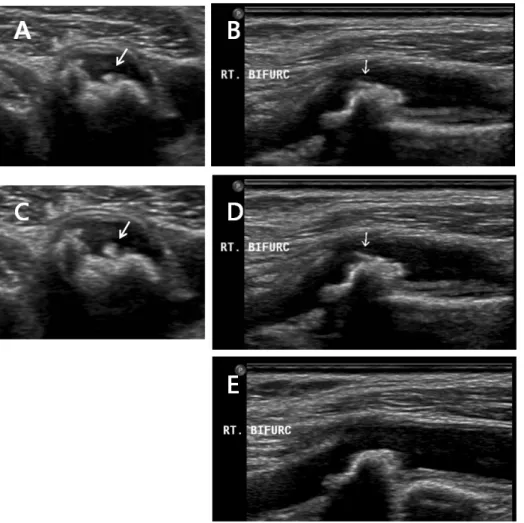
Figure 1. Case 1 (carotid mobile thrombus). (A) B-mode image showed a mobile thrombus during systole in transverse view of right carotid bulb. (B) B-mode image showed a mobile thrombus during systole in longitudinal view of right carotid bulb. (C) B-mode image showed a mobile thrombus during diastole in transverse view of right carotid bulb. (D) B-mode image showed a mobile thrombus during diastole in longitudinal view of right carotid bulb. (E) After 4 months, the mobile thrombus disappeared in longitudinal view of right carotid bulb.
artery, MCA) 영역에 작은 크기의 뇌경색이 일부 관찰되었고, 양쪽 원위부 속목동맥(internal carotid artery, ICA) 에는 심한 협착이 있어 스텐트를 이용한 혈전제거술(stent-retriever thrombectomy)을 시행하였다. 이후 악화가 되거나 재발되지 않아 기존 항혈소판 처방을 유지하고 퇴원시킬 예정이었으나, 이중초음파(IU22, Philips, Washington, USA)에서 심한 협착 없이, 양쪽 온목동맥(common carotid artery, CCA)과 ICA에 서 죽상동맥경화증이 발견되었고, 특이소견으로 우측 ICA와 바 깥목동맥(external carotid artery, ECA) 분기점에서 3.72 mm 크기의 죽상판의 일부가 움직이는 유동성 혈전이 관찰되어, 헤 파린 치료를 시행 후 퇴원하였다. 경두개도플러초음파검사 (transcranial Doppler, TCD; TC8080, Viasys healthcare, Pennsylvania, USA)에서 좌측 MCA가 168 cm/s, 우측 원위부 ICA가 99 cm/s로 높은 평균혈류속도를 보였다. 4개월 후 추적 검사에서 우측 목동맥 분기점에서 발견된 유동성 혈전은 사라 졌으며, 추가적으로 발생한 신경학적 기능손상 및 증상은 관찰 되지 않았다(Figure 1).
2. 자발성 목동맥박리
81세 여성이 가족들과 식사 및 대화를 하던 중 갑자기 의식을 잃고 양팔을 양쪽으로 펼치며 8초간 강직을 보인 후 곧 다시 회 복되어 의식을 되찾는 증상이 하루에 두 차례 관찰되어 응급진 료센터로 내원하였다. MRI와 MRA에서 뇌실 주위 백색 물질 (periventricular white matter)에 급성 뇌경색(acute brain infarction)이 관찰되었고, 우측 CCA와 ICA에서 목동맥박리가 의심되었다. 경두개도플러초음파검사(transcranial Doppler, TCD)에서 모든 혈류는 정방향(anterograde flow)이였으며, 정상적인 혈류속도를 보였다. 이중초음파에서 목동맥박리의 특징적인 소견인 두 개의 속공간이 우측 CCA 전반에 걸쳐 관찰 되었다. 두 개의 속공간 중 하나는 참속공간(true lumen)으로 기존 혈관의 내강(lumen)을 나타내는 것이고, 다른 속공간은 혈관내벽이 찢어진 후 분리된 혈관내막 밑으로 혈류가 새어 들 어가 또 하나의 혈관처럼 보이는 거짓속공간(false lumen)이었 다(Figure 2).
Figure 2. Case 2 (spontaneous carotid dissection). (A) Power Doppler image showed two lumens in transverse view of right common carotid artery (CCA). (B) Power Doppler image showed two lumens in longitudinal view of right CCA. (C) B-mode image showed two lumens in longitudinal view of right CCA.
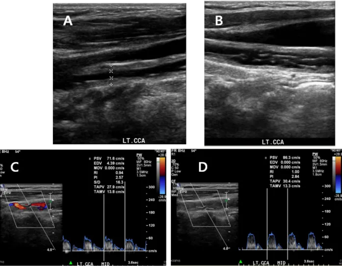
Figure 3. Case 3 (carotid dissection in a patient with Takayasu arteritis). (A) B-mode image showed “Macaroni sign” in longitudinal view
of left distal common carotid artery (CCA). (B) B-mode image showed dissection in longitudinal view of left middle CCA. (C) Doppler
image showed blood flow waveform with a relatively decent pulsatility in a true lumen of left CCA. (D) Doppler image showed blood
flow waveform with a relatively weak pulsatility in a false lumen of left CCA.
3. 타카야수동맥염 환자에서 발생한 목동맥박리
대동맥판역류, 고혈압 및 타카야수동맥염(Takayasu arteritis) 을 진단받고, 대동맥판막대치술(Aortic valve replacement)과 목동맥우회술(carotid artery bypass surgery)이 예정된 52세 여자가 수술 전 신경학적 평가를 위해 협진을 통해 신경과에 내 원하였다. 혈액검사에서 C-반응단백(C-reactive protein)은 1.58 mg/L로 증가되었고, 프로트롬빈시간(prothrombin time), 국제표준화비율(international normalized ratio), 활성 화부분트롬보플라시틴시간(activated partial thromboplastin time), 피브리노겐(fibrinogen), 섬유소분해산물(fibrin de- gradation products), D-이합체(D-dimer) 모두 정상이었다.
이중초음파에서 좌측 목동맥에 심한 협착이 있었으며, 양측 원 위부 CCA의 내막-중막복합체두께(intima-media complex thickness, IMT)가 상당히 두꺼워져 있는 동심형 혈관벽 비후 소견(homogeneous, echogenic, concentric wall thickening) 이 관찰되었다. 이는 타카야수혈관염에서 특징적으로 관찰되 는 macaroni sign으로 알려져 있다. 이와 더불어, 좌측 CCA에 서는 최고수축기혈류속도(peak systolic velocity, PSV)가 251 cm/s, 이완기말혈류속도(end diastolic velocity, EDV)가 21.9 cm/s로 상당히 높은 혈류속도가 관찰되었고, B-mode에서 63% 협착(stenosis)을 확인하였다. 특히, 좌측 CCA의 B-mode 종단면에서 탐촉자 원위부 혈관벽(far wall)과 근위부 혈관벽 (near wall)이 모두 찢어져 떨어져 나와 심박동에 따라 혈관 내 에 펄럭이는 양상을 발견하였고, 참속공간에 비해 거짓속공간 에서 박동성(pulsatility)이 떨어진 혈류파형을 관찰함으로써 목동맥박리를 확인하였다(Figure 3).
고 찰
본 증례는 목동맥에서 매우 드물게 보고되는 유동성 혈전과 전체 허혈성 뇌졸중의 약 2∼3%의 원인으로 알려져 있는 목동 맥박리에 대한 이중초음파 소견이다.
첫째, 유동성 혈전 또는 유동적인 혈전(free–floating throm- bus)은 혈관벽에 일부 부착되어 있는 긴 혈전이 심박동에 따른 반복적인 움직임과 함께 원위부에서 회전성 혈류가 존재하는 경우로 정의할 수 있고, 일과성허혈성발작(transient ischemic attack) 및 진행성 뇌졸중(progressive stroke)과 상당한 관련 성이 있다[2]. 외과적 치료로 내막절제술(endarterectomy)을 통해 좋은 예후를 관찰한 바 있지만[4], 항혈소판제 또는 항응고 제를 이용한 내과적 치료효과는 문헌에 따라 논란의 여지가 있
고, 아직 확실히 정립되지 못한 상태이다[5]. 발병 원인으로는 심인성색전증(cardiogenic embolism), 목동맥박리, 판파열 (plaque rupture) 그리고 판궤양화(plaque ulceration)로 인한 합병증(complication) 등이 포함된다. 이중초음파검사는 순간 찰영(snapshot)한 결과를 보여주는 MRA나 전산화단층촬영술 (computed tomography)과 달리 실시간 움직이는 혈전과 혈 류역학적 정보를 동시에 제공하기 때문에 급성기 뇌졸중 진단 및 예후를 예측하는 데 매우 유용하다. 특히 불시에 떨어져 나가 원위부 대뇌동맥의 폐색을 일으킬 수 있는 유동성 혈전의 경우 에는 지속적으로 혈전의 움직임을 관찰할 수 있기 때문에 진단 에 있어서 필수적이다[5]. 본 증례는 판파열로 인한 유동성 혈전 으로 예상되며, 내과적 치료를 통해 우호적인 결과를 확인함으 로써 목동맥에서 발생한 유동성 혈전의 내과적 치료방침을 정 립하는데 도움이 될 것이다.
둘째, 목동맥박리는 자발성 또는 외상에 의해 3겹의 막으로 구성된 동맥벽(artery walls) 중 가장 안쪽에 위치한 내막 (intima)이 찢어져 내막의 하층으로 혈액이 침투하게 되고, 이 로 인해 막의 분리와 동시에 목동맥의 내경을 감소시켜 원위부 에 허혈증상을 유발하거나 이차적인 색전증(embolization) 및 가성동맥류(pseudoaneurysm)를 일으키는 목동맥 질환이다 [6, 7]. 자발성 목동맥박리의 주된 원인은 고혈압 및 마르판증후 군(Marfan syndrome)과 같은 유전성 결체조직 질환이 대표적 인 발병 원인으로 제기되고 있으며[8], 전체 허혈성 뇌졸중의 약 2∼3%, 청장년층에서는 약 20%로 허혈성 뇌졸중의 원인으로 보고되어 있다[9, 10]. 이외에도 교통사고(traffic accident)나 목부위에 심한 마사지(massage)를 받고 난 후 내막 열상이 일어 나 목동맥박리가 발생할 수 있다. 이중초음파의 박리 소견은 횡 단면(transverse view)과 종단면(longitudinal view) 모두에 서 참속공간과 거짓속공간이라고 불리는 두 개의 속공간이 특 징적으로 관찰되는 것이다[6]. Figure 3에서 제시한 바와 같이, 동일한 위치에 존재하는 참속공간과 거짓속공간에서 각각 측정 한 혈류의 파형을 보면, 참속공간은 기존 혈관의 내강이기 때문 에 상대적으로 정상적인 박동성을 유지하지만, 거짓속공간은 내막 밑으로 흘러 들어가 저류하는 혈류이기 때문에 혈류 속도 가 낮고 박동성 또한 상당히 떨어져 있는 것을 관찰할 수 있다.
또한, 찢어진 내막은 심박동에 따른 유동성을 보이기도 하며, 국 소적으로 발생하는 유동성 혈전과 다르게 보다 넓은 범위에 걸 쳐 관찰된다.
마지막으로, 증례 3은 희귀질환으로 알려진 타카야수동맥염 환자에서 발생한 자발성 목동맥박리이다. 타카야수동맥염은 1908년 일본의 안과의사 타카야수에 의해 보고된 동맥염으로
주로 대동맥이나 그 주요분지를 표적으로 하는 혈관협착질환이 다. 원인은 분명하지 않지만 유전적 요인 및 자가면역성 질환 혹 은 결핵과 같은 만성 감염성 질환에 의해 발생한다고 알려져 있 다[11, 12]. 세계적으로 드문 질병이며, 비교적 우리나라와 일 본을 비롯한 극동 아시아 지역에서 발생 빈도가 높은 것으로 보 고되고 있다. 본 증례는 52세 여성이지만, 주로 40세 이전의 젊 은 여성들에서 발병하는 것이 특징이며, 상ㆍ하지의 맥박이 잘 만져지지 않는 무맥증이나 현기증, 시력장애 등이 주된 증상이 다[13]. 이중초음파에서 타카야수동맥염에 대한 특이적이 소견 은 macaroni sign이다. 이는 넓은 범위에 걸쳐 CCA의 혈관벽 이 균질하고 동심원성으로 비후가 된 것을 말한다[14]. 그 이외 에도 많은 수의 죽상판(atheromatous plaque)이 관찰될 수 있 고, 좌우 상관없이 목동맥 여러 부위에서 협착이 발견될 수 있 다. 타카야수동맥염 환자에서 발생한 목동맥박리에 대한 이중 초음파 소견은 국내에 보고된 바 없다.
목동맥이중초음파검사는 비특이적 증상을 갖고 있기 때문에 진단하기 쉽지 않은 타카야수동맥염을 marcaroni sign을 발견 함으로써 CT와 비교하여 보다 높은 민감도를 가지고 진단할 수 있다고 보고된 바 있다[14]. 또한 목동맥박리에서도 특징적인 소견이 관찰되었을 때 진단적 특이도가 높으며[6], 박리혈관의 재개통 여부 및 재발 여부를 쉽게 추적 관찰할 수 있는 장점이 있 다. 따라서 이중초음파는 유동성 혈전뿐만 아니라, 타카야수동 맥염과 목동맥박리의 진단 및 예후를 파악하는데 매우 중요한 필수검사로 임상에서 활용되어야 할 것이다.
요 약
이중초음파는 비침습적으로 목동맥 질환을 진단하고 추적검 사를 하는데 이용되고 있다. 목동맥 질환 중 유동성 혈전과 목동 맥박리는 심혈관질환 환자에서 매우 드물게 관찰되는 증례이 다. 특히, 목동맥박리는 혈관의 가장 안쪽 내막에 열상이 발생하 여 일어나는데, 목동맥 안에 참속공간과 거짓속공간이라고 불 리는 2개의 속공간이 관찰된다. 본 연구는 유동성 혈전, 자발성 목동맥박리 및 타카야수동맥염에서 관찰된 목동맥박리를 포함 하여 임상에서 드물게 관찰되는 3 증례를 보고하고자 한다.
Acknowledgements: This work was supported by the National Research Foundation of Korea (NRF) grant funded
by the Korea government (MSIP) (2016R1C1B2016028).
Conflict of interest: None
Author’s information (Position): Han M
1,2
, M.T., Graduate student; Choi J1
, M.T.; Seo GS1
, M.T.; Nam HS2,3
, M.D., Professor.REFERENCES