임상병리검사과막외 XI: 저134 권 저12 호,
155-163 , 2002
갑상션질환의 세침흡인도말검사에서 질환별 특징적인 소견에 관한 연구 : 유두상 암종과 다른 질환과의 비교를 중심으로
한양대학JiL 구리병원 해부병리파
· 서울보컨대학 엠상병리과l
백운철·
김성철·
김태곤·
유 숙·
노정환·
김태전 1Study on Characteristic Findings in FNAC of Thyroid Diseases : Cytologic Features of Other Thyroid Diseases in Comparison
with Papillary Carcinoma of the Thyroid
Bac k, Oun Chul., Kim, Sung Chul., Kim, Tae Geun., Yu, Suk., Ro, Joung Wh an. , Kim, Tai Jeon
1Department of
Anatι)micalPathology , Hanyang University
’Kuri Hospital Department of Biomedical La boratory Science , Seoul H ealth Collage
1πllS
study observed appearance existence and non-existence of nuclear groove , intranuclear pseudoinclusion, papi1lae, psammoma body, lymphocyte, multinucleated giant histiocyte and follicular structure that is a feature of cytologic change in FNAC of several thyroid nodules , and analyzed cytologic features of other thyroid diseases in comparison with papillary carcinoma of the thyroid. The FNAC total in 103cases(Benign lesion 40cases , Neoplasm 22cases and Malignant lesion 41cases) through a Hematoxylin-eosin and Papanicolaou stain from cytomorphologic change by nodular lesions was observed and compared. The results were as follows. A papillary structure(88%) and psammoma body(24%) were observed in papi1lary carcinoma , and intranuclear pseudoinclusion was observed in papi1lary carcinoma(94%) and medullary carcinoma(29%) , and nuclear grooves were observed in all nodular lesions(table 5). Appearance of lymphocytes was observed in all nodular lesions except hurthle cell tumor but appearance frequency was highly remarkable in grave ’ s disease (l 00%) , hashimoto ’ s thyroiditis (1 00%) and subacute thyroiditis( 100%).
A multinucleated giant histiocyte was observed in subacute thyroiditis (l oo%) , papillary carcinoma(82%) , adenomatous goiter(33%) and hashimoto
’s thyroiditis(26% ) but expressed a high appearance frequency in subacute thyroiditis(Table 5). A follicular structure was observed in all nodular lesions except hurthle cell tumor and sub acute thyroiditis but expressed a high appearance frequency in follicular neoplasm (l oo%) and grave
’s disease (l oo%) (Table 5).
Conclusively , a papillary structure and psammoma body of cytologic features change showed papi1lary carcinoma in FNAC but it is considered when diagnosing papillary carcinoma because nuclear grooves and intranuclear pseudoinclusion can be observed in other nodular lesions.
Key Words : FNA C.
πlyroiddisease , Papi1lary carcinoma
1 .
서 로갑상선은 인체에서 가장 큰 내분비선으로 신진대 사와 많은 관계를 가지고 있으며 생리적인 측면에 서 가장 민감한 장기이다. 갑상선결절은 전체 인구 의 약 4~7% 에서 발생하는 흔한 내분비 질환으로
(Vander et al , 1968) ,
이러한 갑상선결절은 대부분 양성병변이며 갑상선 암종의 발생빈도는 전체 인구 의 0.1%이 다(Mandreker et al , 1995).
갑상선 결절을 형성하는 질환은 여러 종류가 있으며 이러한 갑상 선 결절을 진단하는 방법 중 세포흡인도말검사 (바leneedle aspiration cytology ,
FNAC)는 결 절 성 병 변 을 가진 환자들 가운데 수술적 치료가 필요한 환자들 을 선별하는데 있어서 많은 도움을 주는 검사방법 으로 간단하고 진단의 정확성이 높아 널리 사용되 고 었 다(DeMay et al , 1996; Ghahrib et al , 1993;
Mandreker
하al , 1995).
갑상선 결절에서의 세침흡인 도말검사의 민감도와 특이도는 83% 와 92% 로 상당 히 높으며 진단의 정확도는 95% 에 달한다 (G뼈hribet al , 1994).
따라서 본 연구는 여 러 갑상선 결절의 세침흡인도말표본에서 세포학적 변화의 특정인 핵 내 능선(nuclear grooves) ,
핵 내 위 봉 입 체(intranu- clear pseudoinclusion) ,
유두(papillae ),
사종체(psam- moma body) ,
림프구Oymphocyte ),
다핵성 거대조직 구(multinucleated giant histiocyte)
및 여 포(fo1licle)
구조의 출현 유·
무를 관찰하였고 특히, 갑상선의 유두상 암종은 전체 갑상선 암종 중 75~80% 를 차 지하는 비교적 좋은 예후를 갖는 암종으로(ostrows- ki et al , 1996)
유두상 암종과 다른 갑상선 질환들의 세포학적 특정을 비교 분석하고자 실시하였다.11.
재료 및 밤법1.
재료본 실험의 겸체는 1999년 1 월부터 2001 년 12월에 걸 쳐 한양대학교 구리병원에 갑상선 질환으로 내원하여 세포흡인천자겸사를 시행한 환자들의 겸체 중 조직학 적 검사로 검증된 표본 103 예를 대상으로 하였다.
2.
방법95% 에탄올에 고정된 세침흡인도말표본을
Hema- toxylin-eosin
(H&E)과Papanicolaou
염 색 을 시 행 하였 다. 광학현미경을 사용하여 염색된 세침흡언도말표 본을 진단하였고 갑상선 질환의 세포학적 진단의 분류를 세 군으로 나누었다. 미 만성 과증식(Grave ‘ s disease) ,
하시 모토 갑상선 염 (Has피moto’sthyroiditis) ,
아급성 갑상선염(Subacute thyroiditis)
그리고 선종 성 종대(adenomatous
goite디는 양성병변, 여포상 신 생 물(follicular
neoplasm) 과 허 들세 포 종 양(Hurthle cell
tumor)은 선 생 물(neoplasm)
그리 고 유두상 암종(papillary
carcinma) 과 수질성 암종 (med띠l따ycarci-
noma)은 악성병변으로 분류하였다. 각각의 진단 분 류된 표본의 전구획에서 핵내 능선, 핵내 위봉입체,유두상, 사종체, 림프구, 거대조직구 및 여포상 구조 의 출현 유
•
무를 관찰하고 각각의 세포학적 특징 변화를 백분율로 비교하였다.III.
결 과1.
세포흘인도말검사의 세포학적 진단의 분포갑상선 질환의 세포흡인검사를 실행하여 조직학 적 검사로 검증된 총 103 예 중 양성병변은 하시모 토 갑상션염 19 예, 미만성 과증식 6 예, 아급성 갑상 선염 3 예, 선종성 종대 12 예로 총 40 예였다. 그리고 신생물은 여포상 신생물 17 예, 허들세포 종양 5 예로 총 22 예였으며, 악성병변은 유두상 암종 34 예, 수질 성 암종 7 예로 총 41 예였다
(Table 1).
2.
갑상선 질환별 세포학적 형태변화1)
양성병변의 세포학적 형태변화(1)
미만성 과증식미만성 과증식 6 예에서 핵내 능선, 림프구의 출 현 및 여포상 구조는 모든 예 (100%) 에서 관찰되었
Table 1. Distribution of cytological diagnosis in 103 thyroid disease Categories
Benign lesion
Noeplasia
Malignant lesion
Total
Cytological diagnosis
Hashimoto
’s thyroiditis Grave ‘ s disease Subacute thyroiditis Adenomatous goiter
Follicular neoplasia Hurthle cell tumor
Papillary carcinma Medullary carcinoma
Table 2. Cytomorphologic
featur않in benign lesion
Diagnosis Case(40) Grooves( % ) In clusion( % ) Papillae( % )
Grave ’ s disease 6 100 0 0
Hashimoto ’ s thyroiditis 19 100 0 0
Subacute thyroiditis 3 100 0 0
Adenomatous goiter 12 75 0 0
Psammoma(% ) 0 0 0 0
No. of case (%) 40 (38.8) 19 (1 8 .5) 6 (5.8) 3 (2.9) 12 (1 1. 6) 22 (2 1. 4) 17 (1 6 .5) 5 (4.9) 41 (39.8) 34 (3 3. 0) 7 (6.8) 103 (1 00.0)
Lymphocyte(% ) Gi ant cell(%)
100 0
100 26
100 100
42 33
Follicle(%) 100
26 0 25
Fig. 1. Gr ave ’ s disease showed follicular structure(A) , nuclear groove and lymphocyte(B , arrow) (H&E X4 (0).
으나
(Fig. 1A , B) ,
다른 세포학적 변화는 관찰되지 않았다(Table 2).
(2)
하시모토 갑상선염하시모토 질환 19 예 중 핵내 능선 과 림프구의 출 현은 모든 예 (100%)관찰되었고 거대 조직구와 여포 상 구조는 5 예 (26%) 에서 볼 수 있었다
(Fig. 2A , B).
다른 세포학적 변화는 관찰되지 않았다
(Table 2).
(3)
아급성 갑상선염아급성 갑상선염 3 예에서 핵내 능선, 렴프구의 출 현 및 거대조직구는 모든 예 (100%) 에서 관찰할 수 있었으나
(Fig. 3A , B) ,
나머지 다른 세포학적 변화 는 관찰되 지 않았다(Table 2).
(4)
선종성 종대선종성 종대 12 예 중 핵내 능선은 9 예
(75%) ,
림프구은 5 예
(42%) ,
거대 조직구는 4 예(33%)
그리고 여 포상 구조는 3 예 (25%)가 관찰되었으며(Fig. 4A , B) ,
다른 세포학적 변화는 관찰되지 않았다
(Table 2).
2)
신생물의 세포학적 형태 변화(1)
여포상 신생물여포상 신생물 17 예 중 핵내 능선이 15 예
(88%) ,
Table 3. Cytomorphologic features in neoplasm
림프구 출현이 5 예
(29%) ,
여포상 구조는 모든 예 (100%) 에서 관찰되었으나(Fig. 5A , B) ,
나머지 세포 학적 형태변화는 관찰되지 않았다(Table 3).
(2)
허들세포 종양허들세포 종양은 핵내 능선만 모든 예
(100%)
에 서 관찰되 었다(Table3) (Fig. 6).
Diagnosis Case(22) Grooves(%) Inclusion(%) Papillae(%) Psammoma(%) Lymphocyte(%) Giant cell(%) Follicle(%)
Follicular neoplasia 17 88 0 0 0 29 0 100
Hurthle cell tumor 5 100 0 0 0 0 0 0
Fig. 2. Hashimoto ’ s thyroiditis showed nu c1 ear groove(A , arrow) and lymphocytic background(B) (Papanicolaou A , H&E ; B X4 (0).
Fig. 3. In subacute thyroiditis , A; the cells showed nu c1 ear groove(arrow). B; lymphocytes and multinu c1 eated giant
histiocyte were present in background(arrow) (H&E X4 (0).
Fig. 4. In adenomatous goiter , A; the cells showed nuclear groove (arrow). B; lymphocytes and multinucleated giant histiocyte were
pr않entin background(arrow) (H&E X4(0).
Fig. 5. Follicular neoplasm showed
tì이licularstructure(A , arrow) , nuclear groove and lymphocyte (B , arrow) (H&E; A ,
Papanicolaou; B X400).
Fig. 6. In hurthle cell tumor , the cells showed nuclear groove(arrow) (H&E; A ,
Pap뼈C이aou;B X4(0).
3)
악성병변의 세포학적 형태변화(1)
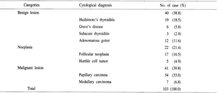
유두상 암종유두상 암종 34 예에서는 모든 세포학적 형태변화 가 관찰되었고 핵내 능선 은 34 예
(1 00%) ,
핵내 위봉 입체는 32 예(94%) ,
유두상 구조는 30 예(88%) ,
사종 체는 8 예(24%) ,
림프구 출현은 29 예(85%) ,
다핵성 거대조직구는 28 예(82%)
그리고 여포상 구조는 4 예(1 2%) 가 관찰되 었다 (Table
4) (Fi g. 7 A, B, C, D).
(2)
수질성 암종수칠성 암종 7 예 중 핵내 능선은 6 예
(86%) ,
핵내 위봉입체는 2 예(29%) ,
림프구 출현은 4 예(57%)
그 리고 여포상 구조는 5예(7 1
%)에서 관찰되었다(Table
4) (Fig. 8A , B).
Table 4. Cytomorphologic features in malignant lesion
Diagnosis Case( 41) Grooves( % ) Inclusion( % ) Papil1ae( % )
Ps빠unoma(%)Lymphocyte(%)
Gi빠cell(%) Follicle(%) Papil1ary carcinoma
Medullary carcinoma 34
7
100 86
94 29
88 0
24 0
85 57
82 0
12 71
Fig. 7. Papi1lary carcinoma showed folliicle(A) , papi1lae(B) ,
야따nmomabody (C , arrow) ,
inπanuclearpseudo-inclusion and nuclear grooves(D ,
aπow)(p apanicolaou; A , H&E; B , C , D X4 (0).
Fig. 8. In medullary carcinoma , A; the cells showed nuclear groove and intranuclear pseudo-inclusion(arrow). B;
lymphocytes and follicular
sσucturewere present in background(arrow) (H&E X4 (0).
3.
세포학적 형태변화의 비교평가 종에서만 관찰되었고, 핵내 위봉입체는 악성병변인 유두상 암종 (94%)와 수질성 암종 (29%) 에서만 관 갑상선 질환들의 세포학적 변화 중 유두상 구조 찰되었으며, 핵내능선은 모든 결절성 병변에서 관 (88%)와 낮은 빈도이지만 사종체 (24%)는 유두상 암 찰되었다(Table 5).
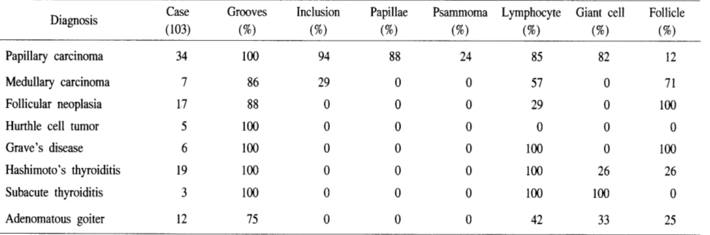
Table 5. Comparison of cytomorphologic features in thyroid disorders
Diagnosis Case Grooves Inclusion
(103) (%) (%)
Papillary carcinoma 34 100 94
Medullary carcinoma 7 86 29
Follicular neoplasia 17 88 0
Hurthle cell tumor 5 100 0
Grave ’ s disease 6 100 0
Hashimoto ’ s thyroiditis 19 100 0
Subacute thyroiditis 3 100 0
Adenomatous goiter 12 75 0
림프구의 출현은 허들세포 종양을 제외한 모든 결절성 병변에서 관찰되었으나 염증성절환인 미만 성 과증식
(1 00%),
하시모토 갑상선염 (100%)과 아급 성 갑상선염 (100%) 에서 출현빈도가 현저히 높았으 며, 다핵성 거대 조직구는 아급성 갑상선염(1 00%),
유두상 암종(82%),
선종성 종대(33%)
및 하시모토 갑상선염 (26%) 에서 관찰되었으나 아급성 갑상선염 일 때 높은 출현 빈도를 나타냈다(Table 5).
여포상 구조는 허들세포 종양과 아급성 갑상선염 을 제외한 모든 결절성 병변에서 관찰되었으나 여 포상 신생물 (100%)과 미만성 과증식 (100%) 일 때 출현 빈도 현저히 높았다
(Table 5).
N.
고찰 및 결론갑상선의 질환에 있어서 세침흡인도말검사는 간 단하면서도 정확성이 높고 안전하며 갑상선에 생긴 결절성 병변을 가진 환자들 가운데 수술적 치료가 필요한 환자들을 션별하는데 있어서 많은 도움을 주는 검사방법 중 하나다
(DeMay et al, 1996).
그러 나 세침흡인도말검사의 특이도, 민감도 그리고 정 확도를 높이기 위해서는 먼저 세침흡인 시술자의 숙련도와 도말표본의 상태가 양호해야하며 가장 중 요한 것은 도말표본을 겸경하는 판독자의 숙련도이 다. 이러한 숙련도는 도말겸사상 세포의 형태학적 변화를 오랜 경힘을 두고 숙지하여야 한다. 본 연구Papillae Psammoma Lymphocyte Giant cell Follicle
(%) (%) (%) (%) (%)
88 24 85 82 12
0 0 57 0 71
0 0 29 0 100
0 0 0 0 0
0 0 100 0 100
0 0 100 26 26
0 0 100 100 0
0 0 42 33 25
는 갑상선 병변에서 세포의 형태학적 변화를 관찰 하였고 갑상션 질환별로 비교 분석하였다.
핵내 능선은 핵막의 접힘 현상으로 유두상 암종 의 핵에서 가장 특징적인 변화이지만
(Chan et al, 1986; Shurba ji et al, 1988)
유두상 암종뿐 만 아니 라 다른 질환에서도 많이 관찰되었다(Table 5).
이러한 결과는 많은 보고서에서 발표되었으며 전자현미경 을 통한 연구에서도 확인되었다(J ohannessen et
떠,1982).
핵내 위봉입체는 세포질이 핵내로 함입되어 나타 나는 현상으로 핵내 능선과 함께 유두상 암종을 진 단하는데 있어서 중요한 특정이다
(Deligeorgi -politi et al, 1987;
Sodersσomet al, 1973).
본 연구에서 핵 내 위봉입체는 악성병변에서만 나타났고, 특히 유 두상 암종에서 많이 관찰되었지만, 수질성 암종서 에 도 29% 나 관찰되 었다(Table 5). Kini
등 (1984) 이 보고한바 의하면 수질성 암종에서 핵내 위봉입체는 일반적으로 나타난다고 하였다(Kini et al, 1984).
진짜 유두상 구조는 유두상 암종에서만 확인할 수 있었고 다른 결절 병변에서는 관찰되지 않았으 나 유두상 구조와 유사한 구조는 관찰되 었다. 조직 학적으로 진짜 유두상 구조는 중심에 섬유성 혈관 구조를 가지고 있으며 유두상 암종에서만 관찰되는 특징적인 세포군집구조이다
(Kini et al, 1987).
사종체는 유두상 돌기의 끝에 존재하는 미세한 혈관의 국소적인 경색에 의해 형성되며, 생화학적 인 메카니즘은 죽어 가는 경색된 세포에 깔숨이 침
착되어 형성된다고 생각하지만 아직은 이해가 부족 하다
(Johannessen et al , 1980; Johannessen et al ,
1982).
사종체는 유두상 암종에서 관찰되는 특정적인 나이테 모양의 감숨 결정처1 로 관찬된 빈도는 낮 았으나 유두상 암종에서만 관찰되었고 다뜬 겸첼성 병변에서는 볼 수 없었다. 그러나
Klinck
등 과Patchefsky
등이 보고한 바에 의하면 미만성 과증식과 하시모토 갑상선염과 같이 양성병변에서도 아주 드문 경우이지만 사종체가 관찰된다는 보고가 있었 다
(Ferenczy et at , 1977; Klinck et al , 1984).
림프구와 다핵성 거대 조직구의 출현은 여러 가 지 원인에 의해서 나타나며 만성적인 염증뿐 만 아 니라 갑상션의 선생물과 암종에서도 나타난다
(Do- niach et al , 1979; Strauss et al , 1983).
마찬가지로 본연구에서의 럼프구의 출현은 허들세포 종양을 제외 한 모든 질환에서 럼프구의 출현이 관찰되었고 특 히, 만성적인 염증첼환일 때 출현빈도가 높게 나타
났으며
(Table 5) ,
다핵성 거대조직구 출현는 아급성갑상선염, 유두상 암종, 선종성 종대 및 하시모토 갑상선염에서 관찰되었고 아급성 갑상션염에서 높 은 출현 빈도를 나타냈다
(Table 5).
여포상 구조는 허틀세포 종양과 아급성 갑상선염 을 제외한 모든 절환에서 관찰되었지만 여포상 신 생물과 미만성 과증식에서 출현 빈도가 현저히 높 게 나타났으며
(Table 5) ,
유두상 구조나 여 포상 구 조는 증식의 형태와 판련이 있다.결론적으로 세침흡인도말검사에서 세포형태학적 특정변화중 유두상 구조와 사종체는 유두상 임-종에 서만 나타났으나, 핵내 능선과 핵내 위봉입체는 다 른 결절성 병변에서도 관찰될 수 있으므로 유두상 암종의 진단시 주의릎 해야할 것으로 사료된다. 또 한 갑상선 잘환에서 임상진단 및 환자의 정보와 함 께 질환별 세포학적 특정변화를 종합하여 진단한다 면 진단의 정확도가 더욱 높아잘 것으로 사료된다.
잠고문헌