Lymphoepithelial carcinoma is an unusual squamous cell neoplasm that is characterized by undifferentiated malignant epithelial cells together with an infiltrate of mature lymphocytes. Lymphoepithelial carcinoma most frequently occurs in the nasopharynx and it has occa- sionally been documented in diverse sites outside the nasopharynx such as the salivary glands, larynx, thy- mus, etc (1-3).
Yet primary lymphoepithelial carcinoma has never been reported in the posterior cervical space in the med- ical literature, whereas several cases of metastasis of lymphoepithelial carcinoma have been reported (4). We report here on an unusual case of lymphoepithelial car- cinoma in the posterior cervical space and we present the MR findings of this tumor and the correlative histopathologic features.
Case Report
A 38-year-old woman had a 5-month history of a pal- pable mass in the left neck. She had been healthy prior to this finding and she had no history of disease. The physical examination performed by a physician re- vealed a painless soft mass in the left neck.
MRI examination was performed on a 1.5T MR unit (Signa Excite, GE Healthcare, Milwaukee, Wisconsin, U.S.A.). The MR image of the neck revealed a 4.3 × 2.6
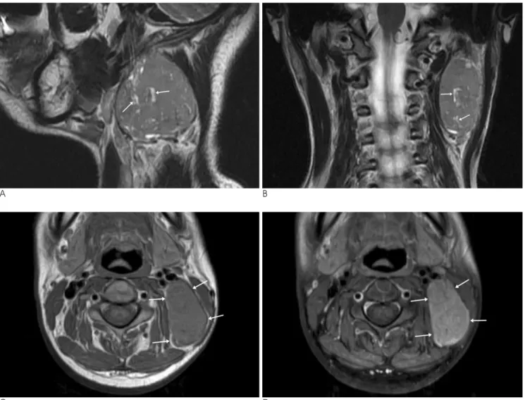
× 4.7 cm, oval, single mass with a somewhat lobulated margin in the left posterior cervical space. The mass was characterized as showing intermediate signal intensity (SI) relative to the surrounding muscle on the spin-echo T1-weighted MR image (T1WI) and slightly higher SI than the surrounding muscle on the fast spin-echo T2- weighted MR image (T2WI) (Figs. 1A-C). Multiple mi- crotubular cystic clefts were demonstrated in the mass on T2WI (Fig. 1B). The gadolinium-enhanced axial fat suppressed T1-weighted spin-echo MR image showed slightly heterogeneous enhancement of the mass with- out central necrosis or an enhancing capsule (Fig. 1D).
There was no significant peri-tumoral soft tissue infiltra-
J Korean Soc Radiol 2010;63:491-494
─ 491 ─
MR Imaging of Lymphoepithelial Carcinoma in the Posterior Cervical Space: A Case Report1
Seok Hoon Lee, M.D., Ki Woong Cheong, M.D., Young Ok Hong, M.D.2
1Department of Radiology, Eulji General Hospital, Eulji University School of Medicine, Seoul, Korea
2Department of Anatomic Pathology, Eulji General Hospital, Eulji University School of Medicine, Seoul, Korea
Received August 5, 2010 ; Accepted August 20, 2010
Address reprint requests to : Ki Woong Cheong, M.D., Department of Radiology, Eulji General Hospital, Eulji University School of Medicine, 280-1, Hagye 1-dong, Nowon-gu, Seoul 139-711, Korea.
Tel. 82-2-970-8290 Fax. 82-2-970-8346 E-mail: kwchung99@eulji.ac.kr
Lymphoepithelial carcinoma is a rare category of malignant neoplasm that consists of undifferentiated malignant epithelial cells admixed with lymphocytes.
Lymphoepithelial carcinoma predominantly occurs in the upper aero-digestive tract and only few cases of metastasis in the posterior cervical space have currently been re- ported. We report here on a case of lymphoepithelial carcinoma in the posterior cervi- cal space.
Index words :Undifferentiated carcinoma Nasopharyngeal Neoplasm Neck
Neoplasm, Metastasis
tion. Several small lymph nodes were found around the mass, but they were too small to characterize. The scanned pharyngeal mucosal space and salivary glands did not show any significant abnormality.
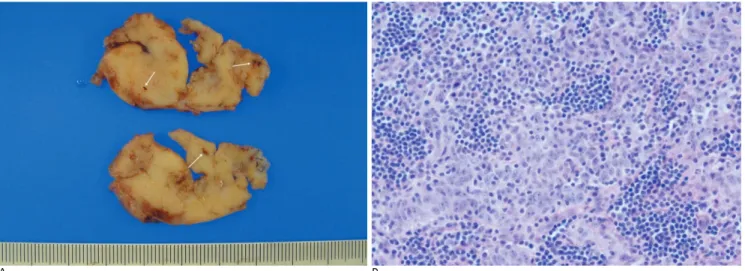
Surgical excision revealed a well-defined grayish tan soft tissue mass with a lobulated contour in the posterior cervical space. Pathologic examination demonstrated is- lands of undifferentiated carcinoma cells mixed with lymphocytes and plasma cells, and this was all consis- tent with lymphoepithelial carcinoma (Figs. 2A, B)
We suspected this tumor to be metastasis of lym- phoepithelial carcinoma because primary lymphoep- ithelial carcinoma in the posterior cervical space has never been reported in the current medical literature.
An endoscopic survey of the upper aero-digestive tract
and whole body PET-CT were performed for evaluating the primary origin of this tumor, but these modalities did not reveal any evidence of a primary tumor.
The patient underwent post-operative radiation thera- py on the operation site and adjuvant chemotherapy for possible hidden primary tumor three times, respective- ly. The patient has been under continuous follow-up at our hospital, and any primary tumor or other metastatic lesions have not developed for approximately 6 months.
Discussion
Lymphoepithelial carcinoma can be divided into two large groups and these are nasopharyngeal and non-na- sopharyngeal lymphoepithelial carcinoma. According to
Seok Hoon Lee, et al: MR Imaging of Lymphoepithelial Carcinoma in the Posterior Cervical Space
─ 492 ─
A B
C D
Fig. 1. The MR imaging reveals a well-defined mass with a lobulated contour in the left posterior cervical space.
A, B. On the coronal (A) and sagittal (B) T2-weighted fast spin-echo MR images, the mass shows slightly higher signal intensity than the surrounding muscle and intra-tumoral microtubular cystic clefts (arrows).
C. The axial T1-weighted spin-echo MR image shows an isointense mass (arrows) relative to the surrounding muscle.
D. The gadolinium-enhanced axial fat suppressed T1-weighted spin-echo MR image shows the slightly heterogeneous enhance- ment of the mass (arrows) without central necrosis or an enhancing capsule.
the World Health Organization classification, nasopha- ryngeal lymphoepithelial carcinoma is the undifferenti- ated subtype of non-keratinizing nasopharyngeal carci- noma. Non-nasopharyngeal lymphoepithelial carcino- ma has been described at diverse sites such as the sinonasal tract, nasolacrimal duct, oral cavity, orophar- ynx, salivary glands, thymus, hypopharynx, esophagus, stomach, larynx, trachea, lung, uterine cervix, urinary bladder and skin (2, 3).
Lymphoepithelial carcinoma of a squamous cell origin is histopathologically difficult to develop in the posterior cervical space because the main contents of this space are lymph nodes, nerves and fat, and there are no soft tissues associated with squamous cells in the posterior cervical space. As a result, lymphoepithelial carcinoma encountered in the posterior cervical space can be gen- erally considered as a metastatic carcinoma (5, 6).
The primary foci of cervical metastatic carcinoma are mostly in the head and neck regions, and the majority of the primary cancers are found by subsequent imaging studies. But they cannot be detected when they are lim- ited to the submucosa of pharyngeal mucosal space, and cervical nodal metastases can only be detected when they reach a certain visible size. The incidence of cervi- cal metastatic carcinoma of an unknown origin is esti- mated to be between 3 and 11% of the cases of head and neck carcinoma. Thus, endoscopy of the upper aero-di- gestive tract should be performed in order to detect the primary tumor in such patients. If possible, blind sys- temic pharyngeal biopsies should also be performed, and CT/MRI or PET-CT should follow (4).
The MRI features of metastasis of lymphoepithelial carcinoma have not been established because of the rari- ty of reported cases. Metastasis of typical squamous cell carcinoma manifests as unilateral multiple masses along the nodal chain and this rarely presents as a single mass.
This tumor generally shows an intermediate SI on T1WI and a slightly higher signal than the muscle on T2WI.
The gadolinium-enhanced MR image frequently shows central necrosis with peripheral irregular enhancement in the tumor, and especially when the size of the mass is greater than 3 cm. But metastatic carcinoma can at times shows variable imaging features according to the type of primary cancer and it can present as a homoge- neous enhancing mass without a necrotic portion (7).
In our case, central necrosis was not shown in the mass despite the large size of the mass, and microtubu- lar cystic clefts were also seen. Microtubular cystic clefts are dilated lymphovascular channels that are seen on the gross photograph of a specimen. We considered that these findings can be a kind of diagnostic clue that favors the diagnosis of metastasis of lymphoepithelial carcinoma rather than metastasis of another primary tu- mor or a solitary tumor such as schwannoma.
The differential diagnosis in this case also can include lymphoma and neurogenic tumors. Lymphoma mani- fests as a large (> 2 cm) solid mass and it typically pre- sents with lymphadenopathy involving multiple other nodal chains. Lymphoma shows homogeneous interme- diate SI on T1WI and high SI on T2WI. Unlike metasta- sis of squamous cell carcinoma, it shows relatively uni- form enhancement and it rarely shows central necrosis
J Korean Soc Radiol 2010;63:491-494
─ 493 ─
A B
Fig. 2. The gross photography of the specimen (A) shows multiple dilated lymphovascular channels (arrows) in the tumor. The mi- croscopic examination (B) shows islands of undifferentiated carcinoma cells admixed with many small lymphocytes and plasma cells (H & E staining, ×400).
or heterogeneous enhancement, except during the post- treatment state (8). Among the neurogenic tumors, schwannoma and solitary neurofibroma occasionally occur on the brachial plexus or cranial nerve 11 in the posterior cervical space. These tumors appear as a well- defined oval or fusiform mass, and they show variable SI from low to high on T1WI and hyperintensity com- pared to muscle on T2WI. Cystic changes frequently can be seen in the tumor, but necrosis is very rare.
Paraganglioma commonly occurs in the carotid body and it very rarely occurs in the posterior cervical space.
This tumor can sometimes show a “salt and pepper” ap- pearance on TIWI if it is bigger than 1.5 cm and the tu- mor shows intense enhancement with internal flow voids on the post contrast image (9).
In summary, we experienced an unusual case of pathologic confirmed lymphoepithelial carcinoma in the posterior cervical space. Although the primary origin of this tumor was not found on following studies, we think that these MR findings such as homogeneous enhance- ment without central necrosis and intra-tumoral micro- tubular cystic clefts can be the points for differentiating metastasis of lymphoepithelial carcinoma from metasta- sis of other primary tumors or solitary tumors.
References
1. Mills. SE, Fechner RE. The nose, paranasal sinuses and nasopharynx.
In: Sternberg SS. Diagnostic surgical pathology. 3nd ed. New York:
Lippincott Williams & Wilkins, 1999:885-924
2. Chan JKC, Bray F, McCarron P, Foo W, Lee AWM, Yip T, et al.
Tumours of the nasopharynx: Nasopharyngeal carcinoma. In: Bames L, Eveson JW, Reichart P, Sidransky D. Pathology and genetics of head and neck tumours: WHO classification of tumours. Lyon: IARC Press, 2005:83-97
3. Wassef M, Le Charpentier Y, Monteil JP, Le Tien K, Galian A.
Undifferentiated carcinoma with lymphoid stroma (undifferentiat- ed carcinoma nasopharyngeal type): optical, electron microscopi- cal and immunofluorescence study. Bull Cancer 69 1982;11-21 4. Haas I, Hoffmann TK, Engers R, Ganzer U. Diagnostic strategies
in cervical carcinoma of an unknown primary (CUP). Eur Arch Otorhinolaryngol 2002;259:325-333
5. Parker GD, Harnsberger HR. Radiologic evaluation of the normal and diseased posterior cervical space. AJR Am J Roentgenol 1991;
157:161-165
6. Som PM, Curtin HD. Head and Neck Imaging, 4th ed. St Louis, Mo:
Mosby-Elsevier Science, 2003:1824-1825
7. Sakai O, Curtin HD, Romo LV, Som PM. Lymph node pathology.
Benign proliferative, lymphoma, and metastatic disease. Radiol Clin North Am 2000;38:979-998
8. Weber AL, Montandon C, Robson CD. Neurogenic tumors of the neck. Radiol Clin North Am 2000;38:1077-1090
9. Kaji AV, Mohuchy T, Swartz JD. Imaging of cervical lym- phadenopathy. Semin Ultrasound CT MR 1997;18:220-249 Seok Hoon Lee, et al: MR Imaging of Lymphoepithelial Carcinoma in the Posterior Cervical Space
─ 494 ─
대한영상의학회지 2010;63:491-494
후경부에서 발견된 임파상피성암종의 자기공명영상 소견: 증례 보고1
1을지대학교 의과대학 을지병원 영상의학과
2을지대학교 의과대학 을지병원 해부병리과 이 석 훈∙정 기 웅∙홍 영 옥2
임파상피성암종은 미분화성 악성 상피세포와 림프구로 구성된 매우 드문 악성종양이다. 임파상피성암종은 대부분 호흡기계 및 상부 소화기계에서 발견되며 후경부에서는 아직까지 일부 전이암의 증례만이 보고되었다. 우리는 후경 부에서 발견된 임파상피성암종의 증례를 보고하고자 한다.