The Journal of the Korean Society for Surgery of the Hand VOLUME 13, NUMBER 4, December 2008
Internal Fixation of Mallet Fractures using a Hook Plate
Kyu Cheol Noh, M.D.
1, Jin Soo Park, M.D.
1, Hong Kyuu Kim, M.D.
1,2, Hui Seong Yu, M.D.
1, Jung Han Yoo, M.D.
1, Kook Jin Chung, M.D.
1, Ji Hyo Hwang, M.D.
1, Soo Bong Hahn, M.D.
2Department of Orthopedic Surgery, Kangnam Sacred Heart Hospital, Hallym University Medical Center, Seoul, Korea1
Department of Orthopedic Surgery, Yonsei University College of Medicine, Seoul, Korea2
Purpose: The purpose of this study is to introduce the result of treatment for bony mallet finger using hook plate
Materials and Methods: From September 2007 to Feburary 2008, 7 patients were treated with hook plate for bony mallet finger. Indications of operative treatment are fractures involving more than 30% of articular sur- face or those with palmar subluxation or fracture frag- ment displaced more than 3 mm. We used hook plate which prebend the 1.2 mm miniplate (Leibinger
�). The mean follow-up period was 7 months. The clinical results were evaluated using Crawford classification.
Results: According to Crawford classification, 6 cases were excellent and 1 case was good. There were margin- al necrosis in one case and nail deformities in 3 cases.
These complications were healed spontaneously.
Conclusion: Internal fixation of mallet finger using
Hook plate is a relatively easy method which can achieve anatomical reduction and firm fixation for earlier interphalangeal joint exercise.
Key Words: Bony mallet, Hook plate
서 론
골성 추지는 원위 수지 기저부의 견열 골절로 신전 기전의 손상이 발생하여 굴곡 변형이 생기거나, 이차 적으로 백조목 변형이 발생할 수 있다1,2. 현재까지 관 혈적 또는 비관혈적 정복술 및 다양한 방법에 의한 고 정술이 소개되어 왔다. 그러나 wire, K-강선, 나사못 를 이용한 해부학적 정복 및 견고한 고정술은 골편의 골절 및 정복의 소실이 발생할 수 있고, K-강선의 위 치가 변하거나, 원위 지절의 장기간 고정으로 인해 원 위 지절의 조기 운동이 불가능하며, K-강선이 피부 밖에 위치하여 감염에 대한 주의가 요구된다3,4. Teoh 와 Lee는 이에 위의 문제점을 최소화할 수 있으면서 해부학적 정복 및 견고한 고정이 가능한 골성 추지의 또 다른 치료 방법을 소개하여 좋은 결과를 얻었기에 저자들도 이 치료법을 좀 더 쉽게 변형하여 그 임상적 결과에 대해서 보고하고자 한다5.
연구 대상 및 방법
2007년 9월부터 2008년 2월까지 골성 추지로 방문 한 환자 중 환측 원위 지절의 수장부 아탈구가 있는 경우, 골편의 크기가 관절면의 1/3이상인 경우, 3 mm 이상의 골편 전위를 보이는 경우를 대상으로 전 향적 연구를 수행하였다6-8. 위의 기준에 맞는 환자 8 명(Table 1)을 대상으로 관혈적 정복술 및 내고정술 을 시행하였고, 추시 기간은 최소 6개월 이상 이었다.
저자들이 사용한 plate는 1.2 mm miniplate (Leibinger�)를 변형시켜서 갈고기 모양의 금속판
H
Ho oo ok k 금 금속 속판 판을 을 이 이용 용한 한 골 골성 성 추 추지 지 골 골절 절의 의 치 치료 료
한림대학교 강남성심병원 정형외과학교실1, 연세대학교 의과대학 정형외과학교실2 노규철1∙김홍균1,2∙유희성1∙유정한1∙박진수1∙정국진1∙황지효1∙한수봉2
통신저자: 김김 홍홍 균균
서울특별시 영등포구 대림1동 948-1 한림대학교 강남성심병원 정형외과학교실 TEL: 02-829-5165, FAX: 02-834-1728 E-mail: [email protected]
* 본 논문의 요지는 2008년도 대한정형외과학회 추계학술대회에 서 포스터 전시되었음.
(hook plate)으로 만들어 사용하였다(Fig. 1). 수술 방법은 원위 지절에 횡절개를 하여, 전층의 피판을 만 들어 충분히 골편을 노출시켜 관혈적 정복술을 시행하 고, 제작된 hook 금속판을 이용해 골편을 고정하였다 (Fig. 2). 수술 시간은 마취후부터 수술방을 나가기전 까지의 시간을 측정하였다. 수술 후 1주간 부목 고정 을 시행하였고, 수술 후 8일째 stack으로 고정하였 고, 매 시간당 10분의 능동적 원위 지절 운동을 시행 하였다. 2주일째 봉합사를 제거하였다. 예기치 않은 사고로부터 보호하기 위해 Stack은 4주까지 잠 잘 때 와 낮 시간에 운동을 하지 않을 때 착용하도록 교육하 였다. 4주 이후에 부목을 완전히 제거하도록 하였다.
추시 기간은 최소 6개월 이상 이었다. 임상적인 평가 는 수술 후 24주(6개월)째 Crawford 판정법(Table 2)을 기준으로 원위지 관절의 신전 소실 및 굴곡 정도 Table 1. Summary of cases
Case No. Age/Sex Cause of Injury Rt./Lt. Injured finger Injury to operation interval (days)
1 31/M Basketball Lt. 2nd 8
2 40/M Hit Rt. 4th 6
3 17/M Football Lt. 4th 8
4 27/F Hit Lt. 4th 8
5 13/M Handball Lt. 3rd 4
6 16/F Basketball Rt. 3rd 7
7 17/F Hit Rt. 4th 9
8 22/M Basketball Lt. 4th 300
Table 2. Crawford classification
Classification Extension loss Flexion Pain
Excellent None Full None
Good 0~10� Full None
Fair 10~25� Any loss of flexion None
Poor >25� Any loss of flexion Persistent pain
Table 3. Operation and postoperative evaluation
Case No. Anesthesia Operation time Extension loss Flexion Pain Crawford classification
1 *BPB* 45 m None 70� None Excellent
2 BPB 40 m 5� 75� None Good
3 BPB 45 m None 80� None Excellent
4 BPB 45 m None 80� None Excellent
5 *0G/A** 40 m None 80� None Excellent
6 BPB 40 m None 85� None Excellent
7 BPB 40 m None 80� None Excellent
8 BPB 40 m None 85� None Excellent
* Brachial Plexus Block
** General Anesthesia
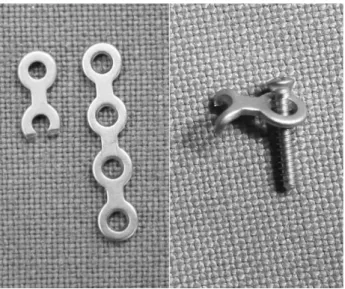
Fig. 1. Hook plate and Mini-screw.
와 동통의 유무에 따라 4단계로 나누어 평가하였다9.
결 과
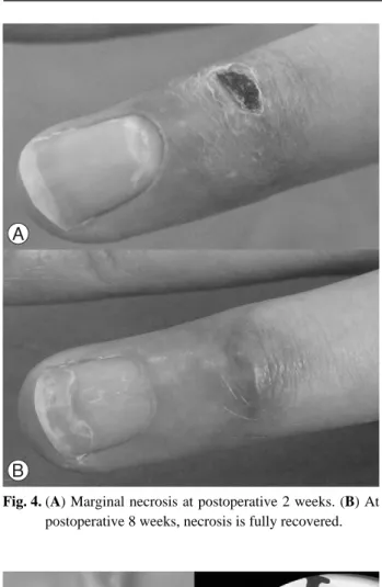
총 8예 중 5도의 신전 소실이 있었던 1예를 제외하 고는 7예에서 신전 소실이 없었고, 골곡 범위는 모든 예에서 정상으로 측정 되었고, 수술부위에 동통을 호 소하는 예는 없었다(Table 2)(Fig. 3). 다만 1예에서 절개 부위 변연부에 괴사가 일어났고, 경과 관찰만으 로도 치료가 되었다(Fig. 4). 또한 3예에서 손톱의 변 형이 관찰되었으나, 수술 후 약 3~4개월째 정상 모습 으로 돌아왔다. 수술 시간은 평균 42분이었다. 환자가 원한 1예를 제외하고는 모두 상완 신경총 마취로 수술 을 진행하였다.
고 찰
골성 추지의 수술적 치료 방법 중 비관혈적 방법으 로는 변형된 신전 차단 K-강선 고정술2,10,11과 원위지간 관절에 손상을 주지 않는 Umbrella Handle tech- nique12 등이 있다. 관혈적 방법으로는 관혈적 정복 후 골편의 K-강선 고정술13,14, 압박나사 고정술, 견인 철사 요법을 이용한 고정술15,Pull-out봉합술1 등이 있 다. 그리고 정복의 소실, 감염, 관절강직, K-강선의 전이, 봉합사의 절단, 신전 제한, 외상성 관절염 등의 합병증이 발생할 수 있다4,16,17. 그러나 저자들이 시행 한 hook 금속판을 이용한 고정술에서는 정복의 소실 이 발생하지 않았으며, 수술 후 조기 운동이 가능하기 때문에 관절 강직이나 외상성 관절염의 결과는 없었 Fig. 2. (A) Skin incision. (B) Illustration of internal fixation. (C) Medical photo of internal fixation with hook plate. (D) Radiograph
showing anatomic reduction and internal fixation using hook plate and screw.
Fig. 3. Radiograph of view of finger and medical Photographs showing full flexion and extension 6 months postoperatively.
다. 또한 장기간 K-강선이나 wire를 피부 밖에 두면 서 발생하는 염증 및 위생문제에 대한 단점도 해결할 수 있었다. 그러나 수술 후 추시 중 손톱 모양의 변형 이 오는 경우가 3예 있었는데 이는 hook 금속판의 위 치가 Germinal matrix와 근접하게 위치하기 때문인 것으로 생각된다. 하지만 특별한 치료없이 약 3~4개 월의 관찰로 손톱 모양은 정상으로 돌아오는 것을 확 인할 수 있었다. 수술 술기 중 금속판이 작아서 골편 과 고정하고 나사못으로 고정하는 것이 쉽지가 않아서 수술시간이 길어질 수 있는데, 우리 연구에 의하면 평 균 42분 소요되어 이전의 다른 방법들의 경험과 비교 해 보면 약 10~20분의 차이가 있는데 이는 갈고리모
양의 금속판 내고정으로 얻을 수 있는 이득을 고려해 볼 때 크게 중요하지 않다고 생각한다. 또 갈고리 모 양의 금속판이 작아서 골편과 압박고정을 하는 것이 쉽지 않았으며, 이를 위하여 저자들은 포겸자(towel clip)를 변형시켜 만든 기구를 이용 골절 정복 후 고정 을 용이하게 하였다(Fig. 5).
결 론
갈고리(hook) 금속판을 이용한 고정술은 해부학적 으로 견고한 고정이 가능하고, 장기간의 원위 지절 고 정이 필요하지 않아 조기 운동이 가능하여 골성추지의 고정방법 중 하나의 방법으로 의미가 있다. 그러나 환 자군이 적고 장기 추기 결과가 부족하므로 앞으로 좀 더 많은 환자 및 장기 추기에 대한 연구가 필요할 것 으로 사료된다.
참고문헌
01) Hahn SB, Kim SH, Park SH, Kang HJ. Treatment of bony mallet finger with extension block technique. J Korean Soc Hand. 2005;10:227-33.
02) Darder-prats A, Fernandez-garcia E, Fernandez-gabada R, Darder-garcia A. Treatment of mallet finger fractures by the extension-block K-wire technique. J Hang Surg Br.
1998;23:802-5.
03) Hahn SB, Kang ES, Kang HJ, Lee WS. Complication of bony mallet fingers after operative treatment. J Korean Soc Surg Hand. 1998;3:10-7.
04) Stern PT, Kastrup JJ. Complications and prognosis of treat- ment of mallet finger. J Hand Surg Am. 1998;13:341-6.
05) Kim YH, Kim KW, Min HJ, Yoon SU, Baek JH. Outcome study on operative treatment of mallet finger. J Korean Soc Surg Hand. 2002;7:34-41.
06) Teoh LC, Lee JY. Mallet fractures: A Novel approach to internal fixation using a hook plate. Journal of Hand surg E. 2007;32:24-30.
07) Niechajev IA. Conservative and operative treatment of mallet finger. Plast Reconstr Surg. 1985;76:580-5.
08) Wehbe MA, Schneider LH. Mallet fractures. J Bone Joint Surg Am. 1984;66:658-69.
09) Crawford GP. The molded polythene splint for the mallet finger deformities. J Hand Surg Am. 1984;9:231-7.
10) Tetik C, Gudemez E. Modification of the extension block Kirschner wire technique for mallet finger. Clin Orthop.
2002;404:284-90.
11) Hofmeister EP, Mazurek MT, Shin AY, Bishop AT.
Fig. 4. (A) Marginal necrosis at postoperative 2 weeks. (B) At postoperative 8 weeks, necrosis is fully recovered.
Fig. 5. (A) Modified towel clip. (B) Fix-ation of fragment with the device.
Extension block pinning for large mallet fracture. J Hand Surg Am. 2003;28:453-9.
12) Rocchi L, Genitiempo M, Fanfani F. Percutaneous fixation of mallet fractures by the “Umbrella handle” technique. J Hand Surg Br. 2006;31:407-12.
13) Badia A, Riano F, A simple fixation method for unstable bony mallet finger. J Hand Surg Am. 2004;29:1051-5.
14) Takami H, Takahashi S. Ando M. 2005;10:162-8.
15) Kang HJ, Shim DJ, Choi CJ, Yoon KH, Lee SY, Hahn SB.
Pulp traction technique for the operative treatment of bony mallet finger. J Korean Soc Surg Hand. 2005;10:162-8.
16) Bischoff R, Buechler U, De Roche R, Jupiter J. Clinical results of tension band fixation of avulsion fractures of the hand. J Hand Surg Am. 1994;1:1019-26.
17) Damron TA, Engber WD, Lange RH. Biomechanical analysis of mallet finger fractures fixation technique. J Hand Surg Am. 1993;18:600-8.