J. of Korean Orthopaedic Research Society Volume 7, Number 2, October, 2 0 0 4
영상분석 시스템을 이용한 골 시멘트 표본의 다공성 검사
가톨릭대학교 성빈센트병원 정형외과, 가톨릭대학교 성모병원 정형외과*
송주현・권순용*・고해석・이한용・이주엽・정진영・강용구・송호욱
= Abstract =
Evaluation of Porosity in Cylindrical Bone Cement Specimen Using Image Analysis System
Joo-Hyoun Song, M.D., Soon-Yong Kwon, M.D.*, Hae Seok Koh, M.D., Han-Yong Lee, M.D., Ju-Yup Lee, M.D., Jin-Young Jeong, M.D., Yong-Koo Kang, M.D., Ho-Wook Song, M.D.
Department of Orthopaedics, St. Vincent’s Hospital, St. Mary’s Hospital*
The Catholic University of Korea
Purpose: The porosity of the bone cement is the most important cause of fatigue failure, the most common mode of failure of bone cement using widely in arthroplasty. It is important to evaluate the porosity of bone cement for improvement or development of bone cement, but the conventional ‘stain, ‘cut, and ‘polish, manu- al method takes long time and efforts. So it is necessary to develop a new technique for evaluation of porosity of bone cement. We tried a technique using computer image analysis system to evaluate the porosity of bone cement specimen and assess efficacy of the method. Simultaneously we evaluated the relationship between the porosity of bone cement and fatigue failure.
Material and Metholds: We made 59, 2.5inch-length bone cement specimens(30 Simplex P, 29 Palacos R) using Simplex P and Palaces R which are widely using in clinical situation and checked radiogram using mam- mography film. After scanning the mammography film, we measured the porosity of the bone cement speci- mens using NIH(National Institute for Health) Image 1.6 version image analysis program. We also, measured the porosity of the bone cement specimens with conventional ‘stain’,‘cut’and ‘polish’method, after then compared the results of two methods. Simultaneously, we evaluated the relationship between porosity &
fatigue failure by loading 9.0, 10.0, 12.5 and 15.0 MPa load with frequency of 10Hz to the bone cement speci- mens under the physiologic condition.
Results: The coefficient of relation of simplex P and palaces R was 0.729 and 0.713 respectively, so there
※ 통신저자: 고 해 석
경기도 수원시 팔달구 지동 9 3 성빈센트병원 정형외과
TEL: 031) 249-7114 FAX: 031) 254-7186 E-mail: hskoh@unitel.co.kr
✽ 본 논문의 요지응 2 0 0 4년 보건 복지부 (바이오 생체 조직 장기 개발 쎈터 No-0405-B001-0204-0006) 연구 과제의 연구비로 이루어졌음.
서 론
John Charnley 경이 인공 고관절 치환술에 골 시멘트를 사용한 이래로 인공 삽입물의 견고한 고정을 위해 골 시멘트가 꾸준히 사용되고 있다3 ). 그러나 골 시멘트 실패에 의한 인공 고관절 재치 환술이 증가하면서 골 시멘트에 대한 소독방법, 혼합방법 그리고 다공성 등에 대한 연구들이 이루 어졌다9 ). Wijin4 )과 G r e e n w a l d5 )는 다공성의 증 가가 피로 실패에 관련이 있다는 보고를 하였고 Burke, Dares그리고 H a r r i s5 )는 골 시멘트 피로 실패의 일차적 원인이 다공성의 증가에 있다고 보 고하였다. 현재 인공 관절 시장에는 많은 새로운 골 시멘트가 소개되고 있는데, 이들 새로운 골 시 멘트는 임상 적용 이전에 피로 실패에 대한 평가 를 거쳐야 한다.
다공성 검사에 대한 기존의 수작업 즉‘s t a i n’,
‘c u t’그리고‘p o l i s h’술기는 시간이 오래 걸리 고 측정자간의 오차가 많아 새로운 다공성에 대한 검사방법이 필요하다8 ). 이에 저자들은 개인 컴퓨 터 프로그램을 이용한 영상 분석 시스템으로 시멘 트의 다공성을 검사하는 방법을 사용하여 그 효율 성을 알아보고 골 시멘트의 다공성과 피로 실패사 이의 연계성을 확인하고자 하였다.
대상 및 방법 1. 골 세멘트 표본의 준비
임상에서 흔히 사용되는 Simplex PⓇ와 P a l a- cos RⓇ을 사용하였고 각 골 시멘트는 제조회사의 안내서 지침에 따라 혼합하였는데, 이때 S t r y k e r
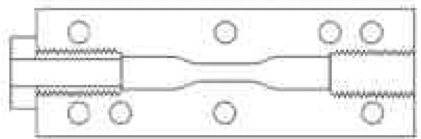
M i x e v a cⓇ II high vacuum system을 이용하 였다. 단량체( m o n o m e r )와 분말이 혼합된 골 시 멘트는 모래시계 모양의 표본을 만들기 위하여 알 루미늄 주형에 주입하였다(Fig. 1). 주형은 P h o s p h a t e가 첨가된 3 7°C의 수조에 담갔다가 2 0분 후에 꺼내어 주형으로부터 모래시계 모양의, 길이 2 . 5 i n c h의 골 시멘트 표본을 분리하였다.
2. 방사선 검사
주형으로부터 표본을 분리 시킨 뒤 다시 3 7°C P B S용액에 담갔고 실험 전 최소 일주일간 수조 에 보관하였다. 각 표본의 방사선 사진은 f a x- itron X-ray machine을 사용하였고 해상도를 높이기 위해 유방 촬영용 필름을 이용하였다. 방 사선 사진상 크기 1 mm 이상의 큰 기공을 가진 표본은 실험대상에서 제외시켰다. 총 7 4개의 표 본(Simplex P 33개, Palacos R 41개)중 3개의 Simplex P와 1 2개의 Palacos R 표본이 1 mm 이상의 기공을 가져 실험 대상에서 제외되어 궁극 적으로 5 9개의 표본(Simplex P 30개 Palcos R was high relationship between the image analysis system method and conventional one. It was easy and took shorter time to measure the porosity of bone cement specimens with image analysis system. There was high correlation between cement porosity and fatigue failure, regardless of level of load.
Conclusion: It was very easy and fast to measure the porosity of the bone cement specimens with image analysis system and there was high correlation between cement porosity and fatigue failure.
Key Words: Cement Porosity, Image Analysis System, Fatigue Failure
Fig. 1. Cross-sectional view of the aluminum specimen mold. A cannulated bolt (left side) allows the easy removal of the specimens from the mold.
Threaded placement holes provide alignment between the two mold halves. The threaded pres- surizer is inserted into the right end of the figure.
2 9개)이 실험에 사용되었다.
3. 피로강도 검사
방사선 촬영을 마친 5 9개의 표본을 3 7°C의 P B S용액에 담근 상태에서 Endura-Tec 3040 Servo-pneumatic mechanical test system에 고정시켰다. 각각의 표본은 9.0, 10.0, 12.5 그 리고 15.0 MPa의 부하를, 10 Hz의 빈도로 sinusoidal tension-compression force를 가하 였다.
4. 영상분석 시스템을 이용한 표본의 다공성 검사
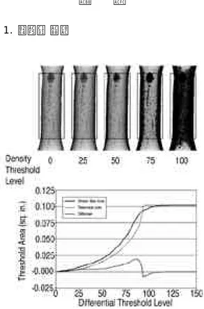
표본 각각의 방사선 사진은 AGFA Arcus II s c a n n e r를 이용하여 s c a n한 뒤 컴퓨터에 입력하 고 NIH(National Institute for Health) image 1.60 version의 마크로 기능을 이용하여 자체적으로 만든 프로그램을 사용하여 다공성을 검사하였다. Step wedge의 i m a g e가 표본 방사 선 사진의 gray level 측정을 위해 사용되었고, 가장 밝은 단계는 1 . 0 0 0으로 하고 각 1 5단계로 나누어 검은색 배경에 일치하는 단계에 도달하면 0의 값을 주었다. 앞서 언급한 바와 같이 N I H image 소프트웨어의 마크로 기능을 이용한 프로 그램을 만들고 각 표본에 대한 명도를 측정하였 다. 이 때 역치(DTL: different threshold level) 내로 포함되는 명도의 영역을 측정하고, 이 영역을 곡선으로 표시하여 기공이 전혀 없는 이상적인 표본의 곡선과 비교하였다(Fig. 2). 이 들 두 곡선 사이의 차이를 표본의 다공성이라고 정의하였다.
5. 수작업에 의한 다공성 검사
방사선 촬영과 피로강도 실험을 거친 각 표본을 Buehler Isomet 11-1180 low speed diamond s a w를 이용하여 1mm 간격으로 절단해 나갔다.
이때 India ink를 바르고 절단면의 기공에만 i n k가 남도록 깨끗이 닦은 뒤 Hitachi HV- C10CLD video camera가 부착된 Nikon dis- secting zoom microscope (5X)에서 촬영하여
단면의 다공성을 측정하였다. 동일한 작업을 1 0 회 반복하여 1 0회의 측정에서 얻어진 수치를 평 균 내어 각 표본의 다공성을 얻었다.
(6) 영상분석시스템을 이용한 다공성 검사치와 수작업에 의한 다공성 검사치의 비교두 검사 방법 에 대한 측정치의 일치정도를 판정하기 위하여 linear regression분석을 이용하였다.
결 과 1. 다공성 검사
Fig. 2. Measurement of porosity. The original x-ray image is calibrated and the lower threshold level set to just above background level. A Density Threshold Level (DTL) of 0 and subsequent increasing threshold levels result in a black area within the test region. This increase in area with increasing threshold level is represented graphi- cally by the solid line in the graph. The specimen density profile is compared to a theoretical curve (dashed line) representing a specimen free of porosity. The maximal value on the resulting dif- ferential curve is recorded as the radiographic porosity value.
영상분석 시스템을 이용한 다공성 값과 수작업 을 이용하여 측정한 다공성 값을 비교하였을 때 상관계수가 Simplex P가 0.729, Palacos R이 0 . 7 1 3으로 positive correlation을 보여( F i g . 3), 새로운 방법인 영상분석 시스템을 이용한 다 공성의 검사법이 기존의 검사방법에 비하여 검사 결과는 큰 차이 없이 빠르고 쉽게 측정할 수 있는 방법임을 알 수 있었다.
2. 피로강도 실험
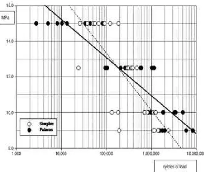
15.0 MPa의 부하에서는 Simplex P가 P a l a- cos R에 비해 뛰어난 피로 강도를 보였으며 반대 로 10.0 MPa에서는 Palacos R이 뛰어난 피로 강도를 보였다. 그러나 생리적인 부하치인 1 2 . 5 M P a에서는 두 시멘트 사이에 유의한 차이를 보 이지 않았다(Fig. 4). 다공성과 피로강도사이에
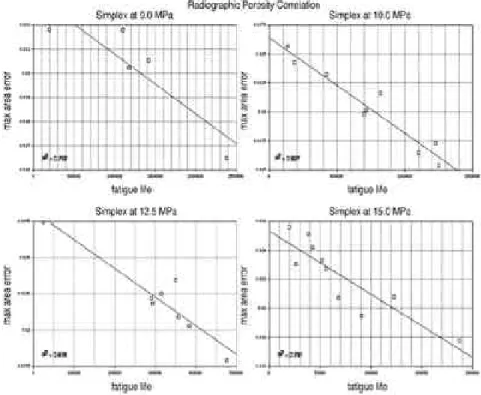
는 부하치에 관계없이 높은 상관관계를 보여 다공 성과 피로 실패 사이에 밀접한 연계성이 있음이 확인되었다(Fig. 5,6).
고 찰
피로 강도에 대한 검사는 현재 사용중인 골 시 멘트와 새로운 골 시멘트의 평가에 있어 매우 중 요한데, 다공성이 혼합된 골 시멘트 내에서 빈공 간의 상대적 부피치이기 때문에 가장 직접적이며 흔히 사용된 측정법은 소위‘s t a i n’, ‘c u t’그리 고‘polish, 방법이었다8 ). 비록 시간이 오래 걸리 며 측정자간의 오차도 있을 수 있지만 많은 연구 소에서 일차적으로 선택되는 방법이다. 그러나 절 단편이 대개 2 ~ 5개 뿐이어서, 작은 검체수로 골 시멘트의 진정한 평균 다공성치를 정확하게 반영 한다고 말하기에는 무리가 있다.
최근에 여러 가지 다공성 측정방법이 소개되고 있는데 water displacement 측정법6 ) r a d i- ographic assessment8, 9, 11), 초음파를 이용한 측
정1 0 ) 그리고 자기공명영상을 이용한 측정1 ) 등이
이에 포함된다. Water displacement 측정법은 주어진 부피의 골 시멘트에서 예상되는 무게의 차 이를 측정하는 방법으로 이론적 무게에서 실제 무 게를 감함으로써 전체 전체의 전위된 양으로 다공 성을 측정한다. 그러나 미세한 기공에 의한 작은 차이를 알아낼 정도의 정확도가 떨어진다. 초음파 그리고 자기공명영상을 이용한 다공성 측정방법은 비용이 많이 들며 일반 연구소에서 쉽게 시행하기 에는 어려움이 있다. 또한 방사선 검사방법은 사 진상 보이는 기공의 면적 측정 시 측정면이 2차원 이라는 한계가 있다.
이번 연구에서는 방사선 사진을 컴퓨터에 입력 하여 영상분석 프로그램을 이용하기 때문에 3차원 에서 다공성을 검사할 수 있으며 기존의 수작업에 의한 검사치와 거의 유사한 결과를 보이면서도 시 간과 노력이 절약되는 장점이 있다.
생체에서 피로강도의 예측치로서 다공성의 의미 에 대해서는 논란이 많다. 그러나 가공이 s t r e s s r i s e r로 작용하여 골 시멘트에 균열을 야기하고, 궁극적인 피로실패를 유발시키는 것은 확실하다.
이번 연구에서도 생체조건과 유사한 상태에 골 시 Fig. 3. Correlation between manual and image analysis
techniques.
Fig. 4. S-N Curve Showing Fatigue relationship between PalacosⓇand SimplexⓇ.
Fig. 5. Correlation curves for PalacosⓇR illustrating the relationship between radiographic Porosity vs fatigue life for all specimens.
멘트 표본을 설치하고 각기 다른 세기의 부하치를 가하였을 때 부하치에 관계없이 다공성이 큰 골 시멘트가 조기에 피로 실패가 일어나 골 시멘트의 다공성과 피로 강도와의 높은 연계성이 있음을 다 시 한 번 확인할 수 있었다.
결 론
영상분석 시스템을 이용한 골 시멘트의 표본의 다공성 검사는 소요시간이 적게 걸리며 사용이 간 편한 방법이며 골 시멘트 표본의 피로강도 예측에 도 정확한 검사법이라고 생각된다. 또한 골 시멘 트의 다공성은 피로강도와 높은 연계성이 있음을 알 수 있었다.
R E F E R E N C E S
01) Borgia GC, Bortolotti V, Dattilo P, Fantazziru P and Maddinelli G : Quantitative determination
of porosity: A local assessment by NMR imaging techniques. Magnetic Resonance Imaging, 14:
919-921, 1996.
02) Burke DW, Gates EI and Harris WH : Cer- trifugation as a method of improving tensile and fatigue properties of acrylic bone cement. J Bone Joint Surg, 66A:1265-1273, 1984.
03) Charnley J: Anchorage of the femoral head prosthesis to the shaft of femur. J Bone Joint Surg, 42B:28-30, 1960.
04) De Wijin JR, Sloff J and Driessens FC: Char - acterization of bone cement. Acta Ortho Scand, 46:38-51,1975.
05) Greenwald AS, Wilde AH and Matejczyk MB:
Clinical applications and properties of acrylic bone cement. Orthopaedics Digest, 5:16, 1977.
06) Hass SS : A characterization of polymethlmetha- cylate bone cement. J Bone Joint Surg, 57A:380- 391, 1975.
Fig. 6. Correlation curves for SimplexⓇP illustrating the relationship between radiographic Porosity vs fatigue life for all specimens.
07) Saha S and Pal S: Mechanical properties of bone cement: A review J Biomed Mates Res.
18:435-462, 1984.
08) Schreus W, Spiering PTJ, Huiskes R and Sloof TJ: Effects of preparation techniques on the porosity of acrylic cements. Acta Orthop Scand, 59(4):403-409, 1988.
09) Smeds S, Goertzen J and Irarsson IL: Influ- ence of temperature and vacuum mixing on bone cement properties. Clin Orthop. 344:326-334,
1997.
10) Strelitzki R, Clarke AJ, Truscott JG and Evans JA: Ultrasonic measurement : An Evalua- tion of three heel bone scanners compared with a bench-top system. Osteoporosis Intenational 6 : 471-479, 1996.
11) Wang JS, Franzen H, Jonsson E and Lidgren L: Porosity of bone cement reduced by mixing and collecting under vacuum. Acta Orthop Scand. 64(2):143-146, 1993.