174
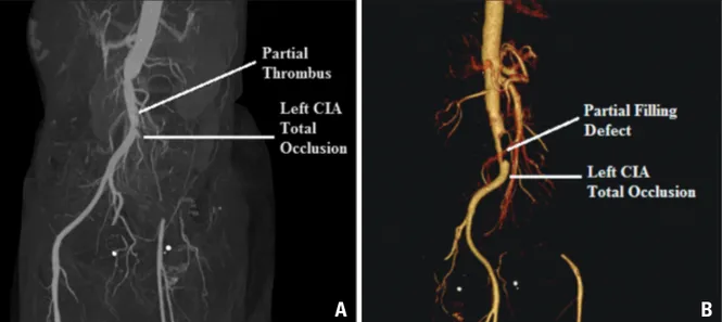
A 50-year-old female presented to our emergency department with complaints of acute onset pain in left lower limb for last 5 days. Her previous history was unremarkable. Physical exami- nation revealed a pulse rate of 90 per minute, blood pressure of 120/76 mm Hg and normal jugular venous pulse. The left lower limb was cold and pale. Her left femoral, left popliteal, left dorsalis pedis and left posterior tibial arterial pulsations were absent. All other arterial pulsations were normally pres- ent. Cardiac and respiratory examinations were within normal limits. Electrocardiogram was within normal limits with normal sinus rhythm. A diagnosis of acute limb ischemia was made and an emergency computed tomographic angiography of infra re- nal aorta with both the lower limbs was done. There was a partial filling defect of the aorta just before the bifurcation along with complete thrombotic occlusion of the left common iliac artery with distal reformation by collateral from the arter- ies of the contralateral limb (Fig. 1). Two dimensional echo- cardiography showed a large (2.0 × 3.0 cm) mobile thrombus at the apico-septal region of the left ventricle along with nor- mal ejection fraction of 60% (Fig. 2A, Supplementary movie 1). The three dimensional (3D) nature of the thrombus was better appreciated on transthoracic 3D echocardiography (Fig.
2B, Supplementary movie 2). Hematologic studies revealed anemia, with hemoglobin level of 10 g/dL, thrombocytopenia (30 platelets/nL) and leucocytocis (240 white blood cells/nL).
Peripheral smear showed 95% blast cells which were peroxi- dase negative (Fig. 3). Bone marrow aspiration confirmed the diagnosis of acute myeloid leukemia (French American British type M2). Aortic with left ileo-femoral thromboembolectomy was done to salvage the limb. Intra venous heparin therapy was started for left ventricular thrombus. Patient was transferred to
pISSN 1975-4612 / eISSN 2005-9655 Copyright © 2016 Korean Society of Echocardiography www.kse-jcu.org
• Received: January 17, 2016 • Revised: April 3, 2016 • Accepted: May 10, 2016
• Address for Correspondence: Rajiv Bharat Kharwar, Department of Cardiology, U N Mehta Institute of Cardiology and Research Centre, Civil Hospital Campus, Asarwa, Ahmedabad, Gujarat 380016, India Tel: +91-990-480-5283, Fax: +91-79-2268-5349, E-mail: dr.rajivkharwar@gmail.com
• This is an Open Access article distributed under the terms of the Creative Commons Attribution Non-Commercial License (http://creativecommons.org/licenses/by-nc/3.0) which permits unrestricted non-commercial use, distribution, and reproduction in any medium, provided the original work is properly cited.
the oncology department where induction phase of chemother- apy was started according to acute myeloid leukemia manage- ment protocol.
Patients with acute leukemia are at an increased risk of both thrombosis as well as bleeding. Severe haemorrhagic compli- cations are seen in acute promyelocytic leukemiausually sec- ondary to disseminated intravascular coagulation. Thrombotic complications are rarely reported. There are very few reports of acute limb ischemia due to large artery occlusion in acute my- eloid leukemia.1) None of the previously reported cases had left ventricular thrombus. To our knowledge, this is the first report of acute myeloid leukemia having left ventricular thrombus and left lower limb thromboembolism on presentation.
The pathology of coagulopathy in acute leukemia is complex.
It is determined by an interplay between various procoagulant factors (like tissue factor, cancer procoagulant factor, prothrom- botic cytokines) and anticoagulant and fibrinolytic factors re- leased or expressed by the leukemic cells.2)
Due to scarcity of occurrence of thrombotic complications in acute myeloid leukemia, no large scale studies are available for guiding management of this complication. Thrombolytic and anticoagulation therapy is not without risk as patient are also at increased risk of bleeding due to simultaneous occurrence of thrombocytopenia and disseminated intravascular coagulation.
Prompt diagnosis of underlying leukemia and initiation of ap- propriate anti leukemia treatment are keys to reducing overall morbidity and mortality.
Supplementary movie legend
Movie 1. Two dimensional transthoracic echocardiography.
There is a large mobile thrombus situated at the apico septal
Acute Myeloid Leukemia with Intracardiac Thrombus Presenting as Acute Limb
Ischemia
Rajiv Bharat Kharwar, MD, Kamal Sharma, MD, and Sharad Jain, MD
Department of Cardiology, U N Mehta Institute of Cardiology and Research Centre, Ahmedabad, Gujarat, India
KEY WORDS: Acute myeloid leukemia · Acute limb ischemia · Thrombus.
IMAGES IN CARDIOVASCULAR ULTRASOUND J Cardiovasc Ultrasound 2016;24(2):174-176 http://dx.doi.org/10.4250/jcu.2016.24.2.174
AML with Intracardiac Thrombus Presenting as Acute Limb Ischemia | Rajiv Bharat Kharwar, et al.
175 Fig. 1. Computed tomographic angiography of aorta with both lower limb. There is a partially occluding thrombus at the lower aorta just before the bifurcation along with complete thrombotic occlusion of the left common iliac artery from the origin. A: The left common femoral artery is being filled by collateral from the contralateral limb. B: The three dimensional computed tomographic reconstruction. CIA: common iliac artery.
A B
Fig. 2. Transthoracic echocardiography. A: Two dimensional echocardiography in apical four chamber view (A) showing normal sized LV with a 2.0 × 3.0 cm thrombus at the apico-septal region. B: The three dimensional (3D) structure of the thrombus is better appreciated on 3D transthoracic echocardiography. LA: left atrium, LV: left ventricle, RA: right atrium, RV: right ventricle.
A B
Fig. 3. Peripheral blood smear at 400 × magnification (A) and 1000 × magnification (B) using Wright stain. A: There are plenty of blast in the peripheral smear which can be identified by hyperchromatic nuclei with a raised nucleus/cytoplasm ratio. Also note the scarcity of platelets. B:
Magnified view of the blast cells.
A B
Journal of Cardiovascular Ultrasound 24 | June 2016
176
region of the left ventricle. The size of the left ventricle is normal with normal left ventricular contractility and ejection fraction.
Movie 2. Three dimensional (3D) transthoracic echocardiog- raphy. The anatomical details of the thrombus are better delin- eated on 3D echocardiography.
References
1. Kafetzakis A, Foundoulakis A, Ioannou CV, Stavroulaki E, Koutso-
poulos A, Katsamouris AN. Acute lower limb ischemia as the initial symptom of acute myeloid leukemia. Vasc Med 2007;12:199-202.
2. Cahill TJ, Chowdhury O, Myerson SG, Ormerod O, Herring N, Grimwade D, Littlewood T, Peniket A. Myocardial infarction with intracardiac thrombosis as the presentation of acute promyelocytic leukemia:
diagnosis and follow-up by cardiac magnetic resonance imaging. Circulation 2011;123:e370-2.