INTRODUCTION
Phyllodes tumors (PTs) are biphasic fibroepithelial neo- plasms, characterized by a leaf-like architecture. They are composed of benign epithelial elements and a cellular, spindle cell stroma. These lesions account for less than 1% of all breast tumors.(1,2)
The behavior of PTs ranges from benign and locally recurrent to malignant and metastatic.(3) The clinical behavior has been difficult to predict based on histologic features. Histologically benign PTs have been known to metastasize, and histologically malignant PTs sometimes neither recur nor metastasize.(4,5) The current (2003) World Health Organization (WHO) classification of tumors of the breast separates PTs into benign, borderline, and malignant categories, mainly based on mitotic activity, margin type, stromal overgrowth, and cellular pleomor- phism.(6) Recently ancillary diagnostic methods to iden- tify PTs with potentially aggressive behavior have been reported. These have included immunohistochemical assessment of p53, Ki-67, and c-Kit.(7-11)
Purpose: Phyllodes tumors (PTs) of the breast have been classified as benign, borderline, or malignant based on their histopathologic features. However, predicting clinical behavior based on these features has proven to be difficult given that local recurrence occurs in both benign and malignant PTs.
Recurrence has been shown to mirror the histologic pattern of the primary tumor or to show dedifferentiation. The aim of this study was to assess the value of the histopathologic parameters, expression or mutation of c-Kit and platelet derived growth factor receptor alpha (PDGFRA) in predicting tumor recurrence. Methods: Representative areas from 39 benign, 16 borderline, and 12 malignant PTs were selected for construction of tissue microarrays. Immunohistochemical analyses for p53, Ki-67, c-Kit, and PDGFRA were performed and SSCP-PCR analysis was carried out to identify mutations in exons 9, 11, 13, and 17 of the c-Kit gene and exons 12 and 18 of the PDGFRA gene. Clinicopathologic features, including tumor recurrence and margin status, were also evaluated. Results: Of the 67 PTs, 11 cases (16.4%) recur-
red from 3 to 92 months following initial diagnosis (4 benign, 2 borderline, and 5 malignant). One benign PT case recurred as a borderline tumor and two borderline PT cases recurred as malignancies. Three patients died of malignant PT. No mutations of the c-Kit or PDGFRA genes were found and there was no statistically significant association of either p53 or p16 immunostaining with recurrent disease (p >0.05).
However, histologic grade (p =0.033), margin status (p <
0.001), Ki-67 (p=0.012), c-Kit (p=0.002), and PDGFRA (p=
0.007) stromal immunopositivity were significantly correlated with recurrence. Conclusion: Even though positive or close margins were significantly associated with tumor recurrence, stromal c-Kit, PDGFRA positivity, and the Ki-67 index were useful for predicting recurrent PTs. Despite this, no c-Kit or PDGFRA mutations were found.
Key Words: Breast, Mutation, Phyllodes tumor, Platelet-derived growth factor receptor alpha, Proto-oncogene proteins c-kit
Breast Cancer
O R I G I N A L A R T I C L E
Mutation-Free Expression of c-Kit and PDGFRA in Phyllodes Tumors of the Breast
Chang-Woo Jung, Kwang-Sun Suh, Jin-Sun Lee
1, Je-Ryong Kim
1, Eil-Sung Chang
1, Hae-Joung Sul
2, Mee-Ja Park
3Department of Pathology, Research Institute for Medical Sciences, 1Department of Surgery, Chungnam National University School of Medicine, Daejeon; 2Department of Pathology, Catholic University Daejeon St. Mary’s Hospital, Daejeon; 3Department of Pathology, Eulji Hospital, Daejeon, Korea
Correspondence: Kwang-Sun Suh
Department of Pathology, Chungnam National University School of Medicine, Munhwa 1-dong, Jung-gu, Daejeon 301-747, Korea Tel: 042-580-8233, Fax: 042-280-7198
E-mail: [email protected]
Received: January 14, 2010 Accepted: July 6, 2010 This work was supported by Chungnam National University Hospital Research Fund, 2007.
257
stromal tumors (GISTs), which have been characterized by the overexpression of c-Kit, show a spectrum of behaviors from benign to malignant, similar to PTs.(13- 15) Recently, expression of both c-Kit and PDGFRA was observed in malignant PTs, although the impact of this recurrence was not addressed.(7-11,16)
The aim of this study was to assess the value of the histopathologic parameters, c-Kit and PDGFRA expres- sion and mutation in predicting tumor recurrence.
METHODS
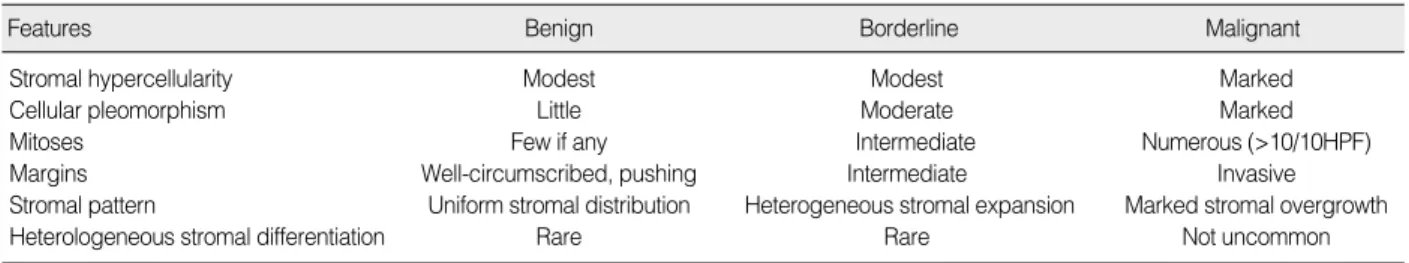
Sixty seven PTs of the breast were diagnosed between January 1998 and December 2006 at the Department of Pathology, Chungnam National University Hospital and Eulji Hospital, Daejeon. This study was approved by the Institutional Review Board of Chungnam National University School of Medicine. According to histologic findings (Table 1), including the degree of stromal hyper- cellularity, cellular pleomorphism, mitotic activity, and tumor border, PTs were classified into benign, borderline, and malignant categories.(1,6) At least two hematoxylin and eosin (H&E) stained tumor sections were reviewed independently by three pathologists for verification of diagnosis. The resection margin was recorded as posi-
construction of tissue microarrays using a 3 mm punch with two punches per case. Samples were arrayed in the recipient blank blocks. A total of 67 PTs were used.
Immunohistochemical analyses for p53, Ki-67, c-kit, and PDGFRA were performed. Further, 4 μm sections were cut from the tissue microarray blocks and fished onto coated slides. The slides were then baked in an oven over- night at 55℃to enhance adhesion. Deparaffinization in xylene and graded alcohol followed. Pre-diluted anti- bodies against Ki-67 (monoclonal; Zymed Lab., San Fran- cisco, USA), p53 (monoclonal; DAKO, Copenhagen, Den- mark), c-Kit (polyclonal; DAKO), p16 (polyclonal; DAKO), and PDGFRA (monoclonal; Santa Cruz Biotechnology Inc., Santa Cruz, USA) were obtained from commercial sources. Immunohistochemical staining protocols for each antibody are summarized in Table 2. Immunolocalization was performed using an LSAB kit (DAKO, Carpinteria, USA). All assays were carried out prospectively in an automated immunostainer (DAKO). Antibody-antigen reactivity was visualized using diaminobenzidine and counterstained with Meyer’s hematoxylin.
The percentage of tumor cells was semiquantified man- ually by counting 500 stromal cells per case. Stains for p53 and Ki-67 were assessed in the cell nuclei, while c- Kit, p16, and PDGFRA were evaluated in the cytoplasm
Table 1. Main histologic features of the 3 tiered grading subgroups for phyllodes tumours(1)
Features Benign Borderline Malignant
Stromal hypercellularity Modest Modest Marked
Cellular pleomorphism Little Moderate Marked
Mitoses Few if any Intermediate Numerous (>10/10HPF)
Margins Well-circumscribed, pushing Intermediate Invasive
Stromal pattern Uniform stromal distribution Heterogeneous stromal expansion Marked stromal overgrowth
Heterologeneous stromal differentiation Rare Rare Not uncommon
HPF=high power field.
and cytoplasmic membrane. p53, c-Kit, p16, and PDGFRA were considered to be positive when more than 10% of stromal cells were positive. A high Ki-67 index was defined
as nuclear staining in more than 10% of tumor cells. An ovarian serous carcinoma known to be positive for p53 and a c-Kit positive case of GIST were used as positive controls. For negative controls, the primary antibody was omitted with each batch run. Overall positive marker expression between tumors that did and did not recur was assessed.
DNA extraction
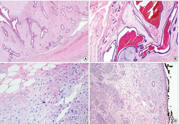
Twenty-eight tumor samples (16 borderline tumors and 12 malignant tumors) were taken from formalin fixed, paraffin embedded (FFPE) tissue samples. Subsequently, H&E-stained 4 μm sections were reviewed under a micro- scope and areas rich in tumor cells were marked. Corres- ponding areas on unstained sections were then scraped from the slides using a scalpel blade. Tumor samples that contained as few non-neoplastic cells as possible (70- Figure 1. A well circumscribed, pushing border of a benign phyllodes tumor (A and B, H&E stain, ×40). An infiltrative border (C, H&E stain, ×400) and a positive surgical margin of a malignant phyllodes tumor (D, H&E stain, ×40).
C D
A B
Table 2. Summary of immunohistochemical staining protocols
Primary antibody
Pretreatment temp (oC), time (min) Reaction
time (min) Dilution Company
(Country)
p53 DakoCytomation 1:120 40 Autoclave (121, 10) (Denmark)
Ki-67 Zymed Lab. 1:250 40 Autoclave (121, 10) (USA)
c-Kit Santa Cruz 1:300 40 Autoclave (121, 10) (USA)
p16 DakoCytomation 1:80 40 Autoclave (121,10) (Denmark)
PDGFRA Santa Cruz 1:250 40 Autoclave (121, 10) (USA)
PDGFRA=platelet derived growth factor receptor alpha.
Mannheim, Germany), 20 mM Tris/HCl, pH 8.3, 5 mM MgCl2, 100 mM KCl, 1% Tween-20, and 1% NP-40). The mixture was boiled for 10 min to inactivate the proteinase K, followed by phenol extraction for purification, and concentration using ethanol precipitation. The isolated DNA solution was quantified spectrophotometrically.
PCR amplification, SSCP analysis, and silver staining
SSCP-PCR analysis for mutations in exons of 9, 11, 13, and 17 of the c-Kit gene, and exons 12 and 18 of PDGFRA was carried out, as previously described.(17,18) PCR amplification was performed in a total volume of 20 μL containing 500 ng of template DNA, one unit of ExTaq polymerase (Takara, Shiga, Japan), 1.25 mM dNTP, 15 pmole primer, and 2 μL of 1×reaction buffer. PCR cycles consisted of 5 min at 94℃followed by 35 cycles for 30 sec at 94℃, 30 sec at 55℃, and 30 sec at 72℃, followed by one cycle for 7 min at 72℃.
Two microliters of PCR product was mixed with 6 μL of sample loading buffer containing 95% formide (deion- ized), 10 mM NaOH, 0.25% Bromophenol blue, and 0.25%
Xylene cyanol. The samples were then denatured for 3 min at 100℃and quickly chilled on ice. They were then loaded onto 12% polyacrylamide gel containing 1×sample buffer (33 mM Tris-sulfate, 7% Glycerol, pH 8.3), and were electrophoresed at 250 V. After electrophoresis the gels were disassembled from the glass plate, then stained using a Silver Stain Plus kit (BIO-RAD, Philadelphia, USA), followed by air drying.
Statistical analysis
Statistical analysis was carried out using SPSS software (PASW Statistics 17.0) (SPSS Inc., Chicago, USA). Histo-
nificant.
RESULTS
Clinicopathological features
A total of 67 patients were diagnosed with PTs during the study period with ages ranging from 14 to 61 yr (mean 37.6±11.3 yr). Fifty-nine patients underwent lumpec- tomy/excisional biopsy, while three underwent vacuum- assisted excision and another five had total mastectomies.
Tumor size ranged from 1.2 to 12.0 cm (mean 5.0±2.5 cm). Histologic classification revealed 39 (58.2%) benign,
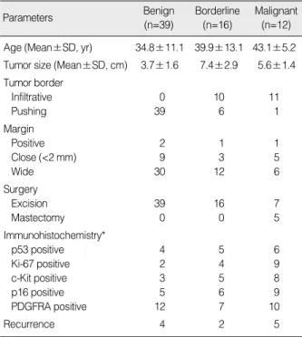
Table 3. Clinicopathologic and immunohistochemical features of phyllodes tumors
Parameters Benign
(n=39)
Borderline (n=16)
Malignant (n=12) Age (Mean±SD, yr) 34.8±11.1 39.9±13.1 43.1±5.2 Tumor size (Mean±SD, cm) 3.7±1.6 7.4±2.9 5.6±1.4 Tumor border
Infiltrative 0 10 11
Pushing 39 6 1
Margin
Positive 2 1 1
Close (<2 mm) 9 3 5
Wide 30 12 6
Surgery
Excision 39 16 7
Mastectomy 0 0 5
Immunohistochemistry*
p53 positive 4 5 6
Ki-67 positive 2 4 9
c-Kit positive 3 5 8
p16 positive 5 6 9
PDGFRA positive 12 7 10
Recurrence 4 2 5
*p53, Ki-67, c-Kit, p16, and platelet derived growth factor receptor alpha (PDGFRA) were considered to be positive when more than 10% of tumor cells were positive.
16 (23.9%) borderline, and 12 (17.9%) malignant PTs. Forty- six tumors (68.7%) were well circumscribed, but 21 pati- ents (31.3%) had infiltrative tumor borders. Four patients
(2 benign, 1 borderline, 1 malignant) had positive surgical margins, and 17 were close to the margin (within 0.2 cm of the margin) (9 benign, 3 borderline, 5 malignant) (Table
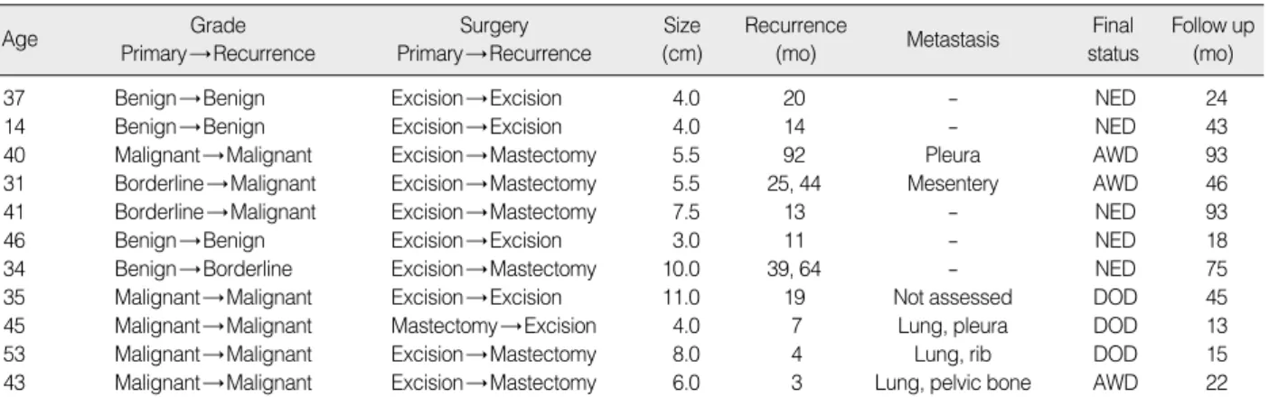
Table 4. Follow up data of patients with recurred phyllodes tumors
Age Grade
Primary→ Recurrence
Surgery Primary→ Recurrence
Size (cm)
Recurrence
(mo) Metastasis Final
status
Follow up (mo)
37 Benign→ Benign Excision→ Excision 4.0 20 - NED 24
14 Benign→ Benign Excision→ Excision 4.0 14 - NED 43
40 Malignant→ Malignant Excision→ Mastectomy 5.5 92 Pleura AWD 93
31 Borderline→ Malignant Excision→ Mastectomy 5.5 25, 44 Mesentery AWD 46
41 Borderline→ Malignant Excision→ Mastectomy 7.5 13 - NED 93
46 Benign→ Benign Excision→ Excision 3.0 11 - NED 18
34 Benign→ Borderline Excision→ Mastectomy 10.0 39, 64 - NED 75
35 Malignant→ Malignant Excision→ Excision 11.0 19 Not assessed DOD 45
45 Malignant→ Malignant Mastectomy→ Excision 4.0 7 Lung, pleura DOD 13
53 Malignant→ Malignant Excision→ Mastectomy 8.0 4 Lung, rib DOD 15
43 Malignant→ Malignant Excision→ Mastectomy 6.0 3 Lung, pelvic bone AWD 22
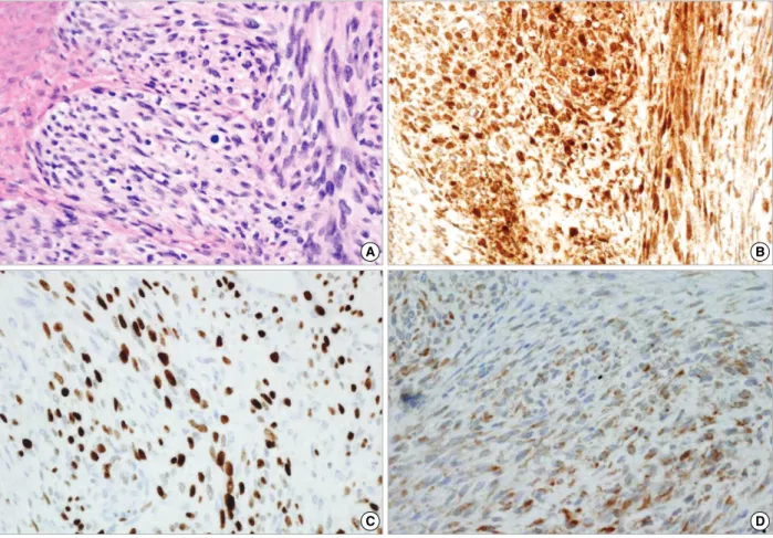
Figure 2. Borderline phyllodes tumor (A, H&E stain, ×200) with a high c-Kit positivity (B, immunohistochemical stain, ×400) recurred as a malignant phyllodes tumor (C, immunohistochemical stain, ×200) with stromal overgrowth and platelet derived growth factor receptor alpha positivity (D, immunohistochemical stain, ×400).
C D
A B
NED=no evidence of disease; AWD=alive with disease; DOD=died of disease.
suffered from metastasis to the lungs or mesentery, and three patients died of malignant PT (Table 4).
Immunohistochemistry
Table 3 shows the overall rate of positivity among benign, borderline, and malignant PTs. p53 stromal cell immunopositivity was observed in 10.3% of benign PTs, 31.3% of borderline PTs, and 50.0% of malignant PTs. The Ki-67 index increased in 5.1% of benign PTs, 25.0% of
SSCP-PCR analysis
Single-stranded conformational polymorphism-poly- merase chain reaction (SSCP-PCR) analysis for mutation in exons 9, 11, 13, and 17 of the c-Kit gene, and exons 12 and 18 of PDGFRA was carried out in 16 borderline tumors and 12 malignant tumors. No mutations of the c-Kit or PDGFRA genes were found.
Figure 3. Malignant phyllodes tumor (A, H&E immunohistochemical stain, ×400) showing c-kit positivity (B, immunohistochemical stain, ×400), a high Ki-67 index (C, immunohistochemical stain, ×400), and platelet derived growth factor receptor alpha positivity (D, immunohistochemical stain, ×400) in stromal cells.
C D
A B
Clinicopathologic correlation
There was no statistically significant association of tumor borders (infiltrative vs. pushing), surgical method (mastectomy vs. excisional biopsy), either p53 or p16 immunostaining with recurrent disease (p>0.05). How- ever, margin status (p=0.000), Ki-67 (p=0.012), c-Kit (p=0.002), and PDGFRA (p=0.007) stromal immunoposi- tivity were significantly correlated with recurrence (Table 5, Figure 4). Multivariate analytical results revealed that only PDGFRA (p=0.040) immunopositivity, among immu- nohistochemical parameters, was correlated with recur- rence (Table 6).
DISCUSSION
The majority of PTs are benign and patient risk focuses on local recurrence rather than metastatic disease. The most powerful predictor of local recurrence is the com- pleteness of excision.(1) Local recurrence occurs in both benign and malignant tumours(6) with a approximately 15-20% of cases developing recurrence,(1) the majority of whom do so within 2-3 yr after initial diagnosis.(19) In this series, 11 of 67 PT cases (16.4%) showed recur- rence between three and 92 months after initial diagnosis.
Among 11 recurrent cases, four were benign, two border- line, and five malignant.
Subsequent recurrence may show increased cellularity, significant nuclear atypia, and increased mitotic activity.(19) Recurrence may mirror the microscopic pattern of the original tumor or show dedifferentiation.(20) According Table 6. Mutivariate analysis of immunohistochemical para- meters of phyllodes tumors with or without recurrence (Binary logistic regression analysis)
Parameters Odds ratio (95% CI) p-value
p53 1.265 0.790
Ki-67 0.589 0.586
C-Kit 1.265 0.175
p16 0.937 0.936
PDGFRA 0.095 0.040
CI=confidence interval; PDGFRA=platelet derived growth factor receptor alpha.
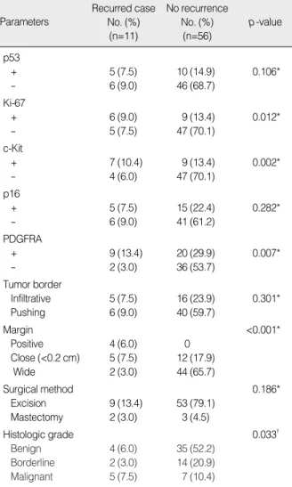
Table 5. Clinicopathologic and immunohistochemical features of phyllodes tumors with or without recurrence
Parameters
Recurred case No. (%)
(n=11)
No recurrence No. (%)
(n=56)
p-value
p53
+ 5 (7.5) 10 (14.9) 0.106*
- 6 (9.0) 46 (68.7)
Ki-67
+ 6 (9.0) 9 (13.4) 0.012*
- 5 (7.5) 47 (70.1)
c-Kit
+ 7 (10.4) 9 (13.4) 0.002*
- 4 (6.0) 47 (70.1)
p16
+ 5 (7.5) 15 (22.4) 0.282*
- 6 (9.0) 41 (61.2)
PDGFRA
+ 9 (13.4) 20 (29.9) 0.007*
- 2 (3.0) 36 (53.7)
Tumor border
Infiltrative 5 (7.5) 16 (23.9) 0.301*
Pushing 6 (9.0) 40 (59.7)
Margin <0.001*
Positive 4 (6.0) 0
Close (<0.2 cm) 5 (7.5) 12 (17.9)
Wide 2 (3.0) 44 (65.7)
Surgical method 0.186*
Excision 9 (13.4) 53 (79.1)
Mastectomy 2 (3.0) 3 (4.5)
Histologic grade 0.033�
Benign 4 (6.0) 35 (52.2)
Borderline 2 (3.0) 14 (20.9)
Malignant 5 (7.5) 7 (10.4)
PDGFRA=platelet derived growth factor receptor alpha.
*Fisher exact test; �chi-square test.
80 70 60 50 40 30 20 10 0
p53 Ki-67* c-Kit* p16 PDGFRA*
Figure 4. Immunohistochemical staining results for the recurrence of phyllodes tumors and phyllodes tumors with no recurrence (*p <0.05).
PDGFRA=platelet derived growth factor receptor alpha.
45.5
Recurred case (%)
17.9 54.5
16.1 63.6
16.1 45.5
26.8 81.8
35.7 No recurrence (%)
According to Kleer et al.,(21) three of seven (42.9%) benign PTs recurred locally, two of which had close mar- gins (within 0.1 cm of the margin), and one had positive margins (tumor at the inked margin). Two of these three tumors recurred as histologically benign PT and one as high-grade malignant PT.(21) Among the four benign PTs which recurred in our study, two cases showed a positive margin and one case had less than 0.2 cm clear surgical margin. Further, two recurred borderline and 4 malignant tumors had positive or close surgical margins at original resection. The importance of adequate margins in the treatment of benign and malignant PTs should be emphasized. A one centimeter margin of surrounding benign tissue is recommended, even though this is not routine in all centers.(1,21)
The reported rate of metastasis and death due to malig- nant PTs has varied from 0% to 40%. The likelihood of metastatic disease has been, to some extent, related to stromal overgrowth and cellularity, as well as cytologic atypia and mitotic activity and, in some series, to the presence of necrosis.(1) In another report, recurrence was observed in 6 of 23 malignant PT cases (26.1%) and the 5-yr disease free survival rate was 48.9%. Frequent mitosis (>10/10HPF) and an invasive margin were the principal determinants of recurrence.(22) In this series, four patients (6.0%) suffered from metastasis to the lungs or mesentery and three (4.5%) died of metastatic malig- nant PTs.
A variety of immunohistochemical markers, including MIB1 monoclonal antibody to cell proliferation-associated Ki-67 antigen, p53, VEGF, CD10, c-Kit, and PDGFRA have been used in an attempt to predict behavior and more accurately classify PTs.(1,7,8,10,16,23) Both Ki-67 and p53 expression correlate with the histologic classifi-
Expression of the CD117 protein within stromal cells has been significantly associated with a myriad of mor- phologic parameters, including grade and recurrent dis- ease.(10) Further, the stromal expression of c-Kit has been reported to be increased in malignant PTs compared to benign PTs.(7,8,16) However, Tse et al.(9) reported that c-Kit was not related to recurrence or metastasis.
Additionally, in a study by Esposito et al.,(11) c-Kit was differentially expressed among tumor grades, but the difference in its expression between borderline and malig- nant PTs was not significant, and c-Kit expression did not significantly correlate with tumor recurrence. Thus, the significance of c-Kit expression in PTs is uncertain.
According to Tan et al.,(10) 13% of PT patients recurred during 30.3 mo of the mean follow-up period. CD117 stromal immunopositivity was correlated with recurrence (p=0.001); however, there was no association with p53 immunostaining. Activating mutations of the c-Kit gene similar to those described for GISTs appear to be rare or absent in malignant PTs.(7,8,16)
Carvalho et al.(16) reported c-Kit expression in 12 of 19 cases (6 of 13 benign cases and all 6 malignant ones) and PDGFFRA expression in 2 of 19 cases (one of 6 malig- nant PT cases and one of 13 benign cases). In our study, PDGFRA immunopositivity was higher than these reported results (33.3% of benign PTs, 43.8% of borderline PTs, and 83.3% of malignant PTs). In this study, >10% immu- noreactivity was considered to be positive. However, in the Carvalho et al.(16) study, they interpreted >25% c-Kit and PDGFRA immunoreactivity to be positive and found that the 2415 c>T alteration in exon 17 of the c-Kit gene was present in two benign PTs. Further, analysis of exons 12 and 18 of the PDGFRA gene showed an intronic inser- tion, IVS17-50T, and an exonic silent alteration, 2866G>
T, in exon 18. However, these alterations have also been found in the normal population, suggesting that they could be population polymorphisms.(16) In this series, no mutations of the c-Kit or PDGFRA genes were found.
Recently, Paulsson et al.(28) reported that high stromal PDGF beta-receptor expression in breast cancer was significantly associated with high histopathological grade, estrogen receptor negativity, and high HER2 expression.
These findings highlight the prognostic significance of stromal markers and should be considered in the ongoing clinical development of PDGF receptor inhibitors.
CONCLUSION
Even though positive or close margins have been signi- ficantly associated with tumor recurrence, we have shown that stromal c-Kit and PDGFRA positivity, and the Ki- 67 index are useful for predicting the recurrence of PTs.
Further, no mutations of c-Kit or PDGFRA were found in the current study. The underlying molecular mechanism of PT progression remains poorly understood and there- fore, further molecular studies are warranted.
ACKNOWLEDGMENTS
The authors would like to thank Sun-Young Na for her excellent technical assistance.
REFERENCES
1. O’Malley F, Pinder SE. Breast Pathology. Edinburgh: Churchill Livingstone Elsevier; 2006. p.118-24.
2. Rowell MD, Perry RR, Hsiu JG, Barranco SC. Phyllodes tumors.
Am J Surg 1993;165:376-9.
3. Jacklin RK, Ridgway PF, Ziprin P, Healy V, Hadjiminas D, Darzi A. Optimising preoperative diagnosis in phyllodes tumour of the breast. J Clin Pathol 2006;59:454-9.
4. Khan SA, Badve S. Phyllodes tumors of the breast. Curr Treat Options Oncol 2001;2:139-47.
5. Inoshita S. Phyllodes tumor (cystosarcoma phyllodes) of the breast. A clinicopathologic study of 45 cases. Acta Pathol Jpn 1988;38:21-33.
6. Bellocq JP, Margo G. Fibroepithelial tumours. In: Tavassoli FA, Devilee P, editors. World Health Organization Classification of Tumours. Pathology and Genetics of Tumours of the Breast and Female Genital Organs. Lyon: IARC Press; 2003. p.99-103.
7. Chen CM, Chen CJ, Chang CL, Shyu JS, Hsieh HF, Harn HJ. CD34, CD117, and actin expression in phyllodes tumor of the breast. J Surg Res 2000;94:84-91.
8. Sawyer EJ, Poulsom R, Hunt FT, Jeffery R, Elia G, Ellis IO, et al.
Malignant phyllodes tumours show stromal overexpression of c-myc and c-kit. J Pathol 2003;200:59-64.
9. Tse GM, Putti TC, Lui PC, Lo AW, Scolyer RA, Law BK, et al.
Increased c-kit (CD117) expression in malignant mammary phyllodes tumors. Mod Pathol 2004;17:827-31.
10. Tan PH, Jayabaskar T, Yip G, Tan Y, Hilmy M, Selvarajan S, et al.
p53 and c-kit (CD117) protein expression as prognostic indicators in breast phyllodes tumors: a tissue microarray study. Mod Pathol 2005;
18:1527-34.
11. Esposito NN, Mohan D, Brufsky A, Lin Y, Kapali M, Dabbs DJ.
Phyllodes tumor: a clinicopathologic and immunohistochemical study of 30 cases. Arch Pathol Lab Med 2006;130:1516-21.
12. Carvalho I, Milanezi F, Martins A, Reis RM, Schmitt F. Overexp- ression of platelet-derived growth factor receptor alpha in breast cancer is associated with tumour progression. Breast Cancer Res 2005;7:R788-95.
13. Sarlomo-Rikala M, Kovatich AJ, Barusevicius A, Miettinen M.
CD117: a sensitive marker for gastrointestinal stromal tumors that is more specific than CD34. Mod Pathol 1998;11:728-34.
14. Miettinen M, Monihan JM, Sarlomo-Rikala M, Kovatich AJ, Carr NJ, Emory TS, et al. Gastrointestinal stromal tumors/smooth muscle tumors (GISTs) primary in the omentum and mesentery: clinico- pathologic and immunohistochemical study of 26 cases. Am J Surg Pathol 1999;23:1109-18.
15. Miettinen M, Sobin LH, Sarlomo-Rikala M. Immunohistochemical spectrum of GISTs at different sites and their differential diagnosis with a reference to CD117 (KIT). Mod Pathol 2000;13:1134-42.
16. Carvalho S, Silva AO, Milanezi F, Ricardo S, Leitão D, Amendoeira I, et al. c-KIT and PDGFRA in breast phyllodes tumours: overex- pression without mutations? J Clin Pathol 2004;57:1075-9.
17. Wardelmann E, Neidt I, Bierhoff E, Speidel N, Manegold C, Fischer HP, et al. c-kit mutations in gastrointestinal stromal tumors occur preferentially in the spindle rather than in the epithelioid cell variant.
Mod Pathol 2002;15:125-36.
18. Yamamoto H, Oda Y, Kawaguchi K, Nakamura N, Takahira T, Tamiya S, et al. c-kit and PDGFRA mutations in extragastrointestinal stromal tumor (gastrointestinal stromal tumor of the soft tissue). Am J Surg Pahtol 2004;28:479-88 .
19. Moinfar F. Essentials of Diagnostic Breast Pathology: A Practical Approach. Berlin: Springer-Verlag; 2007. p.321-3.
20. Grimes MM. Cystosarcoma phyllodes of the breast: histologic features, flow cytometric analysis, and clinical correlations. Mod Pathol 1992;
5:232-9.
21. Kleer CG, Giordano TJ, Braun T. Pathologic, immunohistochemical, and molecular features of benign and malignant phyllodes tumors of the breast. Mod Pathol 2001;14:185-90.
22. Lee HS, Kim HA, Shin DS, Kim YH, Chung SY, Jin MS, et al. Risk factors for recurrence after surgical treatment of a malignant phyl- lodes tumor of the breast. J Breast Cancer 2007;10:248-53.