대한요로생식기감염학회지: 제 6 권 제 2 호 2011년 10월
□ 증 례 보 고 □ Korean J UTII Vol. 6, No. 2, October 2011
206
• 교신저자:김준모, 순천향대학교 의과대학 비뇨기과학교실 경기도 부천시 원미구 중동 1174
우402-767 Tel: 032-621-5464, Fax: 032-621-5016
E-mail: [email protected] Received: August 28, 2011
Revised: September 9, 2011 Accepted: September 12, 2011
폐쇄성 혈전혈관염 (버거씨 병) 환자에서 발생한 음경 괴사
순천향대학교 의과대학 비뇨기과학교실
김준모∙이광우∙김영호∙김민의 [Abstract]Penile Necrosis in Thromboangitis Obliterans (Buerger's Disease) Jun-Mo Kim, Kwang-Woo Lee, Young-Ho Kim, Min-Eui Kim
From the Department of Urology, Bucheon Hospital, University of Soonchunhyang, Bucheon, Korea
Thromboangiitis obliterans is also known as Buerger’s disease, and it usually affects small-to-me- dium-sized arteries and veins of the upper and lower extremities. Although as an atypical course of this disease, bilateral renal artery thrombosis and affection of spermatic cord were reported, there has been no report of penile necrosis due to Buerger’s disease in Korea. We present a case of a 47-year-old man with voiding difficulty and urinary retention due to severely atrophic penis and urethra. The patient’s medical history revealed smoking as much as 50 pack/years, and heavy alcohol consumption. Although the patient had been taking anti-coagulant medication for 2 years after diagnosis of thromboangiitis obliterans, penile necrosis was advanced. The patient could self-void after diversion of the urethra into the perineum.
(Korean J UTII 2011;6:206-208)
Key Words: Thromboangiitis obliterans, Penis, Necrosis
Thromboangiitis obliterans (TAO) is also known as Buerger’s disease.
1This disease has idiopathic, non-atherosclerotic, segmental, vaso-occlusive due to thrombosis, and recurrent inflammatory characteristics.
2It most commonly affects small- and medium-sized
arteries and veins of the extremities, and unusual sys-
temic manifestations include cerebral, mesenteric and
coronary arterial involvement.
3,4Although the preva-
lence of TAO in Korea and Japan was reported as
much as 16-66% of cases of a peripheral arterial dis-
ease,
5this does not reflect the exact prevalence be-
cause the survey was conducted in limited areas from
foreign doctors.
6TAO rarely affects vessels of the
urologic system. This is the first report of urinary re-
김준모: Penile Necrosis in Thromboangitis Obliterans (Buerger's Disease) 207
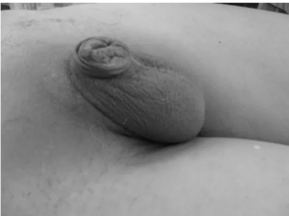
Fig. 1. Penile shaft was severely atrophic and adherent with prepuce, and urethral opening was not identified.
Fig. 2. MRI showed the atrophic change of distal ure- thra and penis (arrow) and the increased bladder wall thickness and trabeculation (arrow head).
tention due to penile necrosis by TAO in Korea. We report our case with a review of the literature.
CASE REPORT
A 47-year-old man presented with frequency and retraction of penis for 2 years. The patient had expe- rienced claudication and coldness on one foot for the previous 3 years and had been diagnosed with Buerger’s disease 2 years earlier at our institution’s Department of Thoracic and Cardiovascular Surgery.
The patient had a history of smoking 2 packs of cig- arettes everyday with a total of 50 packs/year and was a heavy alcohol drinker. Computed tomography angiography performed 2 years previous showed a small amount of deep vein thrombosis from the right popliteal vein to proximal posterior tibial vein.
Although the patient had since been medicated with anticoagulants, wound irrigation and debridement for cellulitis and necrosis of the right third toe had been necessary 1 year earlier. The patient visited our de- partment for complaints of frequency, weak urinary stream and dribbling, urinary incontinence and re- traction of penis which had started 1-2 years earlier and had recently become aggravated. His penile shaft was entirely atrophic, and a lump of penis with at- tached prepuce and urethral opening was not found in
physical examination (Fig. 1). Initial uroflowmetry showed urinary dribbling pattern and large post-void urine, and temporary suprapubic cystostomy was per- formed due to acute urinary retention of more than 700 ml of urine and overflow incontinence 2 days af- ter admission. Pelvic magnetic resonance imaging showed that the length of the penile shaft above ante- rior pelvic wall was <1 cm and displayed severe nar- rowing of distal urethra, and increased thickness of bladder wall and prominent trabeculation (Fig. 2). As definitive treatment, the excision of 2 cm of the tip of atrophic corpus cavernosum and spogiosum, and diversion of new urethral opening to perineum was performed. The patient could self-void well after ure- thral catheter was removed on post-operative day 7.
DISCUSSION
TAO is characterized by segmental inflammatory
thrombotic occlusions of the small and middle-sized
arteries and veins of the upper or lower extremities.
3Although the etiology of TAO has not been eluci-
dated, smoking is a known determinant of its course.
208 대한요로생식기감염학회지: 제6권 제2호 2011년 10월
The prevalence of TAO is widely different according to race and regions, being low in Western Europe (0.5-5.6% of patients with a peripheral arterial dis- ease), and high in Korea and Japan (16-66%).
3,4The most distinctive features of this disease from other forms of vasculitis such as polyarteritis nodosa is preservation of the internal elastic lamina.
7Despite the lack of a unified accepted diagnostic criteria of TAO, Shionoya’s criteria is commonly used: (1) smoking history; (2) onset of symptoms before the age of 50 years; (3) infrapopliteal arterial occlusion;
(4) either arm involvement or phlebitis migrans; and (5) absence of atherosclerotic risk factors other than smoking.5 All five criteria were present in this patient. Although the patient had taken anti-platelet drugs, effectiveness was limited. The patient had been re-admitted to department of vascular surgery after 14 months for cellulitis and necrosis of right third toe, and wound irrigation and debridement was performed.
The patient visited department of urology after 1 year for weak urinary stream and incontinence. The pa- tient’s penile shaft was entirely atrophied and urethral opening was obstructed due to severe attachment with prepuce. Because the voiding difficulty was ag- gravated and acute urinary retention was occurred, temporary suprapubic cystostomy was performed.
After anastomosis between healthy corpus spongiosum into perineum, the patient could self-void well. There has been several case-reports on the penile and scrotal necrosis in Buerger’s disease,
8,9and partial penectomy and scrotal debridement is the usual preferred treatment. However, because the patient visited our hospital after necrosis has advanced across the entire
penile shaft, only diversion into the perineum was considered as a treatment modality. The prognosis af- ter surgical management is generally very good.
REFERENCES