ISSN 2234-3806 • eISSN 2234-3814
https://doi.org/10.3343/alm.2021.41.3.259
Immune Checkpoint Programmed Cell Death Protein-1 (PD-1) Expression on Bone Marrow T Cell Subsets in Patients With Plasma Cell Myeloma
Min Young Lee , M.D., Ph.D.1, Chan-Jeoung Park , M.D., Ph.D.2,*, Young-Uk Cho , M.D., Ph.D.2,*,
Eunkyoung You , M.D., Ph.D.3, Seongsoo Jang , M.D., Ph.D.2, Eul Ju Seo , M.D., Ph.D.2, Jung-Hee Lee , M.D., Ph.D.4, Dok Hyun Yoon , M.D., Ph.D.4, and Cheolwon Suh , M.D., Ph.D.4
1Department of Laboratory Medicine, Kyung Hee University School of Medicine and Kyung Hee University Hospital, Gangdong, Seoul, Korea; 2Department of Laboratory Medicine, Asan Medical Center, University of Ulsan College of Medicine, Seoul, Korea; 3Department of Laboratory Medicine, Inje University College of Medicine, Busan Baik Hospital, Busan, Korea; 4Department of Oncology, Asan Medical Center, University of Ulsan College of Medicine, Seoul, Korea
Background: Plasma cell myeloma (PCM) is caused by immune dysregulation. We evalu- ated the expression of immune checkpoint programmed cell death protein-1 (PD-1) on T cell subsets in PCM patients according to disease course and cytogenetic abnormalities.
This study aimed to find a target group suitable for therapeutic use of PD-1 blockade in PCM.
Methods: A total of 188 bone marrow (BM) samples from 166 PCM patients and 32 con- trols were prospectively collected between May 2016 and May 2017. PD-1 expression on BM T cell subsets was measured using flow cytometry.
Results: At diagnosis, the median PD-1 expression on CD4+ T cells was 24.6%, which did not significantly differ from that in controls. After stem cell transplantation, PD-1 expres- sion on CD4+ T cells was higher than that at diagnosis (P <0.001), regardless of residual disease. PD-1 expression on CD4+ T cells in patients with residual disease after chemo- therapy was significantly higher than that at diagnosis (P =0.001) and after complete re- mission following chemotherapy (P =0.044). PD-1 expression on CD8+ T cells was higher in PCM patients with cytogenetic abnormalities, including monosomy 13, 1q gain, com- plex karyotype, and hypodiploidy.
Conclusions: PD-1 blockade might have therapeutic potential in refractory PCM patients after chemotherapy, especially in those with high- or intermediate-risk cytogenetic abnor- malities.
Key Words: Plasma cell myeloma, Immune checkpoint programmed cell death protein-1 (PD-1), Flow cytometry, T cell subset
Received: April 28, 2020 Revision received: June 22, 2020 Accepted: November 18, 2020 Corresponding author:
Chan-Jeoung Park, M.D., Ph.D.
Department of Laboratory Medicine, University of Ulsan College of Medicine and Asan Medical Center, 88 Olympic-ro 43-gil, Songpa-gu, Seoul 05505, Korea
Tel: +82-2-3010-4508 Fax: +82-2-478-0884 E-mail: cjpark@amc.seoul.kr Co-corresponding author:
Young-Uk Cho, M.D., Ph.D.
Department of Laboratory Medicine, University of Ulsan College of Medicine and Asan Medical Center, 88 Olympic-ro 43-gil, Songpa-gu, Seoul 05505, Korea
Tel: +82-2-3010-4501 Fax: +82-2-478-0884 E-mail: yucho@amc.seoul.kr
* These authors contributed equally to this study.
© Korean Society for Laboratory Medicine This is an Open Access article distributed under the terms of the Creative Commons Attribution Non-Commercial License (https://creativecom- mons.org/licenses/by-nc/4.0) which permits unrestricted non-commercial use, distribution, and reproduction in any medium, provided the original work is properly cited.
INTRODUCTION
Malignant cells evade host immunity and change the tumor mi-
croenvironment milieu through activation of immune check- points. Immune checkpoint inhibitors block the activation of the immune system in the body, usually preventing the over-activa-
2017-03-16 https://crossmark-cdn.crossref.org/widget/v2.0/logos/CROSSMARK_Color_square.svg
tion of T cell responses. Plasma cell myeloma (PCM) is caused by a microenvironment promoting immune escape in the bone marrow (BM) and immune dysregulation through the loss of ef- fector T cell populations. PCM is a genetically complex disease that develops via multistep progression. The cytogenetic catego- ries of PCM are major indicators of prognosis and form the basis for disease risk stratification.
Immune checkpoint programmed cell death protein-1 (PD-1) receptors are normally expressed on T cells, B cells, monocytes, and natural killer T cells. Programmed death-ligand 1 (PD-L1)/
L2 are ligands expressed by antigen presenting cells, pancreatic islet cells, endothelial cells, and epithelial cells. PD-L1/L2 have been reported to be highly expressed on tumor cells [1]. Immune checkpoint inhibitors are effective in managing solid cancers and hematologic malignancies, and are being actively developed and used for treatment. Checkpoint inhibitors are expected to be effective in PCM treatment, and several trials involving PCM patients have been conducted; however, a clear benefit has not been observed. In a phase Ib trial, the PD-1 inhibitor nivolumab did not elicit an objective response in PCM patients [2]. The re- sults of trials examining nivolumab in combination with other agents (NCT01592370 and NCT02726581) are not yet avail- able. Notably, the United States Food and Drug Administration halted two randomized phase-III trials (KEYNOTE-185 trial and KEYNOTE-183 trial) owing to immune-related adverse events in 2017 [3, 4]. The limited effectiveness of immune checkpoint in- hibitors in PCM suggests that the immune checkpoints functions change in different disease states. This study aimed to identify patients who may benefit from treatment with immune check- point inhibitors. We analyzed PD-1 expression on CD4+/CD8+ T cells in PCM patients according to disease course, genetic ab- normalities, disease states, and involved/uninvolved free light chain ratio. To our knowledge, no study has examined PD-1 ex- pression in PCM according to the type of treatment received and genetic variation.
MATERIALS AND METHODS
Sample collection and data acquisitionWe prospectively obtained 192 BM samples from 166 PCM pa- tients at Asan Medical Center, Seoul, Korea, between May 2016 and May 2017; four samples were excluded because of lack of sample volume or patient information, and 188 samples from 162 PCM patients were included. The BM samples of PCM pa- tients included 81 samples obtained at diagnosis and 107 ob- tained at follow-up. The mean value of the follow-up period after
initial diagnosis was 31.8±36.5 months and that after stem cell transplantation (SCT) was 23.3 ±30.5 months. PCM disease stage and treatment response were determined, according to the Revised International Staging System (R-ISS) developed by the International Myeloma Working Group in 2014 [5]. We as- sessed patient characteristics and medical history by chart re- view and performed laboratory investigations, including free light chain, M-peak, FISH, and karyotyping, as routine practice for PCM work-up. Fifty-three patients with available albumin, lac- tate dehydrogenase, karyotype, FISH, and β-2-microglobulin test results at diagnosis were classified using R-ISS index crite- ria; three were at R-ISS stage I; 34, at stage II; and 15, at stage III. Follow-up patients were divided into two groups: those treated with chemotherapy (CTx; N =51) and those receiving autologous SCT (N =56) (Table 1). Cytogenetic abnormalities were categorized as standard-, intermediate- and high-risk us- ing the Mayo stratification for myeloma and risk-adapted ther- apy classification (mSMART) [6].
Control BM samples were collected from 32 patients who had undergone a BM study owing to cytopenia or fever of unknown origin, had no hematologic malignancy of the BM, and were re- ported as having normo/hypo/hypercellular marrow.
This study was approved by the Institutional Review Board of Asan Medical Center (Approval Number: 20161087); the sub- jects’ informed consent to participate in research was waived.
Written informed consent was obtained from all patients for the BM biopsy procedures and genetic testing, and the study was performed in accordance with the Declaration of Helsinki.
Flow cytometric measurement of PD-1 expression on T cell subsets
BM aspirates were collected in EDTA tubes (BD Vacutainer;
Becton Dickinson [BD] Franklin Lakes, NJ, USA). We incubated 100 μL of phosphate-buffered saline (PBS)-diluted BM aspirate (nucleated cell number 10,000–20,000/μL) with fluorescent dye-conjugated antibodies (4–8 μL each of phycoerythrin [PE]- conjugated anti-PD-1 [clone EH12.1, BD Pharmingen PE Mouse Anti-Human CD279], allophycocyanin [APC]-cyanine 7 [Cy7]- conjugated anti-CD3 [clone SK7, BD Pharmingen APC-Cy7 Mouse Anti-Human CD3], APC-conjugated anti-CD4 [clone RPA-T4, BD Pharmingen APC Mouse Anti-Human CD4], fluorescein iso- thiocyanate [FITC]-conjugated anti-CD8 [clone HIT8a, BD Phar- mingen FITC Mouse Anti-Human CD8], and peridinin chloro- phyll protein complex [PerCP]-conjugated anti-CD45 [clone 2D1, BD Pharmingen PerCP Mouse anti-human CD45] antibodies purchased from BD Pharmingen [San Diego, CA, USA]) for 20
min in the dark, at room temperature (20–22°C). After washing with PBS, the samples were lysed with ammonium chloride-based lysing solution for 10 minutes and washed and suspended in PBS. A minimum of 100,000 stained nucleated cells per tube were acquired using a flow cytometer (FACSCanto II; BD Biosci- ences, Sunnyvale, CA, USA) and analyzed using the BD FACS- Diva Software (BD Biosciences). PD-1 expression on CD4+ and
CD8+ T cells was measured as follows: lymphocytes were gated with bright CD45 expression and low side scatter (SSC) on CD45 vs. SSC cytogram, CD3+ CD8+ T cells were regated on a CD3 vs.
CD8 cytogram, and positive PD-1 expression was measured on a CD8 vs. PD-1 cytogram. Positive PD-1 expression on CD3+ CD4+ T cells was measured following the same method, using CD4 instead of CD8. The cut-off values for positivity were as- Table 1. Patient characteristics
Diagnosis Follow-up (N=107) Patient
Total
CTx-CR CTx-RD SCT-CR SCT-RD
Samples, N 81 30 21 29 27 188
Male/female 43/38 16/14 11/10 17/13 18/9 106/86
Age, yr, median (range) 64 (37–86) 62 (38–75) 58 (42–73) 58 (26–66) 61 (47–69) 61 (26–86)
Type of M protein
IgA 16 6 3 6 8 39
IgG 39 14 6 11 13 84
IgD 3 0 4 1 2 10
IgM 0 0 0 1 0
Light chain 20 9 7 8 4 48
Nonsecretory 3 1 1 2 0 7
Plasma cell in BM, %, mean 41.6 1.6 27.4 0.8 29.1 25.5
Treatment
VTD 20 5
TD 3 7
VMP 6 6
HD-Dexa 1 1
TCD 0 1
Palmidronate 0 1
R-ISS
I 3
II 34
III 15
Cytogenetic abnormalities
Monosomy 13 24 5 11 40
1q gain 15 5 9 29
Complex 37 8 14 59
Hypodiploidy 12 3 5 20
Hyperdiploidy 18 3 8 29
t(11;14) 11 4 2 17
del(17p) 2 2 4
Abbreviations: CTx, chemotherapy; CR, complete remission; RD, residual disease; SCT, stem cell transplantation; VTD, vincristine/thalidomide/dexametha- sone; TD, thalidomide/dexamethasone; VMP, vincristine/melphalan/prednisolone; HD-Dexa, high-dose dexamethasone; TCD, thalidomide/cycolophospha- mide/dexamethasone; R-ISS, revised international staging system; CTx-CR, with complete remission after chemotherapy; CTx-RD, with residual disease after chemotherapy; SCT-CR, with complete remission after stem cell transplantation; SCT-RD, with residual disease after stem cell transplantation; BM, bone marrow; t, translocation; del, deletion.
signed according to the isotypic controls, and expression results were presented as the proportion of positive cells (%).
Statistical analysis
Statistical analyses were performed using SPSS version 20 (IBM Corp., Armonk, NY, USA) and Excel 2016 (Microsoft, Redmond, WA, USA). Data were analyzed for normal distribution using the Kolmogorov-Smirnov test and presented as median with range.
Statistical comparisons were performed using the Kruskal-Wallis test and Mann-Whitney test. Spearman’s rank correlation coeffi- cient was used to evaluate the association between PD-1 expres- sion percentages and the involved/uninvolved free light chain ratio and that between PD-1 expression percentages and the follow-up period after SCT. P <0.05 was considered statistically significant.
RESULTS
PD-1 expression on CD4+ T cells
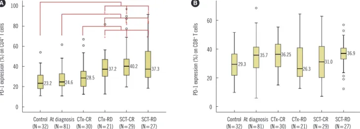
The median expression of PD-1 on CD4+ T cells at PCM diagno- sis did not significantly differ from that in the controls (24.6%
vs. 23.2%). PD-1 expression on CD4+ T cells in patients with re- sidual disease (RD) after CTx was significantly higher than that in patients at diagnosis (P <0.001), patients in complete remis- sion (CR) after CTx (P =0.025), and controls (P =0.001).
PD-1 expression on CD4+ T cells in patients after SCT was
higher than that in patients at diagnosis (P <0.001), patients in CR after CTx (SCT-CR, P =0.006; SCT-RD, P =0.012), and con- trols (P <0.001) (Fig. 1A). Patients who had undergone SCT were divided into early (N=16) and late (N=40) phases based on a cut-off of three months after SCT; no difference in PD-1 expres- sion was observed between the two groups (43.1% vs. 35.3%, P =0.106), and there was no significant correlation between the follow-up period after SCT and PD-1 expression.
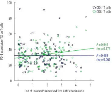
No correlation was observed between PD-1 expression on CD4+ T cells and the log value of involved/uninvolved free light chain ratio (Fig. 2). Moreover, there was no statistical difference in the PD-1 expression on CD4+ T cells according to R-ISS stages (median values of stage II and III were 23.4% and 24.4%, re- spectively).
PD-1 expression on CD8+ T cells
The median expression of PD-1 on CD8+ T cells at PCM diagno- sis did not differ from that in the controls (35.7% vs. 29.3%).
No significant differences in PD-1 expression on CD8+ T cells were observed between clinical states of PCM; however, the SCT-RD patients had a slightly higher median PD-1 expression than the controls (Fig. 1B). There was no difference in the PD-1 expression between the early (40.1%) and late (33.4%) phases after SCT and no correlation between the follow-up period after SCT and PD-1 expression.
The PD-1 expression on CD8+ T cells showed weak positive
Fig. 1. Percentages of PD-1 expression on (A) CD4+ T cells and (B) CD8+ T cells in the controls and PCM patients with various disease states. In each box plot, the median value is reported beside the box. The upper end, lower end, and inner line of the boxes correspond to the 3rd quartile, 1st quartile, and median value, respectively. Error bars denote minimum and maximum values, and circles indicate outlier values. Statistically significant differences are marked with *if P <0.05, with †if P <0.01, and with ‡if P <0.001.
Abbreviations: CTx-CR, complete remission after chemotherapy; CTx-RD, residual disease after chemotherapy; PD-1, immune checkpoint programmed cell death protein-1; PCM, Plasma cell myeloma; SCT-CR, complete remission after stem cell transplantation; SCT-RD, residual disease after stem cell transplan- tation.
100
80
60
40
20
0
Control
(N=32)At diagnosis (N=81) CTx-CR
(N=30) CTx-RD (N=21) SCT-CR
(N=29) SCT-RD (N=27) PD-1 expression (%) on CD4+ T cells
A
60
40
20
0
Control
(N=32)At diagnosis (N=81) CTx-CR
(N=30) CTx-RD (N=21) SCT-CR
(N=29) SCT-RD (N=27) PD-1 expression (%) on CD8+ T cells
B
†
†
‡
‡ ‡
‡
‡
*
23.2 24.6 28.5
37.2 40.2 37.3 29.3
35.7 36.25 26.3
31.0 36.9
correlation with the log value of the involved/uninvolved free light chain ratio (P =0.046, rho=0.176; Fig. 2). However, there was no significant difference in PD-1 expression on CD8+ T cells ac- cording to R-ISS stages (median values of stage II and III, 36.8%
and 32.5%, respectively).
PD-1 expression on T cell subsets according to cytogenetic abnormalities
PCM patients with monosomy 13 (37.7%), 1q gain (37.7%), complex karyotypes (37.5%), and hypodiploidy (39.6%) had higher PD-1 expression on CD8+ T cells than did those with nor- mal karyotypes (32.7%; P =0.020, P =0.011, P =0.041, and P =0.032, respectively). Patients with hyperdiploidy and those with t(11;14) showed no significant difference in the PD-1 ex- pression on CD8+ T cells. PD-1 expression on CD4+ T cells did not differ according to cytogenetic abnormalities (Table 2). Few patients had cytogenetic indicators of high-risk myeloma, such as del(17p) (N=4), t(14;16) (N=0), and t(14;20) (N=0), and significant differences in PD-1 expression among these patients could not be evaluated.
Serial changes in PD-1 expression on T-cell subsets
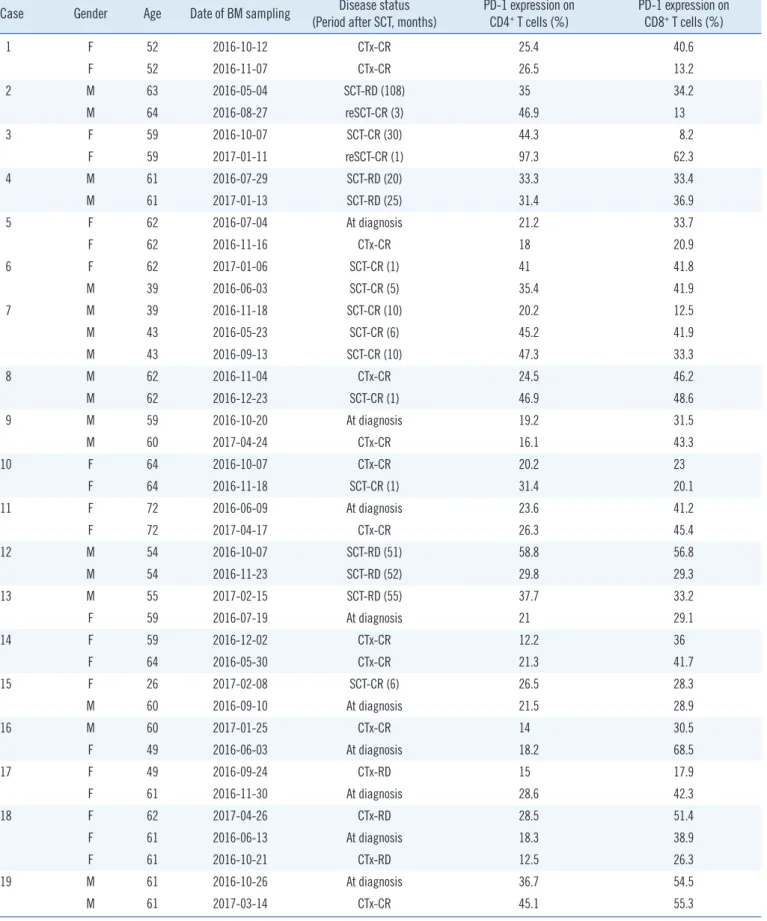
Among the 23 patients with serial data, most showed disease status-dependent changes in PD-1 expression; the results for
eight cases seemed discordant with the expected change in PD-1 expression. Three cases were classified as RD but had decreased PD-1 expression on CD4+ T cells, and five were clas- sified as CR but had increased PD-1 expression on CD4+ T cells (Table 3). In all discordant RD cases, RD was determined based on a follow-up BM examination within five months of diagnosis.
As patients usually require 2–3 cycles of treatment (six weeks per cycle) until CR, they can be considered as transitioning to CR. In four of the five CR cases with increased PD-1 expression on the CD4+ T cells, the M protein reappeared (immunoglobulin isotype switches in two cases) on the protein electrophoresis at the next follow-up. In one case, the reason for the discordance between obtained and expected results could not be determined owing to a lack of follow-up.
DISCUSSION
PCM cases in Korea has rapidly increased, from 5,566 in 2014 to 8,412 in 2019 [7]. Several therapies for PCM have emerged over the past two decades, first CTx, then autologous SCT, and, more recently, the use of proteasome inhibitors and immuno- modulatory drugs; however, many patients still die from disease relapse [8,9].
A higher PD-1 expression on CD4+ T cells was observed to be Table 2. Differences in PD-1 expression between patients with spe- cific cytogenetic abnormalities and those with normal karyotypes in non-CR groups (groups at time of diagnosis, after chemotherapy with RD, and after autologous SCT-RD)
Cytogenetic abnormalities PD-1 expression Difference in PD-1 expression Monosomy 13 (N=40) vs.
NL* (N=59) CD4+ T cells Not significant (P =0.884) CD8+ T cells High (P =0.020) 1q gain (N=29) vs.
NL (N=59) CD4+ T cells Not significant (P =0.214) CD8+ T cells High (P =0.011) Complex (N=59) vs.
NL (N=59) CD4+ T cells Not significant (P =0.657) CD8+ T cells High (P =0.041) Hypodiploidy (N=20) vs.
NL (N=59) CD4+ T cells Not significant (P =0.727) CD8+ T cells High (P =0.032) Hyperdiploidy (N=29) vs.
NL (N=59) CD4+ T cells Not significant (P =0.149) CD8+ T cells Not significant (P =0.162) t(11;14) (N=17) vs.
NL (N=59) CD4+ T cells Not significant (P =0.852) CD8+ T cells Not significant (P =0.251)
*NL is normal karyotype including loss of Y chromosome; loss of Y chromo- some is considered to be related to aging.
Abbreviations: PD-1, immune checkpoint programmed cell death protein-1;
CR, complete remission; RD, residual disease; SCT-RD, with residual dis- ease after stem cell transplantation; t. translocation.
Fig. 2. Correlation between PD-1 expression on T cell subsets and involved/uninvolved free light chain ratio in PCM. The PD-1 expres- sion on CD8+ T cells was weakly correlated with involved/uninvolved free light chain ratio; however, the PD-1 expression on CD4+ T cells was not correlated with involved/uninvolved free light chain ratio.
Abbreviations: PCM, plasma cell myeloma; PD-1, immune checkpoint pro- grammed cell death protein-1.
100
80
60
40
20
0 0 1 2 3 4 5
Log of involved/uninvolved free light chanin ratio
PD-1 expression (%) on T cells
CD4+ T cells CD8+ T cells
P =0.046 rho=0.176 P =0.493 rho=0.061
Table 3. Changes in PD-1 expression on T-cells determined by flow cytometry with serial gating in 23 patients Case Gender Age Date of BM sampling Disease status
(Period after SCT, months) PD-1 expression on
CD4+ T cells (%) PD-1 expression on CD8+ T cells (%)
1 F 52 2016-10-12 CTx-CR 25.4 40.6
F 52 2016-11-07 CTx-CR 26.5 13.2
2 M 63 2016-05-04 SCT-RD (108) 35 34.2
M 64 2016-08-27 reSCT-CR (3) 46.9 13
3 F 59 2016-10-07 SCT-CR (30) 44.3 8.2
F 59 2017-01-11 reSCT-CR (1) 97.3 62.3
4 M 61 2016-07-29 SCT-RD (20) 33.3 33.4
M 61 2017-01-13 SCT-RD (25) 31.4 36.9
5 F 62 2016-07-04 At diagnosis 21.2 33.7
F 62 2016-11-16 CTx-CR 18 20.9
6 F 62 2017-01-06 SCT-CR (1) 41 41.8
M 39 2016-06-03 SCT-CR (5) 35.4 41.9
7 M 39 2016-11-18 SCT-CR (10) 20.2 12.5
M 43 2016-05-23 SCT-CR (6) 45.2 41.9
M 43 2016-09-13 SCT-CR (10) 47.3 33.3
8 M 62 2016-11-04 CTx-CR 24.5 46.2
M 62 2016-12-23 SCT-CR (1) 46.9 48.6
9 M 59 2016-10-20 At diagnosis 19.2 31.5
M 60 2017-04-24 CTx-CR 16.1 43.3
10 F 64 2016-10-07 CTx-CR 20.2 23
F 64 2016-11-18 SCT-CR (1) 31.4 20.1
11 F 72 2016-06-09 At diagnosis 23.6 41.2
F 72 2017-04-17 CTx-CR 26.3 45.4
12 M 54 2016-10-07 SCT-RD (51) 58.8 56.8
M 54 2016-11-23 SCT-RD (52) 29.8 29.3
13 M 55 2017-02-15 SCT-RD (55) 37.7 33.2
F 59 2016-07-19 At diagnosis 21 29.1
14 F 59 2016-12-02 CTx-CR 12.2 36
F 64 2016-05-30 CTx-CR 21.3 41.7
15 F 26 2017-02-08 SCT-CR (6) 26.5 28.3
M 60 2016-09-10 At diagnosis 21.5 28.9
16 M 60 2017-01-25 CTx-CR 14 30.5
F 49 2016-06-03 At diagnosis 18.2 68.5
17 F 49 2016-09-24 CTx-RD 15 17.9
F 61 2016-11-30 At diagnosis 28.6 42.3
18 F 62 2017-04-26 CTx-RD 28.5 51.4
F 61 2016-06-13 At diagnosis 18.3 38.9
F 61 2016-10-21 CTx-RD 12.5 26.3
19 M 61 2016-10-26 At diagnosis 36.7 54.5
M 61 2017-03-14 CTx-CR 45.1 55.3
(Continued to the next page)
Case Gender Age Date of BM sampling Disease status
(Period after SCT, months) PD-1 expression on
CD4+ T cells (%) PD-1 expression on CD8+ T cells (%)
20 F 38 2016-05-09 At diagnosis 29.9 36.9
F 39 2017-04-17 CTx-CR 49.7 51.2
M 54 2016-06-01 SCT-CR (9) 31.4 37.6
21 M 55 2016-08-03 SCT-CR (11) 90 62.8
M 47 2016-05-06 SCT-RD (9) 47.1 20.7
22 M 47 2016-09-13 SCT-RD (13) 23.8 36.8
M 47 2016-11-02 SCT-CR (14) 77.6 47.9
23 F 73 2016-11-09 CTx-RD 34.8 17.9
F 74 2017-02-15 CTx-CR 67.1 36.5
*Classified as RD, but PD-1 expression on CD4+ T cells was decreased; †Classified as CR, but PD-1 expression on CD4+ T cells was increased; ‡Reappear- ance of M protein in the next follow-up protein electrophoresis; §Immunoglobulin isotype switch; llFollow-up loss.
Abbreviations: M, male; F, female; CTx, chemotherapy; CR, complete remission; RD, residual disease; SCT, stem cell transplantation; reSCT; second stem cell transplantation; BM, bone marrow; PD-1, immune checkpoint programmed cell death protein-1; CTx-CR, complete remission after chemotherapy; SCT- RD, residual disease after stem cell transplantation; SCT-CR, complete remission after stem cell transplantation; CTx-RD, residual disease after chemotherap Table 3. Continued
related to CTx refractoriness. However, there was no difference in PD-1 expression on T cells between patients at diagnosis and controls, indicating that PD-1 is involved in disease exacerba- tion or treatment refractoriness rather than disease onset. A pre- vious study reported that PD-1 expression on T cells was increa- sed in minimal RD (MRD)-positive patients compared with that in newly diagnosed or MRD-negative patients [10]; this increased PD-1 expression in RD patients is similar to the results of our study. However, our study differs from the previous one in that PD-1 expression was significantly increased only on CD4+ T cells.
PD-1 signaling in CD4+ T cells enhances the induction of the T regulatory (Treg) cell phenotype from naïve T cells and partici- pates in Treg cell function [11-13]. Treg cells accumulate in the BM microenvironment in PCM, which is increased in relapsed patients. Moreover, increased Treg cells have been associated with vulnerability to disease progression [14]. Therefore, our study shows that increased Treg cells in refractory PCM patients is due to increased PD-1 expression on CD4+ T cells. The in- creased number and activity of Treg cells due to increased PD-1 signaling in CD4+ T cells suppress tumor-specific T cell func- tion, resulting in CTx refractoriness.
Increased PD-1 expression after SCT, regardless of the SCT type, has been frequently reported [15-18]. In our study, PD-1 expression on CD4+ T cells after SCT was higher than that at di- agnosis, as well as that in patients with CR after CTx (CTx-CR) or controls, regardless of RD status. However, there was no sig- nificant difference between PD-1 expression on CD8+ T cells before and after SCT. Simonetta, et al. [17] found that the PD-
1-positive CD4+ and CD8+ T cells proportion increased signifi- cantly during the early phase (three months) after allogeneic SCT, followed by a progressive normalization of PD-1 expression on CD8+ but not on CD4+ T cells. We analyzed PD-1 expression in early and late phases after SCT based on a cut-off point of three months after SCT; however, no statistically significant dif- ference in PD-1 expression was observed between the two phases.
This could be due to the insufficient number of samples for the early phase after SCT. As the patients with follow-up after SCT included in this study were mostly in the late phase after SCT, PD-1 expression on CD8+ T cells before and after SCT may ap- pear not to differ, unlike that on CD4+ T cells.
In a study by Merindol, et al. [18], the PD-1 expression on CD4+ and CD8+ T cells in patients who had undergone umbilical cord blood transplantation (UCBT) for hematologic disease treat- ment was higher than that in healthy controls. Although the PD- 1+ CD8+ T cell proportion in the first three months after UCBT was high in all patients, it was significantly higher in relapsed patients than in non-relapsed patients at two and six months.
Our study also showed a slight, insignificant increase in PD-1 expression on CD8+ T cells in SCT-RD patients. Therefore, pa- tient prognosis after SCT could be more related to PD-1 expres- sion on CD8+ T cells than to that on CD4+ T cells.
One of the hallmarks of high-risk disease is the interaction between myeloma cells and the BM microenvironment to gen- erate a high-risk ecosystem that facilitates both cancer cell sur- vival and immune response failure [19]. The genetic abnormali- ties of malignant plasma cell clones can influence the BM micro-
environment through various mechanisms involving soluble fac- tors, exosomes containing microRNAs, and cell-cell contact [20–
23]. For example, the musculoaponeurotic fibrosarcoma (MAF) protein in t(14;16) plasma cells upregulates integrin β7, thereby altering the adhesion of plasma cells to stromal cells and facilitat- ing their growth and survival [24]. We found that PD-1 expression on CD8+ T cells was higher in patients with cytogenetic abnormal- ities categorized as high or intermediate risk (monosomy 13, 1q gain, hypodiploidy, and complex karyotype). This indicates that high- or intermediate-risk cytogenetic abnormalities affect the T cell expression of immune checkpoints. Additionally, the genetic variation found in high-risk PCM may affect not only the plasma cells themselves but also the BM microenvironment, ultimately leading to poor prognosis or refractoriness to treatment through interactions between stromal cells and plasma cell clones.
Our study had some limitations. This was not a longitudinal study; long-term serial data are needed to determine how PD-1 expression changes according to the disease course in the same patient. Another limitation is that the control group in this study did not include healthy individuals.
In conclusion, high PD-1 expression on CD4+ T cells was as- sociated with CTx refractoriness and that on CD8+ T cells was associated with high- or intermediate-risk cytogenetic abnormali- ties. We hypothesize that PD-1 blockade might have therapeutic potential in refractory PCM patients after CTx, especially for those with high- or intermediate-risk cytogenetic abnormalities.
ACKNOWLEDGEMENTS
We thank Chan Hee Yoon, M.T. and Sang Hee Han, M.T. in the Department of Laboratory Medicine, Asan Medical Center, for technical assistance with flow cytometry.
AUTHOR CONTRIBUTIONS
Park CJ and Cho YU designed the study and Lee MY wrote the paper; Lee MY and You EK carried out data management and analysis; and Jang S, Seo EJ, Lee JH, Yoon DH, and Suh C pro- vided study materials or patients. All authors have agreed with the final version of the paper.
CONFLICTS OF INTEREST
None declared.
RESEARCH FUNDING
This work was supported by a grant (2016-635) from the Asan Institute for Life Sciences, Asan Medical Center, Seoul, Korea.
ORCID
Min Young Lee https://orcid.org/0000-0001-8435-7581 Chan-Jeoung Park https://orcid.org/0000-0003-4396-8348 Young-Uk Cho https://orcid.org/0000-0002-4403-8989 Eunkyoung You https://orcid.org/0000-0002-6836-499X Seongsoo Jang https://orcid.org/0000-0002-0045-1747 Eul Ju Seo https://orcid.org/0000-0002-8247-3746 Jung-Hee Lee https://orcid.org/0000-0002-3127-0068 Dok Hyun Yoon https://orcid.org/0000-0002-8289-3548 Cheolwon Suh https://orcid.org/0000-0002-9178-4431
REFERENCES
1. Patel SP and Kurzrock R. PD-L1 expression as a predictive biomarker in cancer immunotherapy. Mol Cancer Ther 2015;14:847-56.
2. Lesokhin AM, Ansell SM, Armand P, Scott EC, Halwani A, Gutierrez M, et al. Nivolumab in patients with relapsed or refractory hematologic ma- lignancy: preliminary results of a phase Ib study. J Clin Oncol 2016;34:
2698-704.
3. Usmani SZ, Schjesvold F, Rocafiguera AO, Karlin L, Rifkin RM, Yimer HA, et al. A phase 3 randomized study of pembrolizumab (pembro) plus lenalidomide (len) and low-dose dexamethasone (Rd) versus Rd for newly diagnosed and treatment-naive multiple myeloma (MM): Key- note-185. J Clin Oncol 2018;36:8010.
4. Mateos MV, Blacklock H, Schjesvold F, Rocafiguera AO, Simpson D, George A, et al. A phase 3 randomized study of pembrolizumab (Pem- bro) plus pomalidomide (Pom) and dexamethasone (Dex) for relapsed/
refractory multiple myeloma (RRMM): Keynote-183. J Clin Oncol 2018;
36:8021.
5. Palumbo A, Avet-Loiseau H, Oliva S, Lokhorst HM, Goldschmidt H, Ro- sino L, et al. Revised international staging system for multiple myeloma:
a report from International Myeloma Working Group. J Clin Oncol 2015;
33:2863-9.
6. Mikhael JR, Dingli D, Roy V, Reeder CB, Buadi FK, Hayman SR, et al..
Management of newly diagnosed symptomatic multiple myeloma: up- dated Mayo Stratification of Myeloma and Risk-Adapted Therapy (mSMART) consensus guidelines 2013. Mayo Clin Proc 2013;88:360-76.
7. Health Insurance Review & Assessment Service. Healthcare big data hub. http://opendata.hira.or.kr/op/opc/olap3thDsInfo.do (Updated on Jan 2020).
8. Kumar SK, Rajkumar SV, Dispenzieri A, Lacy MQ, Hayman SR, Buadi FK, et al. Improved survival in multiple myeloma and the impact of nov- el therapies. Blood 2008;111:2516-20.
9. Lee HY, Park CJ, Ahn A, Lee MY, Cho YU, Jang S, Seo EJ, Lee KH, Lee JH.Lee HY, et al. Two Rare Cases of Therapy-Related Acute Lympho- blastic Leukemia in Patients With Plasma Cell Myeloma. Ann Lab Med 2019;39:496-8. doi: 10.3343/alm.2019.39.5.496.
10. Paiva B, Azpilikueta A, Puig N, Ocio EM, Sharma R, Oyajobi BO, et al.
PD-L1/PD-1 presence in the tumor microenvironment and activity of PD-1 blockade in multiple myeloma. Leukemia 2015;29:2110-3.
11. Stathopoulou C, Gangaplara A, Mallett G, Flomerfelt FA, Liniany LP, Knight D, et al. PD-1 inhibitory receptor downregulates asparaginyl en- dopeptidase and maintains Foxp3 transcription factor stability in in- duced regulatory T cells. Immunity 2018;49:247-63.e7.
12. Amarnath S, Costanzo CM, Mariotti J, Ullman JL, Telford WG, Kapoor V, et al. Regulatory T cells and human myeloid dendritic cells promote tol- erance via programmed death ligand-1. PLoS Biol 2010;8:e1000302.
13. Amarnath S, Chen H, Foley JE, Costanzo CM, Sennesh JD, Solomon MA, et al. Host-based Th2 cell therapy for prolongation of cardiac al- lograft viability. PLoS One 2011;6:e18885.
14. Muthu Raja KR, Rihova L, Zahradova L, Klincova M, Penka M, Hajek R.
Increased T regulatory cells are associated with adverse clinical features and predict progression in multiple myeloma. PLoS One 2012;7:e47077.
15. Karnell FG, Lin D, Motley S, Duhen T, Lim N, Campbell DJ, et al. Re- constitution of immune cell populations in multiple sclerosis patients af- ter autologous stem cell transplantation. Clin Exp Immunol 2017;189:
268-78.
16. Arruda LCM, de Azevedo JTC, de Oliveira GLV, Scortegagna GT, Ro- drigues ES, Palma PVB, et al. Immunological correlates of favorable long-term clinical outcome in multiple sclerosis patients after autologous hematopoietic stem cell transplantation. Clin Immunol 2016;169:47- 57.
17. Simonetta F, Pradier A, Bosshard C, Masouridi-Levrat S, Dantin C, Koutsi
A, et al. Dynamics of expression of programmed cell death protein-1 (PD-1) on T cells after allogeneic hematopoietic stem cell transplanta- tion. Front Immunol 2019;10:1034.
18. Merindol N, Champagne MA, Duval M, Soudeyns H. CD8(+) T-cell re- constitution in recipients of umbilical cord blood transplantation and char- acteristics associated with leukemic relapse. Blood 2011;118:4480-8.
19. Pawlyn C and Morgan GJ. Evolutionary biology of high-risk multiple my- eloma. Nat Rev Cancer 2017;17:543-56.
20. Corrado C, Raimondo S, Chiesi A, Ciccia F, De Leo G, Alessandro R.
Exosomes as intercellular signaling organelles involved in health and disease: basic science and clinical applications. Int J Mol Sci 2013;14:
5338-66.
21. Roccaro AM, Sacco A, Maiso P, Azab AK, Tai YT, Reagan M, et al. BM mesenchymal stromal cell–derived exosomes facilitate multiple myelo- ma progression. J Clin Invest 2013;123:1542-55.
22. Raimondi L, De Luca A, Amodio N, Manno M, Raccosta S, Taverna S, et al. Involvement of multiple myeloma cell-derived exosomes in osteo- clast differentiation. Oncotarget 2015;6:13772-89.
23. Lemaire M, Deleu S, De Bruyne E, Van Valckenborgh E, Menu E, Vander- kerken K. The microenvironment and molecular biology of the multiple myeloma tumor. Adv Cancer Res 2011;110:19-42.
24. Neri P, Ren L, Azab AK, Brentnall M, Gratton K, Klimowicz AC, et al. In- tegrin β7-mediated regulation of multiple myeloma cell adhesion, mi- gration, and invasion. Blood 2011;117:6202-13.