103
산양유를 섭취한 성인 여성의 분변에서 분리한 Bifidobacteria와 Lactobacilli의 탄수화물 발효특성
최석호1․임영순2․함준상3․정석근3․이승배1*
1상지대학교 동물생명자원학부, 2(주)서울 F&B, 3농촌진흥청 축산과학원
Carbohydrate Fermentation Character of Bifidobacteria and Lactobacilli isolated from Feces of the Adult Women supplied with Goat Milk
Suk-Ho Choi1, Young-soon Lim2, Jun-Sang Ham3, Seok-Geun Jeong3 and Seung-Bae Lee1*
1
Division of Animal Resource and Biotechnology, Sangji University, Wonju 220-702, Korea
2
Seoul F&B Co., Ltd, Hoengseong 225-874, Korea
3
National Institute of Animal Science, RDA, Suwon 441-350, Korea
Abstract
The objective of this study was to investigate the effects of consuming goat milk on the bacterial counts (colony forming units [CFU]) in adult women and to evaluate the carbohydrate fermentation capacity of bifidobacteria and lactobacilli isolated from their feces. Adult women who consumed goat milk (treatment group) had relatively higher CFU of bifidobacteria than did the control group, and the difference was significant (p<0.05) after 8 weeks. In total, 13 strains isolated from the feces of women in the treatment group were identified using 16S rRNA sequencing as Bifidobacterium adolescentis, B. longum, B. pseudocatenulatum, B. dentium, and Lactobacillus sakei. Similarly, 12 strains isolated from the feces of women in the control group included B. adolescentis, B. longum, L. ruminis, L. sakei, and B. pseudocatenulatum. All isolated bifidobacteria and lactobacilli fermented goat milk oligosaccharide and lactulose. All 7 strains of B. adolescentis fermented fructooligo- saccharides, and 3 of the 4 B. pseudocatenulatum strains, 2 of the 3 L. sakei strains, and 1 of the 7 B. longum strains fermented fructooligosaccharides.
Keywords: goat milk, bifidobacteria, lactobacilli, isolation, oligosaccharide
* Corresponding author: Seung-Bae Lee, Division of Animal Re- sources and Biotechnology, Sangji University, Wonju 220-702, Korea. Tel: +82-33-730-0542 Fax: +82-33-730-0503, E-mail:
[email protected]
서 론
최근 질병은 의학적으로 치료되어야 한다는 과거의 사고 방식이 변하여 치료보다는 예방이 우선되어야 한다는 생각 이 지배적이며, 질병의 예방을 위해서는 식생활이 중요하다 는 것을 인식하여 기능성 물질이 있는 식품을 선호하고 있다.
산양유는 다른 유제품과 비교해서 높은 양의 올리고당을
가지고 있으며, 이 올리고당은 기능성 식품 물질로 인체의 건 강을 위해 prebiotics 작용을 가지고 있다(Park et al., 2007).
Prebiotics는 난소화성 식품성분으로 장내 미생물 수를 제한 하거나, 선택적으로 유익균의 성장과 활성을 주어서 숙주에 유익한 영향을 미쳐 숙주 건강을 개선시킨다(De Vuyst, 2002;
Roberfroid, 1998). 기능성 올리고당은 장내 소화효소에 의해 분해되지 않고 장내 미생물에 의해 발효되는 다당류로 beta- glycosidase 결합에 의해 2~9개의 다당류를 포함하며, 감미를 가진 수용성 물질로 알려져 있다(Fiordaliso et al., 1995).
Prebiotics는 사람에서 유산을 생산하는 균주인 Bifidobacterium
과 Lactobacillus와 같은 probiotics 균주들이 이용하여 발효를
할 수 있다(Fooks et al., 1999). 산양유의 올리고당은 prebiotics 의 속성을 갖고 있어 장내 유익균인 비피도박테리아의 성장 을 촉진한다(Reynal-Ljutovac et al., 2008). 비피더스균은 일반 유산균과 달리 담즙에 높은 저항성을 가지고 있어 장내에서 생존이 가능하며, 유기산과 short chain fatty acid(SCFA)을 생 성시켜(Kleessen et al., 1997), 장내의 pH을 낮추어 산에 민감 한 유해균으로 알려진 E. coli, Clostridium perfringens 등과 같은 유해균을 억제시킨다(Koon and Lee, 2002).
산양유(0.25~0.39g/L)의 올리고당의 양은 우유(0.03~0,06g/L) 보다 4~5배가 더 많고, 양젖(0.002~0.004g/L)보다는 10배가 더 많이 들어 있으며(Urashima and Taufik 2010; Silantkove et al., 2010), 산양유에 포함된 다양한 올리고당으로는 6-sialyl-lactose, 3-sialyl-lactose, N-glycolylneuraminyl-lactose, 3-galactosyl-lactose, N-acetylglucosaminyl-lactose 및 높은 분자량의 올리고당이 존재하 여 다른 유제품과도 차이가 많이 있다(Martinez-Ferez et al., 2006).
산양유의 복합올리고당의 종류는 우유와 양젖에 비해 모유 와 대부분 비슷하다(Silanikove et al., 2010). 더욱이 산양유(약 230 mg/kg)는 우유(60 mg/kg)에 비해 sialic acid(N-acetylneuraminic acid)가 4배나 더 많이 포함되어 있다(Raynal-Ljutovac et al., 2008).
유당은 동물 젖의 중요한 탄수화물로써 비록 칼슘, 마그네 슘, 인과 같은 무기물의 장관 흡수와 비타민 D의 이용에 유 당이 중요한 역할을 하지만(Park et al., 2007), 많은 사람들이 유당을 분해하는 효소인 Lactase제가 결핍되어 유당불내증을 일으킨다. 하지만 산양유(4.3%)는 우유(4.7%)보다 유당이 적 게 포함되어 있어 더 잘 소화되며, 유당불내증인사람에게도 거부감이 적은 편이며(Raynal-Ljutovac et al., 2008; Park et al., 2007), 유당불내증으로 고통 받는 사람에게 산양유를 섭취시 키면 비피도박테리아가 유당을 잘 소화시키게 도와준다(Russell et al., 2011). 산양유 내의 유당과 올리고당 이외의 다른 탄수 화물로는 glycopeptide, glycoprotein 그리고 nucleotide 당이 있 다(Park et al., 2007). 특히, 산양유(154 μmol/100 mL)는 우유(68 μmol/mL) 와 양젖(93 μmol/mL)에 비해 상당히 높은 nucleotide 당을 가지고 있으며, 이당은 젖을 합성할 때 glycoprotein, glycolipid 및 올리고당을 위한 전구물질로써 작용한다(Park et al., 2007).
따라서 본 연구는 산양유를 섭취한 성인 여성의 분변 내 bifidobacteria 의 증식 효과 및 분변에서 분리된 bifidobacteria 와 lactobacilli의 당 발효 능력을 조사하였다.
재료 및 방법
1. 산양유 섭취 실험
산양유 시유를 30~50대 중년여성 20명(처리군 10명, 대조 군 10명)에게 1인당 1일 2회(아침, 저녁) 250 mL씩 3달간 섭취 시킨 후 8주, 10주 및 12주에 분변을 채취하여 bifidobacteria,
lactobacilli, clostridia 를 측정하였으며, 산양유의 bifidobacteria 증식, 분리하여 특성을 조사하였다.
2. 분변 내 세균수 측정
사람의 분변을 1% peptone에 희석한 후 각 한천 평판에 도 포하여 37℃에서 48시간 배양하였다. Bifidobacteria, lactobacilli 및 clostridia는 각각 Beerens agar(BD, USA), MRS(deMan, Rogosa and Sharpe) agar(BD, USA, 0.02% NaN
3첨가) 및 TSC(Tryptose Sulfite Cycloserine) agar(BD, USA) 를 사용하였다. Bifidobacteria 와 clostridia는 혐기 배양하였으며, 유산균은 호기 배양하였다.
3. 16s rRNA 염기서열 방법에 의한 분류 균주의 동정 처리군과 대조군 사람들의 분변에서 bifidobacteria를 각각 13균주와 12균주를 0.5% cysteine을 함유하는 MRS agar에 배 양하여 분리하였으며, -70℃ 냉동고에 보존하였다. 분리 균 주의 16S rRNA 염기서열은 Solgent 회사에 의뢰하여 수행하 였다. 염기서열 방법을 요약하면 다음과 같다. PCR 반응액 제조에 사용한 primer는 27F(5'-AGAGTTTGATCCTGGCTCAG- 3')와 1492R(5'-GGTTACCTTGTTACGACT T-3')였으며, Bigdye terminator cycle sequencing ready reaction kit(Perkin-Elmer, USA)를 사용하였다. PCR 조건은 96℃에서 10초, 50℃에서 5 초 그리고 60℃에서 4분의 cycle로 총 30회 반복 진행하였다.
ABI PRISM 3730Xl DNA analyzer로 PCR 반응액을 분석하여 16s rRNA의 염기서열을 결정하였다. 이 염기서열을 미국생명 공학정보센터(NCBI)의 Blast 프로그램을 이용하여 동정하였다.
4. Bifidobacteria 의 당 발효 검사
MRS Fermentation Broth(Scharlau, EU) 21 g, polysorbate 80 1 g, cystein 0.5 g, 증류수 800 mL를 용해한 후 121℃에서 15 분간 멸균하였다. 당 0.6 g을 증류수 24 mL에 용해시킨 후 0.45 μm membrane filter(Advantee MFS, Tokyo, Japan)로 멸 균하였다. 당 용액 20 mL를 배지 800 mL에 첨가하여 시험관 에 3 mL씩 분주하였다. Bifidobacteria를 당이 첨가된 배지에 접종한 후 37℃에서 48시간 혐기 배양하였다. 배지의 색이 적색이 황색으로 변한 균주를 양성으로 결정하였다.
5. 산양유 oligosaccharide의 분리
산양유 50 kg에 Ha-lactase 36 μL를 가하여 4℃에서 2일간
반응하였다. Ultrafiltration membrane(UE4040-PF, Saehan, Korea)
으로 여과하여 여과액을 수집하였다. 여과액 35 kg을 받은 후
농축된 산양유에 증류수 12 kg을 가하였다. 그리고 여과액
15 kg을 추가로 얻은 후 다시 증류수 15 kg을 가하고, 농축액
이 15 kg될 때까지 여과하였으며, 총 여과액의 양은 62 kg이
되었다. Nanofiltration membrane(DL4040F, Osmonics, USA)을
사용하여 여과액을 12 kg이 될 때까지 농축하였으며, 12 kg의 증류수를 5회 농축액에 가하여 농축을 반복하였다. 최종적으 로 농축액 15 kg을 제조하였다. 동결 건조된 농축액 시료 6 g 을 30 mL 증류수에 녹인후, 10 μL의 Ha-Lactase를 가한 후 4 ℃에서 48시간 반응하였다. 농축액을 3,000 rpm에서 10분간 원심분리시킨 후 상등액을 SFCA membrane(Nalgene, USA)에 여과하였다. 여과된 상등액을 5 mM NaCl로 평형화 된 Sephadex G-25 column(5 cm×93 cm) 에 주입하여 chromatography 하였다.
분획된 시료를 페닐-황산법을 사용하여 490 nm에서 흡광도 를 측정하였다.
6. 박층크로마토그래피 분석
분획된 oligosaccharide 시료를 전개하여 TLC로 분석하였 다. 전개용매는 n-butylalchol : 증류수(85 : 15)를 사용하였다.
발색시약으로는 diphenylamine : aniline : phosphoric acid(5 : 10 : 85)를 사용하였다. 표준물질은 glucose, galactose, lactose, siallyllactose를 사용하였다.
7. 통계처리
모든 실험은 3회 반복하였으며, 통계분석은 5% 유의수준 에서 student’s t-test를 행하였다.
결과 및 고찰
1. 산양유 섭취에 따른 분변 중 미생물 조성 변화 Fig. 1은 산양유 250 mL를 1일 2회 섭취한 10명의 여성들 로 구성된 처리군과 섭취하지 않은 대조군의 분변 내 bifido- bacteria, lactobacilli, clostridia의 Log 세균수를 보여주고 있 다. Bifidobacteria는 처리군과 대조군 간에 세균수(log CFU/
mL)가 0.64 차이가 나며, 95% 신뢰한계에서 유의성이 있었 다. 한편, lactobacilli와 clostridia는 각각 0.37과 0.24 차이가 나 며 95% 신뢰한계에서 유의성이 없었다. Langland 등(2004)이 29명의 성인에게 fractooligosaccharide 7.5 g과 inulin 7.5 g을 2주 동안 먹인 후 대장균총을 조사한 결과, 비피더스균은 대 조구 5.2에 비해 처리구 6.4로 크게 증가하여 p=0.01에서 유 의성이 있었으나, lactobacilli의 경우는 대조구 3.1에 비해 처 리구는 3.6으로 약간 증가하여 p=0.04 정도의 작은 유의성이 나타났다고 보고하였다. Fractooligosaccharide와 inulin이 bifido- bacteria와 lactobacili 균의 성장을 촉진하는 효과가 있는 것에 비추어 볼 때 본 연구에서도 산양유에 존재하는 올리고당이 bifidobacteria 균수를 증가시키는 것으로 사료된다.
Fig. 2는 산양유 250 mL를 1일 2회 섭취한 10명의 여성들 로 구성된 처리군과 섭취하지 않은 대조군의 분변 내 bifido- bacteria 의 세균수(log CFU/mL)를 섭취 기간별로 보여주고 있
Fig. 1. Fecal bacterial numbers of the women who consumed goat milk for 8 weeks.
Fig. 2. Bifidobacterial number of feces from the women who consumed goat milk.
다. 8주는 처리군과 대조군 간에 세균수가 0.99 차이가 나며, 95% 신뢰한계에서 유의성이 있었다. 한편, 10주와 12주는 각각 0.39 와 0.43으로 처리군과 대조군 간 상대적으로 차이가 8주보 다 적었으며, 95% 신뢰한계에서 유의성이 없었다. Fig. 1과 Fig.
2를 볼 때 bifidobacteria 세균수가 전체적으로 처리군이 대조군 에 비해서 상대적으로 많이 존재하는 것을 확인할 수 있었다.
2. Bifidobacteria 와 lactobacilli의 분리 및 동정
산양유를 섭취한 여성들의 처리군과 섭취하지 않은 대조군의
분변의 bifidobacteria 세균을 계수하기 위하여 배양한 Beernes
agar로부터 세균을 분리하였다. 이 세균들의 16S rRNA 유전
자의 염기서열로 동정하였다. 처리군에서는 전체 13 균주 중
bifidobacteria가 12 균주로 B. adolescentis(40%), B. longum
(40%), B. pseudocatenulatum(30%), B. dentium(10%)이었으며,
1 균주는 lactobacilli로 L. sake(10%)이었다. 반면에, 대조군에
서는 전체 12 균주 중 bifidobacteria가 7 균주로 B. adolescentis
(30%), B. longum(30%), B. pseudocatenulatum(10%), B. dentium (0%) 이고, lactobacilli가 5 균주로 L. sake(20%), L. ruminis(30%)
Table 1. Bifidobacteria and lactobacilli isolated from the feces of the adult women supplied with goat milk
Isolates
No. of positive samples (% of total) Treatment group
(n=10)
Control group (n=10)
B. adolescentis 4(40) 3(30)
B. longum 4(40) 3(30)
B. pseudocatenulatum 3(30) 1(10)
B. dentium 1(10) 0( 0)
L. sakei 1(10) 2(20)
L. ruminis 0(0) 3(30)
Total 13 12
Table 2. Identification based on 16s rRNA gene sequence and carbohydrate fermentation patterns of bifidobacteria and lactobacilli isolated from feces of the women who consumed goat milk
Identification Isolate Goat milk group
Carbohydrate fermentation
Rib Ara Lac Cel Mel Raf Sor Sta Glu Xyl
B. adolescentis
K1-3 Treated + - + + - + - + + -
K2-2 Treated + + + + - + - + + +
K4-1 Treated + + + + - + + + + +
K6-2 Treated + + + + - + - + + +
C-2 Control + + + + - + - + + +
G-1 Control + + + + - + - + + +
H-1 Control - + + + - + + + + -
B. longum
K3-3 Treated + + + - - + + - - +
K5-2 Treated + + + - - + - - - +
K8-1 Treated - + + - - + + + - +
K9-1 Treated - + + - + + - - - +
F-3 Control + + + - - + - - - +
I-1 Control + + + - + + + - - +
I-3 Control + + + + - + + - - +
B. pseudocatenulatum
K5-3 Treated + - + + - + + + + +
K7-1 Treated + - + + - + + + + +
K7-3 Treated + + + + - + + + + +
B-3 Control + + + - - + + + - +
B. dentium K7-2 Treated + + + + + + - + + +
L. sakei
K10-1 Treated + + + + - - - - - -
D-2 Control + + + + - + - - - -
E-2 Control + + + + - + - - + -
L. ruminis
A-3 Control - - + + - + - - - -
C-3 Control - - + + - + - - - -
H-2 Control - - + + - + - - - -
* Rib: Ribose, Ara: Arabinose, Lac: Lactose, Cel: Cellobiose, Mel: Melezitose, Raf: Raffinose, Sor: Sorbitol, Sta: Starch, Glu: Gluconate, Xyl: Xylose 로 나타났다(Table 1). 이런 결과는 성인의 분변에서는 B. adole- scentis 와 B. longum이 다수를 차지하는 주요한 bifidobaceria 균종으로 발견되며(Biavati et al., 1986; Finegold et al., 1974;
Moore and Holdeman, 1974), 추가적으로 B. catenulatum, B.
pseudocatenulatum, B. angulatum 및 B. dentium이 사람의 장 내에 있는 bifidobcateria로 존재한다고 Scardovi와 Crociani(1974) 및 Scardovi 등(1979)이 보고한 내용과도 일치하는 경향이라고 생각된다. 또한, Matsuki 등(1999)은 48명의 성인의 분변에서 분 리한 bifidobacteria를 보면 B. longum(65%), B. adolescentis (60%), B. bifidum(38%) 및 B. breve(13%)가 많이 나타난 반면, B.
dentium(6.3%) 과 B. angulatum(4.2%)은 적게 나타난 보고에 비추 어 볼 때, 본 연구에서도 B. longum과 B. adolescentis은 많이 나타 나고, B. dentium은 적게 나타난 것은 비슷한 경향이라고 생각된다.
3. Bifidobacteria 와 lactobacilli 당 발효 능력
분변에서 분리한 균주들의 ribose, arabinose, lactose, cellobiose, melezitose, raffinose, sorbitol, gluconate 및 xylose의 당발효 능 력을 조사하였다(Table 2). 이 당들의 발효 능력에 따라 bifido- bacteria 를 동정할 수 있다. B. adolescentis 7 균주는 melezitose 을 발효하지 않았고, lactose, cellobiose, raffinose, starch, gluconate 는 공통적으로 발효하였다. B. longum 7 균주는 gluconate를 공통적으로 발효하지 못하였으며, arabinose, lactose, raffinose, xylose 를 공통적으로 발효하였다. B. pseudocatenulatum 4 균 주는 melezitose는 발효하지 않았고, ribose, lactose, raffinose, sorbitol, starch, xylose를 발효하였다. B. dentium 균주는 sorbitol 은 발효하지 않았고, 모든 당에서 발효하였다. L. sakei는 ribose, arabinose, lactose 및 cellobiose를 공통적으로 발효하였을 뿐 만 아니라, sorbitol, starch 및 xylose도 공통적으로 발효하였 다. L. ruminis는 lactose, cellobiose, raffinose에서 공통적으로 발효하였으며, 나머지 당들은 발효하지 않았다.
4. Sephadex G-25 크로마토그래피에 의한 산양유 올리 고당의 분리
산양유를 유당분해효소(HA-Lactase)로 처리하여 유당을 galac- tose, glucose로 분해하였다. 처리된 산양유를 ultrafiltration을 하여 올리고당, 포도당, 갈락토오스, 염류를 비롯한 저분자 물질이 함유된 여과액을 얻었다. 여과액을 nanofiltration으로 농축하였으며, 증류수를 가하여 농축을 여러번 반복하여서 포도당과 갈락토오스의 농도를 감소시킨 농축액을 얻어 동 결건조하였다. 동결건조된 농축액 시료를 Sephadex G-25 칼럼 에 주입하여 크로마토그래피하였다. 분획된 시료 내 당을 페닐 -황산법으로 측정한 결과, 2개의 peak로 분리되었다(Fig. 3).
Sephadex G-25 크로마토그래피에서 분획을 박막 크로마토 그래피로 분석한 Fig. 4를 보면 첫 번째 peek를 구성하는 분 획 89, 94, 99에서는 표준물질 sialyllactose와 유사하였으며, 두 번째 peak를 구성하는 분획 104, 105, 107, 109, 112는 주 로 glucose와 galactose를 함유하고 있음을 알 수 있었다(Fig.
4). Sephadex G-25 크로마토그래피에서 얻은 분획 89~112번 들을 동결건조한 후, Sephadex G-25 크로마토그래피를 반복 하여 정제하였다. 정제된 올리고당을 박층크로마토그래피로 분석한 결과, 포도당, 갈락토오스, 유당이 없는 올리고당임을 알 수 있었다(Fig. 5).
산양유에 있는 다양한 올리고당은 다른 유제품과는 차이가 있고(Urashima and Taufik 2010; Park et al., 2007), 올리고당 은 산양유(0.25~0.39 g/L)가 우유(0.03~0,06 g/L)보다 4~5배가 더 많고, 양젖(0.002~0.004 g/L)보다는 10배가 더 많다(Urashima and Taufik 2010; Silantkove et al., 2010). 산양유에 포함된 다 양한 올리고당으로는 6-sialyl-lactose, 3-sialyl-lactose, N-glycoly- lneuraminyl-lactose, 3-galactosyl-lactose, N-acetylglucosaminyl-
Fig. 3. Sephadex G-25 chromatography of crude oligosaccharide prepared by using ultrafiltration and nanofiltration from goat milk which was treated with lactase.
Fig. 4. Thin layer chromatography of the fractions from Sephadex G-25 chromatography of nanofiltration concentrate of ultra- filtration filtrate of goat milk treated with lactase. The numbers correspond to fraction numbers in Fig. 3. A:
glucose, B: galactose, C: lactose D: sialyllactose.
Fig. 5. Thin layer chromatography of goat milk oligosaccharide
isolated using Sephadex G-25 chromatography. A: glucose,
B: galactose, C: lactose, D: sialyllactose, E: goat milk
oligosaccharide.
lactose 및 높은 분자량의 올리고당이 존재한다(Martinez-Ferez et al., 2006).
산양유에 있는 복합올리고당의 종류는 우유와 양젖에 비 해 모유와 대부분 비슷하다(Silanikove et al., 2010). 더욱이 산양유는 우유에 비해 sialic acid(N-acetylneuraminic acid)가 4 배나 더 많이 포함되어 있다(산양유 약 230 mg/kg, 우유 60 mg/kg)(Raynal-Ljutovac et al., 2008). 올리고당에 존재하는 sialic acid 는 탄수화물그룹의 재배치, 세균독소와의 mediate 상호작 용, 식물과 동물의 lectin, 인터페론, 어떤 암의 특이성 항체를 형성하는데 관여한다(Higa and Paulson, 1985). Fig. 1과 Fig.
2 에서 볼 때 bifidobacteria 세균수가 전체적으로 처리군이 대 조군에 비해서 상대적으로 많이 존재하는 것을 확인할 수 있 었다. 이런 결과는 Raynal-Ljutovac 등(2008)은 산양유의 올리 고당은 프리바이오틱의 속성을 갖고 있어 장내 유익균인 비 피도박테리아의 성장을 촉진한다는 보고와 Kleessen 등(2003) 이 쥐에 oligofructose/inulin을 섞어 먹인 후 bifidobacteria가 증 가한다고 보고에 비추어 볼 때 산양유에 존재하는 올리고당 이 bifidobacteria의 성장을 증가시킨 것으로 사료된다.
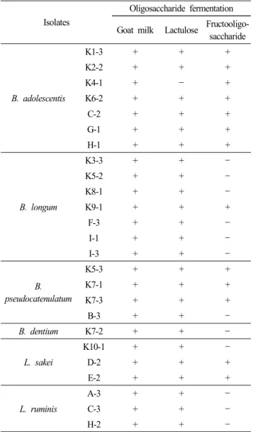
5. Bifidobacteria 와 lactobacilli의 올리고당 발효능력 분변에서 분리한 bifidobacteria와 lactobacilli 균주를 이용하 여 산양유 올리고당, lactulose 및 fructooligosaccharide에 대한 발효 능력을 조사하였다(Table 3). 산양유 올리고당과 lactulose는 모든 bifidobacteria와 lactobacilli가 발효하였으나, fructooligo- saccharide는 B. adolescentis 7 균주 모두와 B. pseudocatenulatum 3 균주, B. longum 1 균주 및 L. sakei 2 균주만이 발효한 것으 로 나타났다.
Perrin 등(2001)에 의하면 probiotics 균주들은 단당류를 섭 취하기 전에 세포에 결합된 glycosidase을 이용하여 올리고당을 미리 가수분해하는 능력을 갖고 있으며, 특히 fructofuranosidase 을 갖고 있는 경우 fructooligosaccharide을 분해할 수 있다(Imamura et al., 1994). Bifidobacteria 는 올리고당을 세포 내로 통과시킨 후 세포안에서 발효하는 방법과 외부에서 올리고당을 분해 한 후 단당류를 섭취하여 이용하는 방법을 이용하는 전략을 가지고 있다고 한다(Perrin et al., 2001). Bezkorovainy(1989) 에 의하면 같은 bifidobacreria 종 내에서도 효소를 발현하지 않고, 속간 내에서도 탄수화물에 대한 선호도의 차이가 있다 고 보고하고 있다. 본 연구에서도 B. longum의 경우 분리된 7 개 균 중 1개만이 fructooligosaccharide을 분해한 결과를 보 면, 같은 균주 내에서도 fructofuranosidase 효소가 발현하지 않은 것으로 사료된다.
Kaplan과 Hutkin(2000)에 의하면 락토바실러스에 의한 프 락토올리고당의 대사의 경우 L. plantarum과 L. hamnosus는 단지 삼당류와 사당류만을 대사하며 오당류는 대사하지 못
Table 3. Oligosaccharide fermentation patterns of bifidobacteria and lactobacilli isolated from feces of the women supplied with goat milk
Isolates
Oligosaccharide fermentation Goat milk Lactulose Fructooligo-
saccharide
B. adolescentis
K1-3 + + +
K2-2 + + +
K4-1 + - +
K6-2 + + +
C-2 + + +
G-1 + + +
H-1 + + +
B. longum
K3-3 + + -
K5-2 + + -
K8-1 + + -
K9-1 + + +
F-3 + + -
I-1 + + -
I-3 + + -
B.
pseudocatenulatum
K5-3 + + +
K7-1 + + +
K7-3 + + +
B-3 + + -
B. dentium K7-2 + + -
L. sakei
K10-1 + + -
D-2 + + +
E-2 + + +
L. ruminis
A-3 + + -
C-3 + + -
H-2 + + -
하고, 단지 삼당류와 사당류을 위한 특별한 전달시스템을 가 지고 있다고 제안했다. 본 연구에서 L. sakei의 경우, 분리된 3균주 중 2개만이 fructooligosaccharide을 분해한 결과는 종 내에서도 왜 이런 차이를 내는지를 더 연구할 필요가 있다고 생각된다.
요 약
산양유을 섭취한 성인의 분변 내 bifidobacteria의 증식 효 과 및 분변에서 분리된 bifidobacteria와 lactobacilli의 당 발효 능력을 조사하였다.
1. 산양유를 섭취한 처리군이 대조군보다 bifidobacteria 세
균수가 상대적으로 높았으며, 8주에서 95% 신뢰한계에서 유
의성이 있었다.
2. 분변에서 분리한 균주들을 13 균주의 16S rRNA 염기서 열로 동정한 결과, 산양유를 섭취한 처리구는 B. adolescentis, B. longum, B. pseudocatenulatum, B. dentium, L. sakei 순으로 분리되었다. 대조군의 12 균주는 B. adolescentis, B. longum, L.
ruminis, L. sakei, B. pseudocatenulatum 순으로 분리되었다.
3. 산양유 올리고당과 lactulose는 모든 bifidobacteria 및 lacto- bacilli 균주가 발효하였다. Fructooligosaccaride는 B. adolescentis 7 균주 모두 발효하였다. 조사한 B. pseudocatenulatum 4 균 주, L. sakei 3 균주, B. longum 7 균주 중에서 각각 3, 2, 1 균 주가 fructooligosaccharide를 발효하였다.
참고문헌