EMG Activities of Core Muscles During Bridging Exercises With and Without a Pilates Resistive Device
Su-jin Kim, B.H.Sc., P.T.
Won-gyu Yoo, M.Sc., P.T.
Min-hee Kim, M.Sc., P.T.
Dept. of Rehabilitation Therapy, The Graduate School, Yonsei University
Chung-hwi Yi, Ph.D., P.T.
Dept. of Physical Therapy, College of Health Science, Yonsei University Institute of Health Science, Yonsei University
Abstract 1)
The purposes of this study were to compare core muscle activities with and without the use of Pilates resistive equipment during bridging exercises and to investigate the efficacy of a Pilates device. Fourteen healthy individuals (6 males, 8 females) between 20 to 26 years of age were examined. They were en- gaged in a bridging exercise with and without a magic circle. Three consecutive repetitions of each ex- ercise were performed. Surface electromyography (sEMG) was used to measure the electrical activities of the right side internal oblique, the adductor longus, the multifidus, and the gluteus maximus muscles.
Normalized EMG activities were compared using a paired t-test and the level of significance was set at
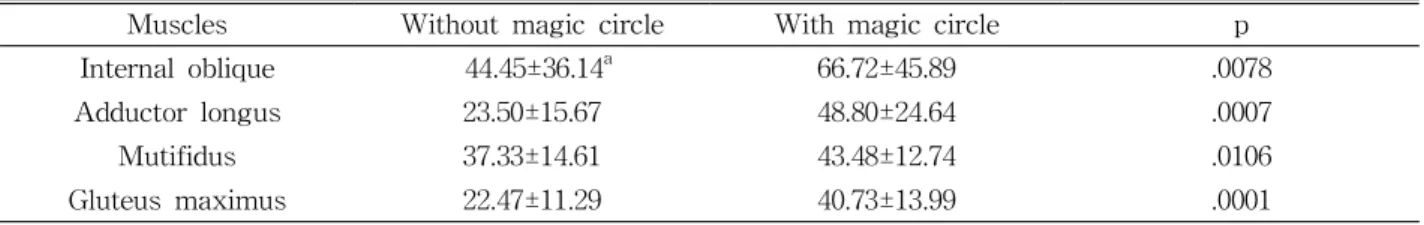
=.05. The results showed that the EMG activities of the internal oblique (p=.0078), the adductor longus (p=.0007), and the gluteus maximus (p=.0001) muscles were significantly higher when using the magic circle during the Pilates bridging exercise. Also, statistically significant change existed in the multifidus muscle (p=.0106). The bridging exercise, combined with hip adduction using the magic circle, may en- hance core stabilization. Therefore, using a magic circle during hip adduction combined with bridging ex- ercise may be recommended usefully for individuals wanting to strength the core muscles. Further re- search is needed to access the nature of motor control of the Pilates mat exercises and to deliver ex- ercise intervention for lower back pain patients.
Key Words: Core muscles; Electromyogram; Lumbar stabilization; Pilates exercise; Resistive device.
Introduction
Along with the development of technology, the numbers of people suffering from lower back pain have increased due to sedentary indoor living and a general failure to exercise (Gallagher and kryza- nowska, 2000; O'Sullivan, 2000). Instability of the spine over the normal range is reported when it oc- curs in lower back pain from many reasons (Hodges and Richardson, 1996). Panjabi (1992) theorized re-
garding spinal instability in terms of laxity around the neutral position of a spinal segment developed by significant decreasing in the capacity of the stabiliz- ing system. This system consists of three systems interacting to maintain the spine in a neutral position. The passive system contains the vertebra and intervertebral discs and ligaments, the active system involves muscles and tendon surrounding the spine, and the neural system includes the nerves and central nervous system. The active system is the
Corresponding author: Chung-hwi Yi [email protected]
This research was supported by Regional Research Center Program which was conducted by the Ministry of
Commerce, Industry and Energy of the Korean Government.
Variable Mean±SD Range
Age (yrs) 23.50±1.65 20∼25
Height (㎝) 166.94±5.03 160∼180 Weight (㎏) 57.86±10.17 45∼80 Table 1. Characteristics of the subjects (N=14) only system capable of voluntary movement so lum-
bar stability under deficit of these stabilizing sys- temsis maintained in vivo by reinforcing the activity of the muscles around the lumbar segment (Cholewicki and McGill, 1996). To protect the spine from trauma, stabilization exercises devised to im- prove function of the trunk muscles activity are en- couraged and are often used in clinical practice (Cholewici and VanVliet, 2002).
The Pilates system of body conditioning contains over 500 stretching and strengthening exercises and these exercises may be classified into mat and appa- ratus exercises. The method provides core strength- ening in both the direct and indirect ways people move their body parts by using the core muscles (Muscolino and Cipriani, 2004; Lange et al, 2000).
Siler (2000) insisted that the areas of the body that are in motion during exercises are the areas in which the mind should be focused. This concept is important in that core muscles have an opportunity to focus on the spinal stability when other areas of the body are not in motion.
In the Pilates mat exercises, exercise has been tried alone and in combination with equipment such as gymball, foam roll, thera-band, and magic circle.
These are often used to enhance the efficiency of the exercises by giving resistance or a modification.
Petrofsky et al (2005) reported muscle activities using the thera-band were higher in efficiency than muscle activities without the thera-band. Also, muscle use was gauged during exercise using conventional weight lifting and was compared to exercise during Pilates with resistive devices. However, there is not any research about measuring activities of the core muscles or the effects of exercises made in compar- ison with and without magic circle during the Pilates mat exercise. Although Pilates is increasingly popular and demand for this exercise is increasing in clinical practice, there has been little research on the effec- tiveness of Pilates exercise and related devices (Herrington, 2005). Therefore, the aims of these study
were to compare activities of core muscles with and without a resistive device during Pilates mat exercise and to examine the utility of resistive devices.
Methods Subjects
Fourteen health young subjects (6 male, 8 female) were recruited from university students. The ex- clusion criteria required that subjects not have past or present neurologic musculoskeletal, cardiopulmo- nary disease and not have pain in the lumbar spine region during the mat exercises. All the subjects provided informed consent after being explained the purpose and method of the study. The subjects had a mean age 23.50±1.65 years, a mean height of 166.94±5.03 ㎝, a mean weight of 57.86±10.17 ㎏. The age, height, and weight of the subjects are summar- ized in Table 1.
Instruments
Surface Electromyographic Recording
2)We collected and amplified electromyographic data
using a Noraxon TeleMyo 2400T
1)and analyzed the
data using MyoResearch Master Edition 1.06 XP
software (Noraxon Inc., Scottsdale, AZ, U.S.A.). The
skin was prepared by shaving hair and rubbing the
skin with an alcohol-water solution to decrease
impedance. Surface electrode pairs were placed in a
bipolar configuration over the fourmuscle sites and
distance between two electrodes were 2 ㎝. The four
sites on the right side were as follows: 1) the in-
ternal oblique abdominal (IO) muscle, approximately
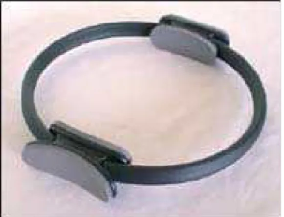
Figure 1. Pilates magic circle.
2 ㎝ medial and inferior to the right anterior superior iliac spine, 2) adductor longus (AL) muscle placed on the medial aspect of the thigh in an oblique di- rection 4 ㎝ from the pubis, 3) multifidus (MF) muscle located in 2 ㎝ part from the L5 vertebra, 4) the gluteus maximus (GM) muscles placed half the distance between the greater trochanter and the sa- cral vertebrae in the middle of the muscle on an obelize angle at the level of the trochanter or slightly above (Cram et al, 1998; Marshall and Murphy, 2003). The raw signal was full wave rectified and filtered using a Lancosh FIR digital filter. The band- pass between 80 ㎐ and 250 ㎐ were used for pre-amplitude and converted to digital date at a sampling rate of 1000 ㎐. The EMG data was proc- essed into the root mean square (RMS) and RMS values from windows of 300 ㎳ data points. For a normalized state, the peak RMS values among the three trials of maximal voluntary contraction (MVC) were calculated for each muscle.
Magic Circle
The resistive device used during Pilates was a round-shaped ring 14 inches in diameter. It consisted of soft rubber material to which handles were at- tached on the inside and the outside of the ring to support or to give resistance on the body part such as thigh and chest (Figure 1).
Procedures
Before starting the study exercises, subjects per- formed four different contractions to obtain a max- imal activity for normalization purposes. Each con- traction was repeated three times, and subjects were given a one-minute rest to prepare for the real study. Two series of experiments were performed. In the first, bridging exercises of the Pilates method were accomplished on the mat. The subjects were taught to perform the bridging exercise by practicing them under the guidance of a Pilates instructor.
Subjects lay supine on an exercise mat, knees flexed and feet flat, hip flexed to 70 as measured by a standard goniometer. The positions were aligned as a tuberosity of ischium, posterior part of the heel and second metatarsal head were placed in the lineal line.
Arms were rested at the sides and buttocks lifted slowly while vertebra moved each segment sepa- rately until the hips were fully extended and the po- sition was held for five seconds. The EMG signal was recorded during these five seconds. A verbal cue was given to correct the posture. In the second experiment, the same subjects performed the same bridging exercise with a magic circle known for re- sistive device in the Pilates. The magic circle was placed in the inner sites of both thighs and was available for bridging exercises with hip adduction.
All of the exercises had three consecutive repetitions, and a short rest of generally one minute was given between each exercise to prevent muscle fatigue.
Statistical Analysis
All data were expressed as the mean and standard
deviation. Paired t-test was used to test for differ-
ences in EMG activities of subjects with and without
the magic circle. The analysis of data was performed
using SPSS version 12.0 program and significant
level set at a=.05.
Muscles Without magic circle With magic circle p
Internal oblique 44.45±36.14
a66.72±45.89 .0078
Adductor longus 23.50±15.67 48.80±24.64 .0007
Mutifidus 37.33±14.61 43.48±12.74 .0106
Gluteus maximus 22.47±11.29 40.73±13.99 .0001
a